Red Eye Flashcards
What are causes of subconjunctival haemorrhage?
- Trauma
- Spontaneous
- Haemorrhagic disorders
- Valsalva pressure spikes
What investigation might you consider doing in somoene with a subconjunctival haemorrhage?
BP check
What is episcleritis?
A benign, self-limiting inflammatory disease affecting part of the eye called the episclera. The episclera is a thin layer of tissue that lies between the conjunctiva and the connective tissue layer that forms the white of the eye (sclera)

What causes episcleritis?
No cause in 70%, but can be:
- Rheumatic fever
- SLE
What are features of episcleritis?
- Pain
- Discomfort
- Sectoral redness

How would you manage someone with episcleritis?
- Systemic/topical NSAIDs
- Topical Steroids
- Lubricants
What is scleritis?
“Vasculitis of the Sclera”
A serious inflammatory disease that affects the white outer coating of the eye, known as the sclera

What are causes of scleritis?
- Autoimmune conditions (Wegener’s, polyangitis)
- Infections
What are symptoms of scleritis?
- Severe Pain
- Redness
- Photophobia
- Decreased vision
What are signs of scleritis?
- Generalised inflammation
- Conjunctival oedema
- Scleral thinning
- Decreased visual acuity
What tests might you consider doing in someone presenting with scleritis?
Check for autoimmune disorder
- ESR
- ANCA
How would you manage someone with scleritis?
Refer to a specialist
- Oral steroids
- Immunosuppression
What is the following?

Corneal foreign body
How would you manage a corneal foreign body?
- Remove foreign body under magnification - Cotton bud or needle
- Remove rust ring
- Treat corneal abrasion
What is the following?

Rust ring
What is a corneal abrasion?
Breach in the epithelium of the eye - occurs without keratitis
What is keratitis?
Inflammation of the cornea - marked by white area on the cornea, indicating a collection of white cells on the corneal tissue

What is important to do when examining someones eyes for a corneal foreign body?
Invert the upper lid to look for additional FBs
What clinical features would point towards a corneal abrasion?
- Pain
- Watering
- Photophobia
- Conjunctival injection
- Swollen lids
How would you investigate for a corneal abrasion?
Stain with flourescin and bright blue light (shone tangentially across the globe)
How would you manage a corneal abrasion?
- Look for conjunctival foreign bodies +Evert eye lid
- Topical antibiotics - chloramphenicol
- Cycloplegics
- Pressure pad and patch
When would you not use a pressure pad and patch in someone with a corneal abrasion?
If there is suspected infection
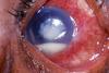
What is a corneal ulcer?
Also known as ulcerative keratitis - an inflammatory, or more seriously, infective condition of the cornea involving disruption of its epithelial layer with involvement of the corneal stroma.
Image - corneal ulcer with hypopyon
What organisms cause of corneal ulcers?
- Bacteria
- Herpes viruses
- Fungi - candidia, aspergillus
- Acanthoemeba
- Vasculitis - RA,
How would you manage a corneal ulcer?
- Bacterial - do not patch
- Topical steroids
- Refer to opthalmologist
What are symptoms of a corneal ulcer?
- Pain
- Redness
- Photophobia
- Watery
- Discharge
What are features of viral keratitis?
- Discomfort
- Foreign Body sensation
- Watering
- Photophobia
What is viral keratitis most commonly caused by?
Herpes simplex/zoster
What are signs of viral keratitis?
- Epithelial dendrites
- Stromal keratitis
- Central ulceration

How would you manage viral keratitis?
- Aciclovir (3%) 5x per day, for 3 weeks
- Topical cycloplegics
- May need topical steroids
- Refer to opthalmologist
What is uveitis?
Inflammation of the uvea, the pigmented layer that lies between the inner retina and the outer fibrous layer composed of the sclera and cornea. The uvea consists of the middle layer of pigmented vascular structures of the eye and includes the iris, ciliary body, and choroid.

What are the different types of uveitis?
- Anterior uveitis
- Intermediate uveitis
- Posterior uveitis
What are symptoms of anterior uveitis?
- Pain
- Blurred vision
- Photophobia
- Redness
- Watering
What conditions is uveitis associated with?
- Positive HLA-B27 - Ankylosing spondylitis
- Arthritis
- Inflammatory bowel disease
- Sarcoid
- Tuberculosis
- Syphilis
- Toxoplasmosis
- Behçet’s syndrome
- Lymphoma
- Viruses - herpes, CMV and HIV infection
What are signs of anterior uveitis?
- Circumcorneal redness - ciliary flush
- Keratic precipitates on corneal epithelium
- Cells/flare in anterior chamber
- Miosis - due to sphincter spasm
- Hypopyon
- Posterior/Peripheral anterior Synechaie/Festooned pupil
- Iris atrophy
- Fibrinous membrane in the pupillary

How would you manage anterior uveitis?
- Topical steroids
- Cycloplegics
- Topical anti-glaucoma meds - if raised IOP
- Steroid ointment - for night
What are cycloplegics?
Used to paralys the ciliary muscle of the eye, resulting in a loss of accommodation. Because of the paralysis of the ciliary muscle, the curvature of the lens can no longer be adjusted to focus on nearby objects.
Often used to prevent synechiae, and reduce pain from ciliary muscle spasm
Which type of uveitis is photophobia and pain most commonly associated with?
Anterior uveitis
What is the pathophysiology of acute closure glaucoma?
Aqueous humour produced by ciliary bodies enters the anterior chamber via the pupil. This drains via the trabecular network at the angle to enter Schlemm’s Canal.
In acute angle closure glaucoma, the iris obstructs the trabecular meshwork, obstructing the drainage of aqueous humour.

What are signs of acute closure glaucoma?
- Hard Eye
- Vision - counts finger -> hand motion visible
- Circumcorneal congestion
- Corneal oedema
- Shallow anterior chamber
- Semidilated pupil
- IOP - 40-70 mmHg

Describe the following features of Anertior Uveitis:
- Pain
- Redness
- Discharge
- Vision
- Photophobia
- Pupil
- Pain - mild/moderate
- Redness - pericorneal/none
- Discharge - minimal/none
- Vision - blurred
- Photophobia - ++
- Pupil - Constricted

Describe the following features of bacterial conjunctivitis:
- Pain
- Redness
- Discharge
- Vision
- Photophobia
- Pain - itching/gritty
- Redness - peripheral/diffuse
- Discharge - yellow
- Vision - normal
- Photophobia - +

Describe the following features of viral conjunctivitis:
- Pain
- Redness
- Discharge
- Vision
- Photophobia
- Pain - itching/gritty
- Redness - peripheral/diffuse
- Discharge - watery
- Vision - normal
- Photophobia - +

Describe the following features of scleritis:
- Pain
- Redness
- Discharge
- Vision
- Pain - severe/boring
- Redness - sectoral/diffuse
- Discharge - none
- Vision - normal

What is the following?

Bacterial conjunctivitis - purulent discharge
What is the following?

Conjunctivits - Most likely viral as no purulent discharge
What are signs of bacterial conjunctivitis?
- Mucopurulent discharge
- Normal vision - after discharge has been blinked clear
- Uniform engorgement of all the conjunctival blood vessels

What are signs of viral conjunctivitis?
Both eyes
- Red with diffuse conjunctival injection
- Clear diacharge
- Small white lymphoid conjunctival aggregations
- May be head and neck lymphadenopathy

What are signs of allergic conjunctivitis?
- Diffusely injected conjunctivae
- Chemosis
- Clear and stringy dsicharge
- Papillae - round oedoematous swelling in the eyelid caused by fibrous septa that tether the eyelid

What is the diagnosis?

Viral conjunctivitis
What is the diagnosis?

Bacterial conjunctivitis
What is the diagnosis?

Corneal ulcer with hypopyon
What is the diagnosis?

Episcleritis
What is the diagnosis?

Scleritis
What is the diagnosis?

Acute angle glaucoma
What is the diagnosis?

Anterior uveitis
What is the diagnosis?

Orbital Cellulitis