Images Flashcards

- This is a barium swallow*
- Demonstrating*:
- food material within the upper thoracic oesophagus which is somewhat dilated.
- Distal to this there is a 5 cm irregular narrowing of the mid thoracic oesophagus with proximal shouldering, giving a classical apple core configuration
- Beyond this there is a short segment of non-strictured oesophagus
- before there is another 5 cm segment of smooth luminal narrowing extending to the gastro-oesophageal junction.
My primary differential would be: oesophageal carcinoma in the mid thoracic oesophagus
Ix: Endoscopy and Bx
AND
A smooth stricture of the lower oesophagus, potentially of peptic origin.
Ix: response to PPIs, may consider pH monitoring
^such pathologies result from long standing GORD.
Other Differentials include other forms of strictures.
Due to the distal narrowing I may also consider achalasia

Oesophageal stricture: benign
This is a barium swallow
Demonstrating: a tight smooth stricture in the mid thoracic oesophagus with proximal dilation and non-strictures oesophagus distally.
Primary differential would be: a benign strictures, most likely peptic
Benign lesions: smoother borders with little irregularity, although some larger lesions may ulcerate as they outgrow their vascular supply.
Other differentials of an oesophageal stricture, resulting in a filling defect include obstruction that may be either luminal, mural or extramural.
- Luminal
- FB
- Large food bolus
- Mural
- Benign stricture
- Web (e.g. Plummer-Vinson)
- Oesophagitis
- Trauma (e.g. OGD)
- Malignant stricture
- Pharynx, oesophagus, gastric
- Pharyngeal pouch
- Benign stricture
- Extra-Mural
- Retrosternal goitre
- Rolling hiatus hernia
- Lung Ca
- Mediastinal LNs (e.g. lymphoma)
- Thoracic aortic aneurysm

Oesophageal Cancer
Image
- Irregular, shouldered stricture of the oesophagus
- An “apple-core” lesion
- What type of Ca?*
- Epidemiology?*
- Pathophysiology*
- Major RF*
- Presentation*
- Ix*
- Management*

Where?
Distal third: adenocarcinoma (commoner)
Proximal third: SCC
Epidemiology
- M>F = 5:1
- ↑ in Transkei and China
Pathophysiology
- 65% adenocarcinoma
- Lower 3rd
- GORD → Barrett’s → dysplasia → Ca
- 35% SCC
- Upper and middle 3rds
- Assoc. c¯ EtOH and smoking
- Commonest type worldwide
- Spread
- Local → LNs → blood
Major Risk Factors
- GORD → Barrett’s
- EtOH and smoking
- Achalasia (SCC)
- Plummer-Vinson: 20% risk of SCC
Presentation
- Progressive dysphagia
- Wt. loss
- Upper 3rd: hoarseness + bovine cough
Ix
- Dx: OGD + biopsy
- Staging: CT, EUS, laparoscopy, mediastinoscopy
Mx
- MDT
- Oesophagectomy: 25% have resectable tumours
- Ivor-Lewis 2 stage
- McKeown 3 stage
- 15% 5 year survival
- Palliation: 75% of pts.
- Analgesia
- Laser coagulation
- Stenting
- 5% 5 year survival

This is a barium swallow
Demonstrating:
food material within the upper thoracic oesophagus which is somewhat dilated.
Proximal dilatation of the oesophagus w food particles visible and smooth distal tapering – giving the bird’s beak appearance.
(Significant negative) I note that there are no apple-core strictures seen to suggest the presence of oesophageal SCC. (~3% of pts w achalasia)
My primary differential would therefore be: achalasia.

Achalasia
- Proximal dilatation of the oesophagus c¯ smooth distal tapering – the bird’s beak appearance.
- May be food particles visible.
- Significant negative: apple-core stricture suggestive of oesophageal SCC.
- Occurs in ~3% of pts c¯ achalasia
- Define*
- Cases*
- Presentation*
- Ix*
- Mx*

Definition: Focal motility disorder of the oesophagus caused by degeneration of the myenteric plexus of Auerbach
Causes
- Usually idiopathic
- May be 2O to Chagas disease
Presentation
- Dysphagia to liquids then solids
- Retrosternal cramps
- Regurgitation: esp. @ night ± aspiration pneumonia
- Wt. loss
Ix
- Ba swallow
- CXR: wide mediastinum + double RH boarder
- Manometry: failure of relaxation + ↓peristalsis
- OGD: exclude oesophageal SCC
Mx
- Med: CCB, nitrates
- Int: botox injection, endoscopic balloon dilatation
- Surg: Heller’s cardiomyotomy (open or lap)

- Present the image*
- Pathophysiology*
- Presentation*
- Management*

Image
- This is a barium swallow
- Demonstrating… Contrast filling of blind-ended pouch adjacent to and in
- continuity with the oesophagus.
- This is consistent with… a pharyngeal pouch, also known as Zenker’s diverticumun
Pathophysiology
- Out-pouching between crico- and thyro-pharyngeal components of the inf. pharyngeal constrictor.
- Pulsion diverticulum
- Area of weakness = Killian’s dehiscence
Presentation
- Dysphagia
- Swelling on left side of neck
- Regurgitation
- Halitosis
Mx
- Excision
- Dohlman’s Procedure: endoscopic stapling

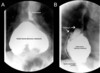
This is a Barium Swallow
Demonstrating... a dilated pouch (the hernia) below a smooth, short,
symmetric, ring-like constriction (the GOJ) - Schatzki ring.
May be assoc. c¯ smooth area of concentric narrowing in the distal oesophagus: reflux-stricture.
I note (Significant negative) that there are no apple-core strictures seen

Hiatus Hernia
- Classification*
- Presentation*
- Ix*
- Mx*
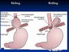
Classification
- Sliding (80%)
- Gastro-oesophageal junction slides up into chest
- Often assoc. c¯ GORD
- Rolling (15%)
- Gastro-oesophageal junction remains in abdomen but a bulge of stomach rolls into chest alongside the oesophagus
- LOS remains intact so GORD uncommon
- Can → strangulation
- Mixed (5%)
Presentation
- Often asymptomatic
- Sliding hernias → GORD: dyspepsia
- Rolling hernias → strangulation
Ix
- CXR: gas bubble and fluid level in chest
- Ba swallow: diagnostic
- OGD: assess for oesophagitis
- 24h pH + manometry
- Exclude dysmotility or achalasia
- Confirm reflux
Mx
- Conservative: ↓wt., raise head of bed, stop smoking
- Medical: PPIs, H2RAs
- Surgical
- If intractable symptoms despite medical Rx.
- Should repair rolling hernia (even if asympto) as it may strangulate.


Barrium Swallow
What is it?
Indications
Contraindications
- Barium swallow is a dedicated test of the pharynx, oesophagus, and proximal stomach
- It may be performed as a single or double contrast study.
- It is used to evaluate the upper GI pathway from the lips to the gastric fundus.
- Upper GI endoscopy has largely replaced the barium swallow for the assessment of peptic ulcer disease and the evaluation of haematemesis.
Indications:
- high or low dysphagia
- GORD
- hiatus hernia (assessment)
- generalised epigastric pain
- globus hystericus
- persistent vomiting
- assessment of fistula
- inability to pass the endoscope during UGIE
Contraindications:
Patients at risk of aspiration
Water-soluble contrast agents should be used instead of barium in the following cases:
- suspected perforation
- post-operative assessment for leak
Image

Image
This is an abdonminal x-ray
Demonstrating: a curvilinear calcification extending in parralel with the lumbar spine, representing calcification of the wall of the abdominal aorta. It is double the dimension of the vertebral bodies at its maximal diameter.
Thus my primary differential is a calcified fusiform abdominal aortic aneurysm.
Defined as an Abnormal dilatation of the abdominal aorta to >50% of its normal diameter = ≥3cm

Calcified Abdominal Aortic Aneurysm
Define
Risk Factors
Ix
Mx
Complications

Definition: Abnormal dilatation of the abdominal aorta to >50% of its normal diameter = ≥3cm
Risk Factors
- Male
- Age >60yrs (prevalence: ~5%)
- Smoking
- HTN
- FHx
Ix
- Abdo US: used for surveillance and screening
- CT/MRI: Ix of choice
- Angio: useful to delineate relationship of renal arteries
Management: When to Operate
- Repair aims to avoid complications
- Operate when risk of complications, esp. rupture, > risk of surgery.
Indications
- Symptomatic aneurysms
- Asymptomatic
- ≥5.5cm
- Expanding >1cm/yr
Complications
- Death
- MI
- Renal failure
- Spinal or mesenteric ischaemia (Beck’s syndrome)
- Distal trash from thromboembolism
- Anastomotic leak
- Graft infection
- Aortoenteric fistula
Operative Mortality
- Open
- Emergency: 50%
- But only 50% reach hospital alive
- Elective: 5% (lower in specialist centres)
- ↑ if IHD, LVF, CRF, COPD
- Emergency: 50%
- EVAR: 1%
EVAR
- ↓ perioperative mortality (1% vs. 5%)
- No mortality benefit after 5yrs
- Significant late complications: e.g. endoleaks
- EVAR not better cf. medical care in unfit pts.
General Aneurysms:
Definition
Classification (3)
Definition: Abnormal dilatation of a blood vessel > 50% of its normal diameter.
Classification
- True Aneurysm
- Dilatation of a blood vessel involving all layers of the wall and is >50% of its normal diameter
- Two different morphologies
- Fusiform: e.g AAA
- Saccular: e.g Berry aneurysm
- False Aneurysm
- Collection of blood around a vessel wall that communicates c¯ the vessel lumen.
- Usually iatrogenic: puncture, cannulation
- Dissection
- Vessel dilatation caused by blood splaying apart the media to form a channel w/i the vessel wall.
- Used to be classified as “dissecting aneurysms” but not technically correct as represents a different pathology.

This is an arteriogram
Demonstrating bilateral superficial femoral artery occlusion
…and collatrals filling in the popliteals
This is thus indicative of a chronic progressive narrowing of the femoral artery consistent with chronic peripheral vascular disease

Angiogram showing: narrowing of the left SFA with complete occlusion more distally, collaterals filling the distal segment
Remind me of the branches of the femoral artery


Image
Pathophysiology
Presentation
Management

Image
- This is an abdominal radiograph
- demonstrating a sigmoid volulus emerging from the LIF c.20cm at it’s largest diameter
- ^coffee bean sign (caecal volvulus-embryo sign)
Pathophysiology
- Long mesentery c¯ short base predisposes to torsion
- Vascular supply may be compromised → strangulation
- ↑ risk in psychogeriatric pts.: disease and Rx
- Typically a long Hx of constipation
Presentation
- Often elderly pts. c¯ comorbidities
- Grossly distended, tympanic abdomen
Mx
- Resuscitate: drip and suck
- Sigmoid
- Detorse c¯ flatus tube
- May need sigmoid colectomy
- Often recurs
- Caecal
- ~ 10% can be detorsed by colonoscopy:
- therefore often needs surgery
- Caecostomy
- Right hemi c¯ primary ileocolic anastomosis
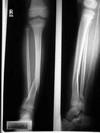
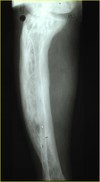
Tibia and fibula fractures
(lateral and AP radiograph of the leg of Mr XX, XXyo, taken on XX date at XX time)
Pattern: complete, spiral
Location: extra-articular fracture of the distal diaphysis of both the tibia and the fibula
(Pieces: simple, 2 fragments)
displacement/angulation: valgus and mild dorsal angulation of the distal fragment
Other: Immature skeleton? of note this is a skeletally immature leg, as suggested by the incomplete fusion of the epiphysial plates
ABC+ resus
unstable fracture , treated best with flexible nails.

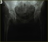
Intracapsular hip fracture
Radiograph of the pelvis showing an intracapsular subcapital fracture of the left neck of femur
-Undisplaced Fracture:
—-any age
——–internal fixation with screws (hemiarthroplasty if unfit)
-Displaced Fracture: (25-30% risk of AVN)
—- <55 years
——– internal fixation
—- >65 NILL comorbidities, independent, mobile
——– THR
—- >65 immobile/comorbidities
——– Hemiarthroplasty

Extracapsular hip fracture: minimal risk of AVN
Extracapsular Neck of Femur #
-normal
——–dynamic hip screw
-reverse oblique, transverse or subtrochanteric
——–intramedullary device

Colles: Low energy, dorsally displaced, extra-articular fx
(lateral and AP radiograph of the distal forearm of Mr XX, XXyo, taken on XX date at XX time)
Pattern: showind a complete, transverse
Location: extra-articular fracture of the distal diaphysis of the radius
(Pieces: simple, 2 fragments)
displacement/angulation: with dorsal displacement and angulation of the distal fracture
- and sparing of the ulnar bone.*
- this is consistent with a colles fracture*
ABC Resus (&NV examination) + reduce + hold (commonly closed reduction and cast imobilisation in extra-articular #) + Rehabilitation (with home exercises)

Total hip arthroplasty

Dynamic Hip Screw

AC joint dislocation
C6 fracture


Osteomyelitis
- regional osteopaenia
- periosteal reaction/thickening (periostitis): variable, and may appear aggressive including formation of a Codman’s triangle
- focal bony lysis or cortical loss
- endosteal scalloping
- loss of bony trabecular architecture
- new bone apposition
- eventual peripheral sclerosis
https://radiopaedia.org/articles/osteomyelitis


x-ray: early changes include widening of joint space, later changes include decreased femoral head size/flattening


- Complete fracture: Both sides of cortex are breached
- Toddlers fracture: Oblique tibial fracture in infants
- Plastic deformity: Stress on bone resulting in deformity without cortical disruption
- Greenstick fracture: Unilateral cortical breach only
- Buckle fracture: Incomplete cortical disruption resulting in periosteal haematoma only


Colles: Low energy, dorsally displaced, extra-articular fx
(lateral radiograph of the distal forearm of Mr XX, XXyo, taken on XX date at XX time)
Pattern: showing a complete, transverse
Location: extra-articular fracture of the distal diaphysis of the radius
(Pieces: simple, 2 fragments)
displacement/angulation: with dorsal displacement and angulation of the distal fracture
and sparing of the ulnar bone.
this is consistent with a colles fracture
ABC Resus (&NV examination) + reduce + hold (commonly closed reduction and cast imobilisation in extra-articular #) + Rehabilitation (with home exercises

Tibia and fibula fractures
(lateral and AP radiograph of the leg of Mr XX, XXyo, taken on XX date at XX time)
Pattern: complete, spiral
Location: extra-articular fracture of the distal diaphysis of both the tibia and the fibula
(Pieces: simple, 2 fragments)
displacement/angulation: valgus and mild dorsal angulation of the distal fragment
Other: Immature skeleton? of note this is a skeletally immature leg, as suggested by the incomplete fusion of the epiphysial plates
ABC+ resus
unstable fracture , treated best with flexible nails.

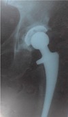
Radiograph of the pelvis demonstrating
a Hemi arthroplasty of the left hip

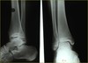
isolated lateral malleolus fracture
(lateral and AP radiograph of the distal leg of Mr XX, XXyo, taken on XX date at XX time)
Pattern: complete, spiral
Location: intra-articular fracture of the distal diaphysis of both the fibula
(Pieces: simple, 2 fragments)
displacement/angulation: without displacement or angultion of the distal fragment
Other: Immature skeleton? of note this is a skeletally immature leg, as suggested by the incomplete fusion of the epiphysial plates
and I would therefore like to rule out a salter harris # in this circumstance
ABC+ resus (NVs) + Open reduction and internal fixation if >3mm displacement/talar shift due to the importance of accurate redction in the ankle to maintaom the ankle mortise + rehab


- part of initial ATLS evaluation
- Mechanism typically high energy blunt trauma
- Mortality rate 15-25% for closed fractures, as much as 50% for open fractures
- hemorrhage is leading cause of death overall (commonly venous injury)
- Anatomy
- ring structure made up of the sacrum and two innominate bones
- stability dependent on strong surrounding ligamentous structures
- displacement can only occur with disruption of the ring in two places
- neurovascular structures intimately associated with posterior pelvic ligaments
- high index of suspicion for injury of internal iliac vessels or lumbosacral plexus
Classified according to stability e.g. Tile classification or young burgess
- Management
- Resuscitation PRBC:FFP
- non-surgical External Fixation: Pelvic binder/sheet: for stability
- later open reduction internal fixation may be considered


May be linear, drepressed or diastatic (transvers 1 or more sutures, therefore widen the suture), basilar, compound (laceration or dermis and meninges)
associated with: bone fragments under the #. other penetrating injuries, intracranial H’gge

Total hip arthroplasty:
Traumatic: NOF#
Degenerative: OA/RA
Vascilar: osteonecrosis/AVN
Infection: sever septic arthritis?


(AP radiograph of the left GHJ of Mr XX, XXyo, taken on XX date at XX time)
Pattern: complete, spiral
Location: extra-articular fracture of the proximal humerous across the surgical neck
(Pieces: simple, 2 fragments)
displacement/angulation: ?secondary view required to fully assess displacement
- mechanism
- low-energy falls
- elderly with osteoporotic bone
- high-energy trauma
- young individuals
- concomitant soft tissue and neurovascular injuries
- low-energy falls
- nerve injury
- axillary nerve palsy most common
- fracture-dislocations more commonly associated with nerve injuries
- Request 3 view complete trauma series
- Management: 85% of proximal humerus fractures are minimally displaced and can be treated nonoperatively
- sling imobilisation + progressive rehab
- if stable then early ROM (range of motion)
- if unstable then period of immobilization followed by ROM once moves as a unit
- If anatomical neck: ORIF in young + potential hemiarthroplasty in elderly
- sling imobilisation + progressive rehab
nb vascularity of articular segment is more likely to be preserved if ≥ 8mm of calcar is attached to articular segment…ie risk of vasc necrosis if articular segment is <8mm of calcar attached
ABC+ resus
unstable fracture , treated best with flexible nails.


- accounts for up to 15% of acute wrist injuries
- most common mechanism of injury is axial load across hyper-extended and radially deviated wrist
- transverse fracture patterns are considered more stable than vertical or oblique oriented fractures
- Risk
- Risk of SNAC (Scaphoid Nonunion Advanced Collapse) A condition characterized by advanced collapse and progressive arthritis of the wrist that results from a chronic scaphoid nonunion
- Risk of AVN varies with # location: proximal 5th AVN = 100% !
- major blood supply is dorsal carpal branch (branch of the radial artery)
- enters scaphoid in a nonarticular ridge on the dorsal surface and supplies proximal 80% of scaphoid via retrograde blood flow
- minor blood supply from superficial palmar arch
- Physical exam
- anatomic snuffbox tenderness dorsally
- scaphoid tubercle tenderness volarly
- pain with resisted pronation
- recommended views
- AP and lateral
- scaphoid view: 30 degree wrist extension, 20 degree ulnar deviation
- 45° pronation view
- if radiographs are negative and there is a high clinical suspicion should repeat radiographs in 14-21 days
- Management:
- thumb spica cast immobilisation
- stable nondisplaced fracture (majority of fractures)
- if patient has normal xrays but there is a high level of suspicion can immobilize in thumb spica and reevaluate in 12 to 21 days
- Unstable #
- ORIF or percutaneous screw fixation
- thumb spica cast immobilisation


Osteolytic bone metastases
Pb-Ktl i.e. lead kettle
Prostate (blastic/sclerotic)
Breast (mixed)
Kidney (lytic)
Thyroid (lytic)
Lung (lytic)


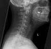
- All trauma patients have a cervical spine injury until proven otherwise
- High energy accident e.g. fall from great height
- Mechanism of accident e.g. hyperextension
- Condition at the scene e.g. degree of consciousness
- associated comorbidities
- Cervical spine clearance defined as confirming the absence of cervical spine injuryimportant to clear cervical spine and remove collar in an efficient manner
- Missed cervical spine injuries: may lead to permanent disability
- cervical clearance can be performed with
- physical exam
- remove immobilisation
- examine face and scalp for evidence of direct trauma
- inspect for angular or rotational deformities in the holding position of the patient’s head
- palpate posterior cervical spine looking for tenderness along the midline or paraspinal tissues
- log roll patient to inspect and palpate entire spinal axis
- perform careful neurologic exam
- physical exam
- Removal of cervical collar WITHOUT radiographic studies allowed if
- patient is awake, alert, and not intoxicated AND
- has no neck pain, tenderness, or neurologic deficits AND
- has no distracting injuries
- radiographical clearance
- Xray: multpole views e.g. AP, lateral odontoid view
- CT

Boxer’s #
base of the 5th metacarpal
Colles #
Colles’ fracture
- FOOSH
- Described as a dinner fork type deformity
- Classical Colles’ fractures have the following 3 features:
- Transverse fracture of the radius
- 1 inch proximal to the radio-carpal joint
- Dorsal displacement and angulation

Smith’s fracture (reverse Colles’ fracture)
Volar angulation of distal radius fragment (Garden spade deformity)
Caused by falling backwards onto the palm of an outstretched hand or falling with wrists flexed

Bennett’s fracture
- Intra-articular fracture of the first carpometacarpal joint
- Impact on flexed metacarpal, caused by fist fights
- X-ray: triangular fragment at ulnar base of metacarpal

Monteggia’s fracture
- Dislocation of the proximal radioulnar joint in association with a proxima/mid third ulna fracture
- Fall on outstretched hand with forced pronation
- Needs prompt diagnosis to avoid disability

Galeazzi fracture
- Radial shaft fracture with associated dislocation of the distal radioulnar joint
- Occur after a fall on the hand with a rotational force superimposed on it.
- On examination, there is bruising, swelling and tenderness over the lower end of the forearm.
- X Rays reveal the displaced fracture of the radius and a prominent ulnar head due to dislocation of the inferior radio-ulnar joint.

Barton’s fracture
- Distal radius fracture (Colles’/Smith’s) with associated radiocarpal dislocation
- Fall onto extended and pronated wrist