Cancers of the female reproductive tract and breast structure (7.1) Flashcards
Define the following terms:
Metaplasia Dysplasia
Neoplasia Cancer
Atypia Anaplasia
Carcinoma Carcinoma in situ
Intraepithelial neoplasm
Differentiation
• Metaplasia: Change in cell type e.g. cuboidal epithelium to squamous
• Dysplasia: Disorganised cell growth e.g. pleiotrophic
• Neoplasia: New uncontrolled cell growth
• Cancer
• Atypia
• Anaplasia: Undifferentiation of cells (loss of specialised characteristics)
• Carcinoma
• Carcinoma in situ
• Intraepithelial neoplasm
• Differentiation: Specialisation of cells (changes in gene expression)
Outline the normal epithelium seen…
- Covering the ovary
- Endocervix
- Ectocervix
- Endometrium
- Myometrium
- Covering the ovary: Epithelium continuous with the pelvic peritoneum
- Endocervix: Mucus secreting columnar epithelium
- Ectocervix: Stratified squamous epithelium
- Endometrium: Cuboidal-columnar epithelium
- Myometrium: Smooth muscle cells

Cervical Cancer
- Tumour type, incidence, prevalence and prognosis*
- (presenting symptoms, detection, risk factors)*
Tumour type:
- 90 % Squamous carcinoma, 10 % adenocarcinoma
Incidence:
- Dramatically reduced since the incidence of screening
- CIN incidence peaks at 30 y/o
- Invasive carcinoma often seen between 45 - 50 years (long onset between cell changes and carcinoma)
Prevalence:
Prognosis: Dependent upon the stage and tumour differentiation of the invasive carcinoma
5 year survival at:
Stage I - 90 %
Stage II - 82 %
Stage III - 35 %
Stage IV - 10 %
Presenting symptoms: Post-coital/unexpected bleeding, dyspareunia, dysuria (more advanced cases)
Detection: Cervical screening
Risk factors: Sexual intercourse, early age of first intercourse, STD history, smoking, low socioeconomic status, HPV infection (detected in majority of CIN and invasive neoplasms, HPV 16 and 18 integrate into genome and produce E6 → p53 and E7 →Rb)
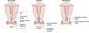
Outline the normal cellular changes of the cervix
When observed? Changes seen? Consequences?
Changes to the cervix are observed during puberty, pregnancy and cyclically
The squamo-columnar junction is repositioned. The cervical columnar cells are exposed to the low pH environment of the vagina. The exposed cells take red appearance.
The cells in this transformation zone take a red appearance.
The cells within the transformation zone become vulnerable to dysplasia and neoplasia.
Define C.I.N. and the characteristics of CIN cells
C.I.N. (Cervial Intraepithelial Neoplasm):A pre-cancerous condition where abnormal cells grow on the surface of the cervix.
Cellular characteristics: > Nuclear:cytoplasmic ratio, clumped chromatin, clear zone around the nucleus
Cellular changes of this nature are often observed within the transition zone.
Ectocervical squamous cells are the most likely to undergo changes and dysplasia. Excessive growth (neoplasia) is seen, leading to an ‘abnormal’ smear test result.
Neoplasia of the endocervical cells, mucus secreting cuboidal epithelium, can also be seen. This is known as CGIN.
State how abnormal cellular changes of the cervix are detected and how these changes are classified
Detected by: Pap (papanicolau stain) smear of desquamated cells
Classification: According to degree of CIN (cervical intraepithelial neoplasm).
Stages:
CIN I: Mild dysplasia (lower 1/3 epithelial thickness)
CIN II: Moderate dysplasia (lower 2/3 epithelial thickness)
CIN III: Severe dysplasia and carcinoma in situ (upper 1/3 and full thickness BUT basement membrane still intact)
Uterine Cancer
- Tumour type, incidence, prevalence and prognosis*
- (presenting symptoms, risk factors)*
Tumour type: Adenocarcinoma
Incidence:
7 % of all tumours in women.
Most common cancer of the female reproductive tract
Peak incidence is senn between 55 and 65 y/o
Prevalence:
Prognosis: Older women presenting with oestrogen independent tumours have a worse prognosis than those presenting with oestrogen dependent tumours.
5 year survival rate:
Stage I - 90 %
Stage II - 50 %
Stage III - 30 %
Stage IV - 10 %
Presenting symptoms: Postmenopausal bleeding
Risk factors: Obesity (↑ oestrogen from adipose tissue), diabetes, hypertension, infertility
Uterine neoplasms: Benign and malignant
Benign: Polyps, leiomyoma (fibroids)
Malignant: Uterine carcinoma (endometrial hyperplasia may preceed this - caused by oestrogen stimulation. Also known as EIN (endometrial intraepithelial neoplasm))
Ovarian Cancer
- Tumour type, incidence, prevalence and prognosis*
- (risk factors, presenting symptoms, diagnosis and management)*
Tumour type: ⋆ Complex pathology due to the abundance of pluri- and totipotent cell types ⋆
70 % from Surface/germinal epithelium (simple cuboid)
10 % from stroma
20 % from follicles
May also be metastases from else where (colon/stomach, breast, uterus and cervix)
Incidence:
5th most common cancer in women
Germinal epithelium neoplasm seen 20 + years
Teratoma neoplasm seen 0 -25 years
Prevalence:
Prognosis: Not improved significantly
Risk factors: Nuliparity, family Hx of BRCA1/2 mutations
Presenting symptoms: None until at advanced stages.
Advanced stage symptoms include: Pain, GI/urinary problems, hormonal effects, ascites
Diagnosis: Ultrasound (TVS and TAS), CA125 and CEA, CXR (check for metastasis), AFP (anti-fetal protein) and HCG in younger patients
RMI (risk of malignancy index) is used to determine the chance of malignancy and whether surgery is required. Based upon 3 domains: CA125 level, ultrasound score and menopausal status
Define toti-, pluri- and multipotent
-
Ovarian Cancer
Surface/germinal epithelium cell tumours: Types and how neoplasia arises
Surface/germinal epithelial cell tumours make up approximately 70 % of ovarian tumours
Derived from coelomic mesothelium
Types: Serous, mucinous (better prognosis), endometroid, Brenner (rare)
How neoplasm arises:
- Ovulation and subsequent scarring pulls bit into the cortex
- These bits form cysts that can undergo metaplasia
- They may then undergo neoplasia, forming tumours
Ovarian cancer
Teratoma: From what do they originate? What types of tissue may be found in them? What indicates malignancy of these neoplasms?
Originate from germ cells
Contain a mixture of mature tissues: Most commonly hair, skin and teeth
Malignancy is indicated by the presence of less differentiated/immature tissues. This is rare. Usually seen in younger girls, 20 y/o.
Describe the structure of normal breast tissue
Key terms: Lobe, lobule, (lactiferous) duct, lactiferous sinus, Cooper’s ligaments, adipose tissue, connective tissue
A lobe is a group of lobules (the cloud-like structure at the ‘end’ of the duct)

Describe the different causes of benign breast lumps
-
Fibrocystic changes (40 %)
- Types include: Fibroadenosis, epithelial hyperplasia and cysts
Fibroadenoma is the most common: Lobule epithelium and connective tissue proliferation, seen in younger women, clinically mobile (breast mobile)
- No disease (30 %)
- Misc benign (13 %)
- Cancer (10 %)
- Fibroadenoma (7 %)
- Duct papilloma
Explain the pathogenesis and development of breast cancer, including the local signs and symptoms
Pathogenesis:
Development:
Carcinoma in situ → invasive (adeno)carcinoma
Adenocarcinoma = Epithelial cells of ducts and glands
Invasive malignant tumours are known as invasive ductal or lobular carcinoma.
Local signs and symptoms:
Palpable lump, mammograhy findings (opaque area seen. These are easier to detect in the breast tissue of older women due to them having less radio-opaque tissue)