Skin Flashcards
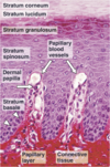
Epidermis
keratinized stratified squamous epith
Epidermis derived from ectoderm
Dermis
dense CT
Dermis derived from mesoderm
Hypodermis

subcutaneous tissue
not considered part of skin
Contains adipose
Epidermis Characteristics

Has deep invaginations- epidermal pegs
which interdigitate w/ dermis projections called dermal paipple
Thick & thin skin
Thick skin on palms of hands & soles of feet- no hairs
Thin skin has hairs & found everywhere else
Thickness depending on epidermis
Epidermis Breakdown
4 layers in thin skin
strat squamous

Keratinocytes
begin life @ bottom layer & as move up
cease dividing, diff, die & sloughed off (desqamation)
Stratum Basale
1 layer @ BM at dermal-epidermal junction
Contains stem cells w/ intense mitotic activity
As cells progress upwards, number of keratin intermed filaments increases until keratins represent half the total protein in stratum corneum
Stratum spinosum
Prickle cells- polyhedral keratinocytes; few layers
Desmosomes give them spiny or prickly appearance
Stratum Germanitivum
cells of lower stratum spinosum & stratum basale
only keratinocytes that divide!
Tonofilaments
another word for keratin intermed filaments in skin
Stratum granulosum
most superficial layer in which nuclei are still prsent
Cytoplasm w/ basophilic granules called keratohyalin granules!
Substance in keratohyalin granules binds w/ keratin filametns
Stratum granulosum & stratum spinosum

Both contain lamellar granules (lamellar bodies)
Lamellar granules- small rodlike structures formed by lipid bilayers
Discharge their content into IC space of stratum granulosum to be deposited as lipid sheets.
Barrier to penetration by foregin mat= sealing effect of skin
“waterproof” skin
LIKE INTERCELLULAR CEMENT
Stratum lucidum

clear layer just superficial to stratum granulosum
Can be observed only in palmar & plantar thick skin
Has keratinocytes w/o nukes or organelles just keratin filaments
stratum corneum
15-20 layers of dead cells
Non viable & scale like= squames
OUtermost layer is shed by desqamation
Much thinner in thin skin than thick skin
Changes of Keratinocytes
- mitotically active- statum bsale & lower spinosum
- nuke & organelles- up to stratum granulosum
- keratin/tonofilaments- get more as you move until top layer
- waterproof- granulosum & above
- desmosome- almost to top but not in stratum corneum
Thick skin
lines palms of hands & soles of feet
lack hair follicles
lack sebaceous glands
Thin Skin
Has epidermis
all over body
contains hair follicles & sebaceous glands
In thin skin

indiv cells of stratum granulosum & stratum lucidum are scattered at interface of stratum spinosum & stratum corneum.
Even though these 2 layers are absent or not well defined!
Callous
thickening of stratum corenum from P on A of skin
Most often in thick skin
Wart
benign epidermal growth due to papilomavirus infection of keratinocytes
Salicylic acid is Rx
- dissolves keratin (keratolytic)
Nonkeratinocytes in epidermis

melanocytes- syn dark brown pigment melanin
langerhan’s cells- APCs
Merkel cells- sensory mechanoR w/in nuero endocrine f
Melanocytes
Derived from NC cells
scattered among basal cells of stratum basale
Melanin begins to be degraded by lysosomes soon after it enters keratinocytes
Can replicate throughout their life, although much slower than keratinocytes

Melanocytes

syn melanin from tyrosine then transfer pigment into keratinocytes
Tryosinase syn in RER & accumulates in vesicles in Golgi & then as free vesicles= mealnosomes
Melanin syn begins in stage II melanosome & forms stage III melanosomes
Melanin granule is after stage III loses its tyrosinase activity
transferred to keratinocytes of malpighian layer (stratum basale & stratum spinosum) from melanocyte’s processes
Tyrosinase activated by UV light, this is why you tan when exposed to sun
Melanin

melanin lies between sun & nuke
melanin protects DNA from UV radiation of sun
This explains why people with lighter skin have higher incidence of skin cancer
Cells of darker skinned people tend to contain more melanosomes & these can be found throughout cytoplasm
pic of stratum spinosum w/ melanin deposits
Skin color
determined by ratio of melanocytes to keratinocytes
not by number of melanocytes
dark skinned & light skinned have same number of melancoytes
but dark skinned make more melanin
light skinned people degrade melanin faster in keratinocytes
light skinned have most of melanosomes in basal layer keratinocytes
Freckle
due to increased melanin production & accumulation in basal area of epidermis
No increase in melanocytes
Mole
group of melanocytes in skin
Dendritic nature of Langerhan’s cells

Dermal papillus
contains capillaries that provide O2 & nutrients for the overlying AVASCULAR epidermis= NO BLOOD VESSLES!!!

Pacinian corpuscles
deep P R for mech & vibratory P
In deeper dermis & hypodermis
Contain mech gated ion channels

Meissner’s corpuscle
touch R
Dermal papillae below epiderm basal lamina
contain mech gated ion channels
lips, external genitalia, nipples

Hair

Glassy mem- thick BM separates dermis from epith of hair follicle
Hard keratin from hair
soft keratin from epidermis of skin
Melanoctyes give hair its color, transfer melanin to epith cells like in skin
Hair

prolif of matrix cells accounts for growth of hair
homologous to stratum basale
blood supply very important
Hair follicle & arrector pili m.
contraction of arrector pili m. causes gooseflesh
Cutis anserina
smoth m.

Eccrine sweat glands

sweat is product
simple coiled, tubular glands
Secretion of merocrine
Very important for thermoregulation
Sweat pore allows for duct to pass from secretory portion of gland across dermis & epidermis
Not associated w/ hair follicles!
Secretory unit of eccrine sweat gland

dark cells- line lumen of gland, contain mucinogen rich granules
clear cells- underlie dark cells, watery & electrolyte rich material
myoepith cells- contract & aid in expressing gland’s secretions into duct **
Apocrine sweat glands

Ducts open into hair follicles
Secrete phermones in animals but in humans ??
Very wide lumen comapred to eccrine (b/c store product in lumen)
Sebaceous glands

sebum- oily & degent epith cells
Sebum prevents H2O loss from skin & lubricates hiar but weak antibac & antifungal properites
Found in face & scalp in abundance- acne contributor
Starts @ puberty
holocrine secetion- prod of secretion released w/ remnants of dead cells
Stem cells in base of gland prolif to replace lost cells
Nail

nail or nail plate hard keratin plate growing out of nail be d
Nail bed is special epith with same epidermis layers
Nail plate= stratum corneum
Nail plate hard, tight, keratinized epidermal cells
Nail body, free dedge into dermis to form nail root/groove
Prolif in nail root form nail matrix & nail elongates
Wound healing

- Injury
- Coagulation - both after wounding & blood clots
- early inflammation, neutrophils dom cell type
- late inflammation, macrophages dom cell type & being replaced by fibroblast to secrete collagen
- remodeling, fibroblasts repair
Burns
tissue is burnt, kills all cells
