Lab 6 Flashcards
Pepsin
……………………. is a proteolytic enzyme secreted in an inactive form by the gastric glands.
activity is most effective at a pH of 2.0.
Pancreatic Lipase
digests triglycerides into monoglycerides and free fatty acids.
Emulsification
The separation of large aggregates of fat into smaller droplets is called _______________________ and is the primary function of bile salts.
__________________ _________________ will break polysaccharides down into the disaccharide ________________.
Salivary Amylase/Maltose
_____________ ______________ is a standard test for starch and it will turn dark blue in the presence of starch.
Lugol’s Iodine
_____________________ _________________ is a standard test for sugar and if a colored precipitate forms after boiling this is a positive test for sugar.
Benedict’s solution

Taste Buds

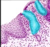
Salivary Gland

Salivary Gland Excretory ducts

Salivary Gland serous ancini
What is the whole picture?
What is 1?
What is 2?
What is 3?
What is 4?

stomach
stomach mucosa
stomach submucosa
stomach muscularis
stomach adventitia

Stomach rugae
Stomach gastric pits
What is the whole picture?
what is the purple area?
what is the yellow area?
what is the red area?
What is the blue area?

The layers of the duodenum
the mucosa of the duodenum with villi and microvilli
the submucosa of the duodenum
the muscularis of the duodenum
the adventitia of the duodenum
What is I?

Intestinal glands (aka crypts of lieberkuhn)
What is the red box pointing to?

Duodenal (brunner’s) glands
What is the whole picture?
What is the purple area?
What is the lighter pink area?
What is the magenta area?
What is the area at bottom of slide?

The colon
The mucosa of the colon
the submucosa of the colon
the muscularis of the colon
the adventitia of the colon

goblet cells in the colon
What is the whole picture?
What is the pointer pointing to?
What is the hole?
Liver
Liver Sinusoids
Liver central vein
What is 1?

Pancreatic ancini
What are 9 and 10?
What is 18?
What are 1 and 8?
What is 15?
What are 2 and 12?
What is 5?

upper and lower lips
Gingiva
The vestibule (superior and inferior)
The tongue
hard and soft palate
uvula

Parotid salivary gland

Sublingual salivary gland

Submandibular salivary gland

pharynx

Esophagus

lower esophageal sphincter
Locate:
fundus, cardia, body, pyloric antrum, rugae, and greater/lesser curvature


Locate:
duodenum, jejunum, ileum, ileocecal valve, cecum, vermiform appendix


What 3 things are in this pic?

pancreas, pancreatic duct, major duodenal papilla
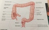
Locate:
ascending colon, hepatic flexure, transverse colon, splenic flexure, descending colon, sigmoid colon, haustra, taenia coli, rectum, anal canal

Locate:
R and L lobes, Falciform ligament, gall bladder, cystic duct, R and L hepatic ducts, Common bile duct


Locate:
crown, neck, root, enamel, dentin, pulp cavity, root canal, gingiva

crown = I, neck = H, root = G, enamel = A, dentin = B, pulp cavity = C, root canal = F, gingiva = D

Superior mesenteric artery

Inferior mesenteric artery

celiac trunk

gastroepiploic artery

Gastroduodenal Artery
The red one

cystic artery
Locate:
R and L Hepatic Arteries, and the Proper Hepatic Artery


hepatic vein
Locate:
Superior and Inferior mesenteric veins, hepatic portal vein, cystic vein

