15 - Hemoglobin Disorders Flashcards

1
Q

What are the 2 classes of hemoglobin disorders?

A
  1. structural variants
    (abnormal globin chain structure due to globin gene mutation; varied clinical effect depending on location and nature of mutation)
  2. thalassemias
    (under-production of structurally normal globin chains; generally microcytic/hypochromic anemais of varying severity)
2
Q

What are the two main globin gene clusters?

A
  1. alpha cluster on chromosome 11
    (2 alpha, 1 theta, 2 psi, and 2 zeta genes)
  2. beta cluster on chromosome 16
    (1 beta, 1 delta, 2 gamma, 1 epsilon gene)
3
Q

What are the 3 normal hemoglobin species? What are the percentages in a normal adult?

A
  1. Hb A (2alpha2beta)
    *96% in adults
  2. Hb A2 (2alpha2delta)
    ​*3% in adults
  3. Hb F (2alpha2gamma)
    *dominates during fetal life, 1% in adults
4
Q

What is the incidence of abnormal hemoglobin? What are the usual clinical symptoms?

A
  • 500+ structural hemoglobin variants (mostly single amino acid replacements, occasionally deletions/insertions)
  • mostly clinically silent with no functional consequences
5
Q

What are some examples of clinical consequences caused by abnormal hemoglobin?

A
  • sickling
  • Hb instability
  • altered oxygen affinity (increased or decreased)
  • increased susceptibility to oxidation (to methemoglobin)
  • under-production of globin chains
  • combinations of the above
6
Q

What are the 2 main laboratory techniques for diagnosing abnormal hemoglobin?

A
  • electrophoresis (gel or capillary)
  • HPLC (high performance liquid chromatography)
7
Q

Describe hemoglobin electrophoresis

A
  • typically performed in parallel with alkaline and acid buffers
  • HbA has isoelectric point of 6.8
    • neg charge in alkaline buffers, migrates towards anode (+)
    • pos charge in acid buffers, migrates towards cathode (-)
8
Q

Describe HPLC

A
  • fully automated cation exchange chromatography method
  • whole blood method:
    • Hb adsorbed onto resin particles
    • different species differentially eluted based on affinity for resin by gradually changing ionic strength of elution buffer
    • some correlation with migration on alkaline electrophoresis
9
Q

Describe sickle cell disease

A
  • homozygous abnormality of the beta globin chain
    • more common in african americans (1 in 600 homozygous)
  • Glu -> Val substitution at AA 6 of the beta chain (beta6val)
  • heterozygous HbS “S trait” confers protection against malaria
10
Q

Describe the pathophysiology of sickle cell disease

A
  • deoxygenated HbS forms long polymers that distort the shape of the cell into an elongated, sickled form
  • extend of polymerization is time and concentration dependent
  • initially reversible but after multiple sickling/unsickling cycles, membrane damage produces irreversibly rigid sickled cell
  • RBC lifespan decreased to 20 days
11
Q

What affects the concentration of HbS?

A
  • percentage of HbS of total Hb:
    • homozygous or heterozygous
    • presence of other Hb species (e.g. Hb F)
  • total Hb concentration in the red cells (MCHC; Mean corpuscular hemoglobin concentration)
    • concentration increased in cellular dehydration
    • concentration decreased when co-existent thalassemia
12
Q

What factors influence the time dependence of sickling?

A
  • transit time of red cells through low oxygen tension microvasculature
  • sickling enhanced in anatomic sites with sluggish flow (e.g. spleen and bone marrow)
  • blood flow thorugh microvasculature retarded in certain pathologic states (e.g. inflammation)
13
Q

What clinical settings predispose a patient to sickling?

A
  • hypoxia
  • acidosis (shift dissociation curve to the right causing increased deoxygenation of HbS)
  • dehydration (hypertonicity causing RBC dehydration)
  • cold temperatures (sluggish blood flow)
  • infections
14
Q

What are 2 major effects of RBC sickling?

A
  1. chronic hemolysis (correlates with the number of irreversibly sickled cells)
  2. microvascular occlusion with resultant tissue hypoxia and infarction (related to increased “stickiness” of SS red cells because of membrane damage)
15
Q

When do symptoms usually begin for a patient with sickle cell?

A
  • newborns clinically fine because of high HbF
  • hematologic manifestations begin by 10-12 weeks of age
  • clinical severity variable from patient to patient
16
Q

What are 6 common clinical manifestations of sickle cell disease?

A
  1. severe anemia
  2. acute pain crises (from vaso-occlusion)
  3. auto-splenectomy (from repeat splenic infarction)
    *increases infection risk
  4. acute chest syndrome (major cause of death from pulmonary infections or fat emboli)
  5. strokes (first usually when 2-8 years old)
  6. aplastic crisis (acute decrease in RBC production usually from parvovirus B19 infection “fifths disease”)
17
Q

What are the laboratory findings in sickle cell disease?

A
  • chronic anemia (Hb 5-11 g/dl)
  • increased bilirubin
  • sickled cells, target cells, polychromasia
  • increased reticulocytes
  • normal MCV
18
Q

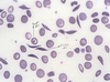
What does this blood smear show?
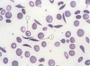
A

**sickle cell disease

  1. target cell
  2. sickled cell
19
Q

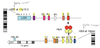
What would the gel electrophoresis results look like for sickle cell disease?

A

In class she stressed to KNOW THIS!

**bands for S and F hemoglobin (along with the normal A and A2 bands)

20
Q

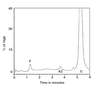
What would the HPLC results look like for sickle cell disease?

A

Large spikes for Hb S and F (note lack of A spike)

21
Q

What are the other 2 main sickling disorders?

A
  • Hb SC disease
    • compound heterozygous state
    • Hb C from glu6lys substitution of the beta globin
    • generally milder than SS but highly variable
  • Hb S/beta thalassemia
    • heterozygous Hb S with trans beta thalassemia allele, resulting in decreased or absent production of normal beta chains
    • asymptomatic to nearly identical to SS
    • lab findings= Hb S > Hb A
22
Q

How do you manage a patient with sickle cell disease?

A
  • newborn screening
  • infection prophylaxis
  • supportive care for acute manifestations
  • hydroxyurea (chemo to reduce RBC counts and increase HbF levels)
  • regular red cell transfusions
  • allogeneic stem cell transplant (the only curative therapy but it’s only used in very sick patients)
23
Q

What is the outcome for a patient with sickle cell disease?

A
  • median age of death= 45 for males, 48 for females
    • although much better than <20 year expectancy in the 1970’s
    • gains due to decreased child mortality (infection prophylaxis and comprehensive care)
  • major causes of death:
    • liver dysfunction
    • pulmonary HTN
    • stroke/vaso-occlusive crisis
    • acute chest syndrome
24
Q

What is S-trait?

A
  • clinically benign carrying of sickle cell gene (in 8% of african americans)
    • no anemia/normal RBC survival
    • NO sickling
    • may be mild, sub-clinical kidney damage
  • lab values= 60% Hb A, 40% HbS
25
Q

What would the HPLC results look like for S-trait?

A

Large Hb A and S spikes (small F spike, unlike sickle cell disease)

26
Q

What is HbC disease?

A
  • homozygous for Hb C
    • glu6lys substitution of the beta globin
    • cells abnormally rigid/dehydrated
    • NOT a sickling disorder
  • mild to moderate hemolytic anemia
    • RBC lifespan shortened to 30-35 days
  • often asymptomatic
  • splenomegaly (occasional abdominal pain)
  • 1/6000 african americans
27
Q

What are the lab findings for HbC disease?

A
  • Hb levels 8-12 g/dl
  • numerous target cells
  • mild microcytosis
  • spherocytes
  • occasional C crystals
28
Q

What would the HPLC results look like for HbC disease?

A
  • >90% Hb C
  • NO Hb A!
  • <7% Hb F
29
Q

Describe Hb C trait

A
  • 2% of african americans
  • NO anemia (no effect on patient but important for genetic counseling)
  • few target cells
  • 50-60% HbA, 30-40% HbC
30
Q

What are thalassemias?

A

A group of inherited disorders characterized by decreased production of structurally normal globin chains

(highly heterogeneous both clinically and genetically)

31
Q

What are the 2 main types of thalassemias? Where are they commonly seen?

A
  • beta-thal (wide distribution in the mediterranean, middle east, parts of india/pakistan, and southeast asia)
  • alpha-thal (occurs throughout africa, mediterranean, middle east, and southeast asia)
32
Q

Describe the main features of thalassemias

A
  • typically microcytic/hypochromic anemias (due to decreased Hb) of varying severity
  • severity directly related to the degree of chain imbalance
    • excess normally produced globin chains accumulate and cause intramedullary cell death and/or decreased RBC survival
33
Q

Describe beta thalassemia. What are the 3 main types?

A
  • decreased beta globin chain production from affected alleles
    • 250+ known mutations
    • most common= splicing errors
    • gene deletions=rare
  • clinically classified:
    • beta-thal major (Cooley’s anemia)
    • beta-thal intermedia
    • beta-thal minor
34
Q

Describe beta-thal major

A
  • absence or marked decrease in beta chain production on BOTH beta alleles
    • excess of normal alpha chains (unable to form tetramers and precipitate in normboblasts and erythrocytes)
    • intramedullary cell death and decreased RBC lifespan
35
Q

What are the symptoms of beta-thal major?

A
  • infants well at birth (anemia develops over the first few months of life)
  • severe anemia (Hb 2-3 g/dl; recall normal is 12-15)
    • virtually all Hb F
    • bizarre red cell morphology (hypochormia, targeting)
  • transfusion dependent
  • severity dependent on adequacy of transfusion program and efficacy of iron chelation
36
Q

What are some symptoms of an inadequately transfused patient with beta-thal major?

A
  • stunted growth and bony abnormalities
  • frontal bossing (mongoloid facies)
  • increased skin pigmentation
  • fever
  • wasting
  • hyperuricemia
  • spontaneous fractures (expanded marrow spaces)
  • hepatosplenomegaly
  • infections
  • folate deficiency
  • death in childhood
37
Q

What are some features of an adequately transfused patient with beta-thal major?

A
  • essentially normal development (avoidance of classic complications)
  • without adequate iron chelation therapy:
    • absence of pubertal growth spurt/menarche
    • endocrine disturbances (DM, adrenal insufficiency)
    • death from cardiac iron deposition by 30s
  • with aggressive iron chelation therapy:
    • less severe cardiac disease/endocrine disturbances
    • significantly improved life span
38
Q

Describe beta-thal minor

A
  • heterozygous form
    • mild or no anemia, Hb > 10 g/dl
    • microcytosis, scattered target cells
    • basophilic stippling
    • elevated HbA2 (3.5-7%) **KNOW
  • discovered incidentally on CBC (usually asymptomatic)
  • incidence:
    • common in mediterranean and asian populations
    • 1.5% of african americans
39
Q

Describe beta-thal intermedia

A

Heterogeneous group (everything between the extremes of major and minor)

40
Q

What are the clinical subtypes of alpha thalassemia?

A

**usually a result of gene deletion (in contrast to beta-thal), recall we have 4 alpha genes; 2 on each chromosome 11

  1. silent carrier (1 gene deleted; asymptomatic)
  2. alpha-thal trait (2 genes deleted)
  3. Hb H disease (3 genes deleted)
  4. hydrops fetalis (4 genes deleted; infants stillborn or die within hours)
41
Q

Describe alpha-thal trait

A
  • 2 genes deleted
  • mild microcytic anemia (hematologically similar to beta-thal minor)
  • discovered incidentally
42
Q

Describe Hb H disease

A
  • 3 genes deleted
  • mild to moderate; chronic hemolytic anemia
  • Hb H= beta tetramer that doesn’t effectively transfer oxygen (recall alphas cannot combine in beta thalassemias so this doesn’t happen)
  • Hb H is soluble (doesn’t initially precipitate in normoblasts) but becomes unstable over time, precipitating in circulating RBCs **causes hemolysis