Intraocular Inflammation and Uveitis Flashcards
Test that is indicator of active infection in syphilis
serum rapid plasma reagin (RPR)
does not confirm that patient has syphilis
Confirmatory: fluorescent treponemal antibody absorption (FTA-ABS) assay and microhemagglutination assay for T pallidum antibodies (MHA-TP) (treponemal antigent tests that do not correlate with disease activity, may be positive for a lifetime)
What topical corticosteroid medication is most likely to cause a corticosteroid-response elevation in IOP?
prednisolone acetate 1%
greatest at reducing intraocular inflammation with greatest effect on IOP
(Fluorometholone is very potent but penetrates poorly into eye)
Equivalent of difluprednate 0.05% to PF1%
dosing 4 times daily is equivalent of 8 or more toal drops of PF1% but higher rise in IOP than PF1%
MOA of methotrexate (in reducing inflammation)
extracellular release of adenosine
folic acid analogue and inhibitor of dihydrofolate reductase inhibits DNA replication but its anti-inflammatory effects result from extracellular release of adenosine
Tx Lyme dz
doxycycline
alt: ceftriaxone, amoxicillin, cefuroxime
unilateral stellate KP, cataracts, increased risk for glaucoma
Fuchs heterochromic uveitis
(AKA: fuchs heterchromic iridocyclitis or Fuchs uveitis syndrome)
other s/s: elevated IOP, diffuse iris atrophy, TID, 5% bilateral, rubella
Another name for chronic anterior uveitis
iridocyclitis
2-3% of patients referred to uveitis clinics have this dz
Fuchs heterochromic uveitis
Signs of Fuchs heterochromic uveitis
diffuse iris stromal atrophy with variable pigment epithelial layer atrophy
small, white, stellate KPs scattered diffusely over entire endothelium
cells in AC and anterior vitreous
glaucoma + cataracts (PSC)
RARE/ABSENT: macular edema, synechiae, fundus scars, retinal periphlebitis
chronic indolent intraocular inflammatin s/p cataract, think..
propionibacterium acnes
Candida parapsilosis
others implicated: aspergillus flavus, torulopsis candida, paecilomyces lilacinus, verticillum
Uveitis associated CME that fails topical corticosteroids. Next steps?
20-40 mg triamcinolone sub-Tenon (superotemporal posterior) q monthly
delivers juxtascleral corticosteroid closest to macula
if still fails…
2-4 mg of intravitreal preservative-free triamcinolone
uveitis tx CI in uveitis a/w MS
tumor necrosis factor-alpha inhibitors
etanercept and infliximab - aw exacerbations of CNS demyelination
% of patients with MS that have uveitis and pars planitis
30% uveitis in patients with MS
15% pars planitis in patients with MS
Preferred tx for MS-associated
IV and intravitreal corticosteroids
topical cycloplegics
ocular inflammatory disorder aw activation of mast cells via IgE antibodies
vernal conjunctivitis
how long does 0.59-mg fluocinolone acetonide implant release therapeutic levels of corticosteroids to the vitreous cavity?
30 months
Positive RPR, next step?
serum FTA-ABS (since RPR may be falsely positive) if positive
Lumbar puncture with
CSF VDRL (diagnostic) and
CSF FTA-ABS
Antibiotic therapy - series of PCN IV or IM dosing
Percentage of blindness in the US attributed to inadequately treated uveitis
10%
Clinical triad to diagnose ocular histoplasmosis syndrome
peripapillary atrophy (pigment changes)
multiple white, atrophic punched-out chorioretinal scars (histo spots)
choroidal neovascular membrane causing maculopathy
NO vitreous cells
If histo spots are seen in the macula, what is % chance of macular CNV developing in 3 years
if no spots are observed
25%
2% (no spots)
What would active CNV look like in ocular histoplasmosis syndrome (OHS)
yellow-green subretinal membrane typically surrounded by a pigment ring
overlying neurosensory detachment
subretinal hemorrhage
frequently border of histo scar in disc-macula area
Useful clinical feature suggests Vogt-Koyanagi-Harada syndrome?
Alopecia
Poliosis
(is the decrease or absence of melanin (or colour) in head hair, eyebrows, eyelashes or any other hairy area)
Vitiligo (loss of pigment in skin)
3 seen in 30%
Usefulness (positive predictive value) of ANA screen in uveitis
prevelance of lupus is less than 0.47% in patients with uveitis so the PPV of ANA for diagnosis is less than 3%
weight loss, cough and findings in image. Test for granulomatosis with polyangiitis

c-ANCA antibody (anti-proteinase 3 antibody)
most specific
(picture: anterior scleritis)
How to differentiate progressive outer retinal necrosis from acute retinal necrosis
Host immunosuppression
ARN - intact immune function
PORN - immunocompromised
HLA Aw33
increased RR of ARN in Japanese patients
virus aw conjunctival papillomas
human papillomavirus (HPV) 6 and 11
65 yo M with unilateral vision loss (photophbia, floaters, pain), occlusive retinal arteriolitis, vitritis, multifocal yellow-white peripheral retinitis. Best Tx?
systemic ACYCLOVIR (IV 10mg/kg q8 hrs x 10-14 days)
after 24-48 hours antiviral add prednisone 1mg/kg/day then taper
Dx: ARN
Note: presentation above is classic triad seen within 2 weeks for ARN
Feared complication of ARN
75% of patients will develop a combined tractional-rhegmatogenous retinal detachment
immune response to mycobacterial infection
conversion of macrophages to giant cells that form caseating granulomas
Causative organisms in healthy 40 year old man

herpes simplex and varicella-zoster virus
ARN AKA necrotizing herpetic retinitis
Diagnosis

ARN
Diagnosis

ARN
Diagnostic criteria for this disease

One or more foci of retinal necrosis with discrete borders, located in the peripheral retina
Rapid progression in absence of antiviral therapy
Circumferential spread
Occlusive vasculopathy, affecting arterioles
Prominent vitritis and/or anterior chamber inflammation
Diagnostic characteristic of this syndrome

Usually unilateral
MEWDS
Goldmann-Witmer (GW) test needs these 2 tissues
Aqueous and serum
GW = (xIgG in aqueous/total IgG in aqueous)/(xIgG in serum/total IgG in serum)
systemic complication of prolonged NSAID use in patient with non-necrotizing anterior scleritis
myocardial infarction
others: HTN, stroke (COX-2), GI ulceration, GI bleeding, nephrotoxicity, hepatotoxicity
most likely cause of uveitis in HIV patient with CD4+ <50/uL who responded to HAART
immune recovery uveitis
Condition can lead to elevated IOP due to blockage of angle by inflammatory cells and debris
TOXOPLASMA retinochoroiditis
others: ARN, CMV, sarcoid, Fuchs heterochromic iridocyclitis
27 year old bilateral anterior and intermediate uveitis, vitreous cell, KP, vitreous cellular aggregates near vitreous base. Test provides most definitive evidence of this disease?

histopathology of conjunctival nodule, lung secimen or other tissue involved demonstrating noncaseating granuloma
Other tests:
spiral CT (higher sensitivity)
CXR (plain PA and lateral)
ACE (least specific, moderate sensitivty)
Diagnosis.
Active or recurrent?
Serpiginous choroiditis
lesions involve the outer retina, RPE, choriocapillaris, and large choroidal vessels.
Patients present with acute geographic or serpentine lesions that are gray or gray–yellow (due to disruption of the RPE or outer retina).
Fundus photo showing recurrence of serpiginous choroiditis near the edge of a scar.

Only presenting feature. Diagnosis?

Granular pigment changes in the fovea
Multiple Evancescent White Dot Syndrome (MEWDS)
pathognomonic
Diagnosis
Expected finding on FA?

MEWDS
Fundus photo:
- subtle numerous white spots at the level of the RPE/deep retina.*
- There are also blurred disc margins.*
- There is foveal granularity.*
FA: subtle early and late hyperfluorescence of the white dots in a wreath-like pattern

HLA-A29 with fundus and FA
Diagnosis?

Birdshot chorioretinopathy
multiple creamy, subretinal lesions radiating fom ON
FA: CME
Diagnosis

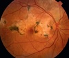
left posterior pole showing a hazy view and the distribution of deep creamy oval lesions
Birdshot chorioretinopathy
27 yo AA M with unilateral photophobia and reduced VA. Grade 3+ vitritis with peripheral retinal whitening. Possible PMHx?

Previous viral meningitis
Younger patients with HSV-1 and HSV-2 retinitis have a higher RR of previous viral encephalitis or meningitis
(Image: ARN)
Histological finding in Vogt-Koyanagi-Harada (VKH) syndrome
Dalen-Fuchs nodules
epithelioid hisiocytes and lymphocytes between Bruch membrane and RPE

Risk factors for this disease

Candida chorioretinitis or endophthalmitis
Hospitalization with h/o recent GI surgery
bacterial sepsis
systemic abx
indwelling catheter
hyperalimentation
debilitating Dz eg, DM
immunomodulatory therapy
prolonged neutropenia
organ transplantation
NOT Immunodeficiency
Diagnosis

Block early, stain late
APMPPE
Acute posterior multifocal placoid pigment epitheliopathy
23 yo M with recurrent unilateral anterior uveitis, KP, IOP 32 and 1+ cell and flare with mixed white and pigmented clels in AC. Normal fundus exam.
Diagnosis?

Herpes simplex virus keratouveitis
stellate KP
Findings expected
Tx

Sclerosing keratitis from onchocerciasis
intraocular microfilariae (in AC)
anterior uveitis
SPK
sclerosing keratitis
scleritis
chorioretinitis
optic neuritis and atrophy
cataract
PAS
glaucoma
Tx: ivermectin, suramin, diethylcarbamazine (DEC)