Week 6- Nervous System Flashcards
What is the structure of a neuron?
1) Has cell body
2) Contains dendrites that composes impulses towards the cell body (short, branched fibres)
3) Contains axons that send impulses away from the cell body. (Long, single process)

What are the three different neurons?
1) Multipolar neuron
2) Bipolar neuron
3) Unipolar neuron
What is the function of the neuroglia?
1) Support
2) Segregate/ insulate neurons
3) Protection
4) Promotes health and growth
What is the myelinated neuron?
Cells that surround axons with layers of plasma membrane (lipid & protein)
what is the function of the myelin shealth?
Electrically insulate axon that increase the speed of nerve impulse conduction.
What is the ganglion?
A collection of cell bodies of the neurons outside the CNS.

What is the nerve?
- Bundle of neuron fibres outside the CNS.
- A collection of many axons.
What is grey matter?
- Grey matter is the nerve cell bodies inside the CNS.
What is white matter?
Neuron fibres within CNS

What is tract?
- A bundle of neuron fibres in the CNS.
- Runs from brain-spinal cord (vice versa)
- White in colour due to myelin sheaths covering the axons.

Voluntary contration decisions happens where?
The brain
AKA CNS
The decisions for voluntary movements are transferred to the muscle from what?
Motor neurons
(Brain-motor neurons)
What is the organization of the peripheral nervous system?
-Peripheral Nervous system
(divides into 2 categories)
- Somatic/automic nervous systems
(automic nervous system divides to 2 sections)
- Sympathetic division / parasympathetic division

What is the different of afferent and efferent nerves in the PNS?
- Afferent nerves carry electrical impulses from receptors in the body to the CNS.
- Efferent nerves carry electrical impulses from the CNS to the muscles and glands.
What is the function of the somatic nervous system?
- Controls voluntary movements
- Deals with the parts of the body that you can move voluntarily
What is the function of the automic nervous system?
- Regulates the functions of internal organs such as the heart, stomach, intestines and some muscles.
- Unaware of the automic nervous system because it’s involuntary.
Automic nervous system facts?
- Divided into sympathetic and parasympathetic divisions
- Many organs receive efferent neurons from both these divisions
- divisions are often antagonistic
- Neurotransmitters secreted by nerve ending usually different between the two divisions
- Hypothalamus – control centre
What is the difference of sympathetic/parasympathetic ?
- Sympathetic divison is arousing
- Parasympathetic division is calming
What is the sympathetic and parasympathetic division for eyes?
- Sympathetic = pupils dilate
- Parasympathetic = Pupils contract
Sympathetic/parasympathetic division for salvation
Sympathetic- Decreases
Parasympathetic- Increases
Sympathetic/Parasympathetic division for skin
Sympathetic - Perspires
Parasympathetic - Dries
Sympathetic/Parasympathetic division with Respiration
Sympathetic- Increases
Parasympathetic- Decreases
Sympathetic/Parasympathetic division for the heart
Sympathetic- Accelerates
Parasympathetic- Slows
Sympathetic/Parasympathetic division for Digestion
Sympathetic- Inhibits
Parasympathetic- Activates
Sympathetic/Parasympathetic division for Adrenal Glands
Sympathetic- Secretes stress hormones
Parasypathetic- Decrease secretion of stress hormones
Sympathetic/Parasympathetic division for Immune system functioning
Sympathetic- Reduced
Parasympathetic- Enhanced
When does the action potential occur?
When a neuron sends information down an axon, away from the body.
Resting membrane potential facts
- Present in all cells
- Inside of the cell membrane is negative compared to the outside
- Has large quantities of negatively charged protein molecules inside the cell that cannot leak out.
What are the three key factors for the resting membrane potential?
1) Sodium / potassium pump
2) The prescense of leaky potassium channels
3) Large quantities of negativelycharged protein molecules inside the cell that cannot leak outside
Leaky potassium ion channels facts
- More of them inside than outside of cell
- potassium (k+) leak channels that lets potassium leave the cell body by facilitated diffusion through concentration gradient
- Exit of potassium carries a positive charge to outside of cell
- Contributes to the inside of the cell being more negative than the outside

What are the types of plasma membrane ion channels?
1) Passive / leakage channels- always open
2) Ligand- gated channels- open with a binding of a specific neurotransmitter
3) Mechanicallly- gated channels- opens/closes with stimulation (vibration, touch, etc.)
4) Volted- gated channels- Open/closes in response to membrane potential
What is the resting state of the neuron?
Phase 1
- All voltage-gated channels are closed from NA+/K+ to pass through.
- The axon plasma membrane is at its resting membrane potential.
AKA: equal buildup of negative and positive ions in inside/outside of membrane surface.

What is th depolarizing stage of the neuron?
phase 2
- NA+ channel activation gates open when membrane potential of axon reaches threshold.
- When the na+ rushed through this channel, the inside of the cell becomes depolarize

What is propagation?
When an action potential occurs at one side of the membrane and then follows all the way down to the end (like a chain reaction)

What is the repolarization phase of the neuron?
phase 3
- Happens when about 30mV of action potential is reached
- The NA+ gates close and the K+ gates open.
- The membrane starts to become polarized again due to k+ resurfacing to outside of cell, leaving negative charge inside cell.

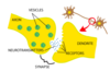
What is the synapse transmission process?
1) Nerve impulses arrive at axon bulbs
2) Volted gated calcium (Ca2+) gates open, rushing Ca2+ into the bulb
3) Ca2+ causes vesicles to move to presynaptic membrane, undergoing exocytosis, releasing neurotransmitters into synaptic cleft
4) Neurotransmitters diffuse across clefts
5) Neurotransmitters fit into receptors of ligand-gated Na+ gates, opening them
6) Na+ rushes into cell, causing action potential to develop
Acetylcholine neurotransmitters facts
- Released at many synapses in the brain and peripheral nervous systems
- Released at the neuromuscular junction
- Can have either excitatory or inhibitory action, depending upon which part of the nervous system is using it
- Its effects are terminated through the action of acetylcholinesterase

What are the three types if meninges?
1) Dura mater
2) Arachnoid mater
3) Pia mater

What is the cerebrum/ functions?
- the principal and most anterior part of the brain in vertebrates, located in the front area of the skull and consisting of two hemispheres, left and right, separated by a fissure.
- Function:
1) Perception or interpretation of sensory impulses
2) Emotional and intellectual processes
3) Control of motor movements

What is the Diencephalon?
- Central core of the forebrain
- Consists of 2 major structure:
1) Thalamus
2) Hypothalamus - Encloses the third ventricle

What are the thalamuc functions?
- Afferent impulses from all senses converge and synapse in the thalamus
- Impulses of similar function are “sorted out,” edited, and relayed as a group
- All inputs ascending to the cerebral cortex pass through the thalamus
- Plays a key role in mediating some sensation
(e. g. pain, temperature, light touch and pressure) - Role in limbic system – pain and pleasure interpretation

What is the brain stem / functions?
- the central trunk of the mammalian brain consisting of:
1) midbrain
2) pons
3) medulla oblongata - continues downward to form the spinal cord
- Function:
1) Controls automatic behaviors necessary for survival
2) Provides the pathway for tracts between higher and lower brain centres

What is the midbrain/ functions?
- Located between the diencephalon and the pons
- Function: Reflex center for head and eye movements in response to sight and sounds

What are pons/ functions?
- Bulging brain stem region between the midbrain and the medulla oblongata
- Functions:
1) Also forms bridge between cerebellum and brain stem
2) All sensory and motor fibres pass through the pons
3) Pneumotaxic centre regulates respiration

What is the medula oblongata/ functions?
- Most inferior part of the brain stem
Functions:
1) Cardiac centre – regulates heart rate
2) Respiratory centre- regulates rate and depth of breathing
3) Vasomotor centre – regulates blood pressure
4) Regulates swallowing, sneezing, vomiting

What is the cerebellum/ functions?
- the part of the brain at the back of the skull in vertebrates
- Functions:
1) Provides precise timing and appropriate patterns of skeletal muscle contraction
2) Maintains posture
3) Maintains equilibrium using sensory input from inner ear

What is the spinal cord?
- a series of 31 segments, each giving rise to a pair of spinal nerves
- Begins at occipital bone and ends at vertebral disc between first and second lumbar vertebrae (bottom of ribs)
- Nerves for lower lumbar area and below, arise from end of spinal cord and make up the “cauda equina”

What are the functions of thr spinal cord?
- Provides a two way conduction pathway to and from the brain
- Conveys sensory impulses from the periphery to the brain, and conducts motor impulses from the brain to the periphery
- Provides a means of integrating reflexes **photo**

How does a reflex arc work?
1) Sensory receptor
2) Sensory neuron
3) Integrating centre
4) Motor neuron
5) Effrector

What is the pathway of olfactory impulses from receptors to the cerebrum?
1) Axons of olfactory receptor cells unite in bundles to form olfactory nerves
2) Synapse with olfactory bulb neurons inside olfactory bulbs
3) Axons of olfactory bulb neurons unite to form olfactory tract, which goes to the olfactory cortex in temporal lobe
4) The only sensory information that does not go through thalamus

Describe the sense of taste with respect to the structure of gustatory receptors
- Each gustatory cell has a single long microvillus projecting through a taste pore
- Sodium ions enter via sodium channels, causing depolarization (salty)
- Hydrogen ions enter via hydrogen channels, causing depolarization (sour)
- Chemicals that cause sweet, bitter and umami tastes fit into receptors, that then cause depolarization
- Taste impulses travel to medulla, then to thalamus, then to parietal lobe.
- The pattern of reception of the five different tastes create the sensation of many different tastes
- Taste buds are specialized for one taste, but are spread evenly over buccal surface
