MSK2 Flashcards
What are the terminal nerves of the brachial plexus and are they innervating flexors or extensors?
Flexors: musculocutaneous, median, ulnar
Extensors: radial, axillary

What are the terminal nerves of the lumbar plexus and are they innervating flexors or extensors?
Flexor: obturator (adductors of hip flexors) Extensor: femoral (extensors of knee)

What are the terminal nerves of the sacral plexus and are they innervating flexors or extensors?
-sciatic nerve then splits at knee Flexor: tibial nerve (plantar flexors) Extensor: common fibular nerve (dorsiflexors and extensors)

What deficits do you expect with an proximal injury to a nerve that forms a plexus?
- partial paralysis (paresis) in the muscle
- 1/3 of the dermatome that is supplied by that spinal level being affected
What deficits do you expect with a distal injury in a plexus?
- nerves have joined together from spinal levels already
- complete paralysis of the muscle and dermatomes all affected
What deficits do you expect with a proximal injury to a peripheral nerve?
- before the division of the muscular and sensory branches
- both sensory and motor loss
What deficits do you expect with a distal lesion to a peripheral nerve after it has given off its deep branches?
-sensory loss only
How are bones developed?
- develop from cartilage
- cartilage models in upper and lower limb develop into the bones that make up both limbs between 5 and 6 weeks in utero
- connective tissue on outside of cartilage- perichondrium
- ossification centres: diaphysis and epiphyses
- diaphysis ossification centre begins in the middle and moves towards the epiphysis while the inside of the epiphysis also forms bone
- eventually these will fuse together to give you one bone

What is the epiphyseal plate?
- where diaphysis and epiphysis meet
- after the age of 20, it disappears
- after that you can see the epiphyseal line which is the remnant of the cartilaginous plate

What cavity is found in the diaphysis? What type of marrow is found here?
- completely hollowed out forming the medullary cavity with cortex of bone on the outside
- filled completely with fat
- important storage site for fat and energy
- yellow marrow

What type of marrow is found in the epiphysis? What is the role of this area of the bone?
- in the epiphysis, it is not completely hollowed out
- pillars of bone with spaces in between filled with blood
- spongy bone
- red marrow
- very good blood supply
- role is to form new blood cells

What is periosteum? What does it originate from?
- perichondrium on the cartilage models becomes periosteum once the bone is formed
- connective tissue around the bone
- 2 layers: outer fibrous layer made of collagen fibres running in many directions which allows for the attachment of ligaments/tendons, inner layer where mesenchymal stem cells are located (important for formation and healing of bones)

What do periosteum mesenchymal cells turn into?
- osteoprogenitor cells turn into osteoblasts
- osteoblast secretes ECM (made up of collagen and hydroxyapatite)
- some osteoblasts differentiate into osteocytes

Describe the arrangement of osteocytes and osteoblasts in the compact and spongy bone
- osteocytes have projections off of them- they sit in wells of bones surrounded by bone
- channels run longitudinally around the bone and osteocytes are located around the channel
- spongy bone- osteoblasts located on the surface of bony pillars that are creating the spongy bone

Describe where osteoclasts originate from
-bone marrow derived macrophages fuse to form osteoclasts
What happens during bone reabsorption?
- osteoclasts use acid and collagenase to dissolve bone and release calcium
- under certain circumstances with lots of osteoclastic activity, lots of spongy bone can be removed
- this can happen under the influence of some hormones

Describe the changes to calcium homeostasis when calcium levels are too high
- thyroid releases calcitonin (comes from thyroid gland)
- increases osteoblastic activity and deposition of calcium in the bone
- decreases calcium uptake in the intestines
- decreases calcium reabsorption from the urine
- mobilizes calcium from blood to deposit into the bone

Describe the changes to calcium homeostasis when calcium levels are too low
- parathyroid gland releases PTH
- stimulates osteoclastic activity
- increases calcium release from the bones, uptake in the intestines, and reabsorption from the urine
- calcitriol- skin and GI


-places where a joint is formed remain as cartilage (articular cartilage)

What is a comminuted fracture?
- break causes it to splinter
- found in extreme trauma
- happens when older person breaks a bone (less cartilage and collagen so bone is more brittle)

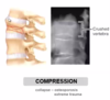
What is a compression fracture?
- osteoporosis and extreme trauma
- vertebrae become smaller and more prone to being crushed
- can happen from jumping off of high things
What is a spiral fracture?
- lots of collagen in bones in children and it hasn’t been completely ossified
- active people are moving and rotating body

What is a depression fracture?
- blunt force trauma
- bone depresses inwards

What is a greenstick fracture?
- in children, bone is not completely ossified so still a bit pliable
- multiple splits but bone remains together

What is a simple vs compound fracture?
- simple: injury only to the bone
- compound: bone pierces the skin

What are the steps of bone healing?
- Bone breaks and has good blood supply so it begins to bleed into medullary cavity and area outside of the bone. Hematoma is retained by the periosteum- see a bulge around the break.
- Newly formed fibrous tissue and cartilage come in as blood vessels invade the broken area. Connective tissue external and internal callus forms.
- Cartilage then becomes ossified- turns into a bony callus.
- Osteoclasts need to come in and remove the excess bone to remodel it to pretty much how it was before (no internal callus and small remnant of external callus)


What type of ligament runs in spaces between bones?
- interosseus membrane
- makes bone a bit lighter so you aren’t carrying around extra weight

What kind of injury results from a ligament being torn?
- sprain
- get bruise from blood vessels breaking when the collagen of the ligament breaks as well

What are tendons?
- have contractile tissue in the middle
- connection between skeletal muscle and periosteum
- as muscle approaches the bone, the contractile units start to go away and you are left with the epimysium attaching to the periosteum

What happens in muscle strain?
- separating muscle from the connective tissue (muscle is being torn)
- results in bleeding and hematoma formed in the injured area
- medial head of the gastrocnemius muscle is a common area for injury

What are fibrous joints?
- some areas of the body you need strong supportive joints
- these bones don’t have much movement with respect to each other
- eg. in skull, distal end of tibia and fibula

What are cartilagenous joints?
- some joints you want a bit of movement
- eg. rib cage where it articulates at the sternum
- piece of cartilage is bendable so it allows you to be able to breathe deeply (in rib cage example)

What are synovial joints?
- maximum amount of movement
- fluid contained within the joint
- inside of joint capsule is lined with epithelium that is richly vascularized
- epithelium secretes fluid created from the blood plasma
- lots of protein in this fluid so it is sticky
- articular disc in complex joint helps the bones fit together (have this in the jaw)

What is rheumatoid arthritis?
- inflammatory disease involving the immune system
- happens all over the body
- small joints affected first
- joints swell then we get bone remodelling to try to limit the amount of painful movement

What is osteoarthritis?
- wear and tear of joints
- removes the articular cartilage
- bone spurs form to limit movement
- usually found unilaterally
