Mediastinum Flashcards
Describe the contents of the superior mediastinum?
- thymus (before puberty)
- Branches of the superior vena cava
- Arch of aorta and its branches
- Trachea and oesophagus
- Vagus and phrenic nerves
Describe the thymus and its function
What are its neurovascular structures
Thymus is a structure important for T lymphocyte development, posterior to the manubrium of sternum It regresses when people reach puberty.
NVS:
- Internal thoracic artery and internal thoracic vein
Clinic:
- source of ectopic PTH as it develops from the same third pharyngeal pouch

Describe the branches of SVC?
How is it used in the clinic?
- SVC branches into the right and left brachiocephalic veins that branch into internal jugular vein and subclavian vein respectively
- Left brachiocephalic vein also receives the left superior intercostal vein that drains the 2nd, 3rd, 4th posterior intercostal vein.
In the clinic
- SVC is used to gain access to IVC to put inferior vena cava filter (DVT to prevent PE); transjugular liver biopsy
- Via axillary, subclavian or internal jugular veins, venous access is gained into SVC for central or dialysis lines to administer large amount of fluid and blood

Describe the branches of the arch of aorta?
Where is the ligamentum arteriosum and what did it used to be?

Aorta branches off to form ascending aorta (in middle mediastinum); arch of aorta (superior mediastinum); thoracic aorta (posterior mediastinum)
Arch of aorta has 3 main branches before terminating at TIV:
- Right brachiocephalic trunk-right common carotid (rt side of head and neck), right subclavian artery (right upper limb)
- Left common carotid (lt head and neck)
- Left subclavian artery (lt upper limb)
Ligamentum arteriosum is between arch and pulmonary trunk. It used to be patent ductus arteriosus in fetus to bypass pulmonary circulation (lungs filled with fluid)
Describe the course of the trachea and oesophagus in superior mediastinum?
- Trachea starts at CVI before dividing into rt/lt main bronchi at TIV
- Oesophagus continues from superior mediastinum to posterior mediastinum
Describe the courses of the vagus nerves?
Describe the courses of phrenic nerves?
Thinking about this what can give you hoarseness of voice?
Vagus nerves
Supply PNS information to CNS (about physiological processes.
- Right vagus nerve starts between the brachiocephalic vein and brachiocephalic trunk and goes down, behind root of lung, to give off pulmonary, oesophageal and cardiac plexuses
- Left vagus nerve starts between left common carotid and left subclavian artery and goes down behind root of lung, to give off pulmonary, oesophageal and cardiac plexuses. In addition, it gives off left recurrent laryngeal nerve the loops around aortic arch to supply all laryngeal muscles except cricothyroid.
Phrenic nerves
- Right phrenic nerve is lateral to vagus nerve and goes in front of root of lung to pierce diaphragm
- Left phrenic nerve is lateral to left vagus nerve and goes in tront of root of lung to pierce diaphragm.
Clinic:
Anything that damages the left recurrent laryngeal nerve can give rise to hoarseness of voice. Like pancoast tumour or lymph node enlargement in aortopulmonary window.
Describe the contents of the posterior mediastinum?
- oesophagus
- Thoracic aorta and its branches
- Azygos system of veins
- Thoracic duct
- Sympathetic trunks and splanchnic branches
Describe the oesophagus in the posterior mediastinum?
What is its neurovascular supply?
What are the constrictions in the oesophagus that contents often get stuck in?
Oesophagus is the food pipe between the pharynx and the stomach. It starts from behind the cricoid cartilage at CVI, goes through the oesophageal hiatus in the diaphragm at TX, and ends at the stomach at TXI.
NVS supply:
- Arterial supply-Oesophageal arteries from thoracic aorta, bronchial arteries and left gastric artery
- Venous drainage-thoracic vein, bronchial vein and left gastric vein
- Supply from vagus (PNS) and sympathetic trunks which converge to form oesophageal plexus that divides into anterior vagal trunk-in front; posterior vagal trunk before oesophageal hiatus. Vagus (PNS) relay information about physiological processes to CNS; sympathetics relay information about pain.
Constrictions in the oesophagus
- between oesophagus and pharynx
- between arch of aorta and oesophagus in superior mediastinum
- between bronchi and oesophagus in posterior mediastinum
- Between oesophageal hiatus and oesophagus in posterior mediastinum
Describe the course and branches of the thoracic aorta?
- Thoracic aorta starts off from TIV and courses down to give off branches, before going through aortic hiatus in diaphragm at TVII
Branches (7)
- Pericardial branches
- Bronchial branches- 2 on left; 1 on right indirectly branching off from posterior intercostal arteries
- oesophageal branches
- mediastinal branches
- Posterior intercostal branches supplying the last 9 ICS spaces. First 2 supplied by supreme intercostal artery from costocervical trunk of subclavian artery.
- Superior phrenic artery
- Subcostal artery-rib VII
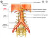
Describe the azygos system of veins? and what is it useful for?
Azygos system of veins consist of a series of longitudinal vessels on each side of body. If IVC block, azygos is useful as it is connected. Consists of:
- Azygos vein on right
- starting from LII at junction between ascending rt lumbar vein and rt subcostal vein; towards the SVC. Receives tributaries from:
- right superior intercostal vein (2,3,4 posterior intercostal veins)
- 5-11th posterior intercostal veins
- other veins like oesophageal, mediastinal, pericardial etc.
- Accessory hemiazygos vein on left
- starts from superior portion of posterior mediastinum and ends at TVIII. Receives from 4th-8th posterior intercostal veins on left
- Hemiazygos vein on right
- starts from junction between left ascending lumbar vein and left subcostal vein. Drain the lower posterior intercostal veins
Describe the thoracic duct system?
- Starts off from cisternal chyli at LII (drain lymph from pelvis and perineum; abdomen; lower limbs) and then at TV joins left brachiocephalic vein (between left internal jugular vein and left subclavian vein)
- Joined by left jugular trunk-drains left side head and neck
- and left subclavian trunk-drains left upper limb

Describe the sympathetic trunks and the sphlanchic nerves which they give off?
- Sympathetic trunks relay pain to CNS. They have 12 ganglion which are connected to spinal nerve via white (pre-ganglionic) and gray (post-ganglionic) rami.
- They give off 3 sphlanchic nerves for the abdomen:
- greater splanchnic nerve from 5th-9th/10th nerve to the celiac ganglion
- lesser splanchnic nerve from 9th/10th to aortorenal ganglion
- least splanchnic nerve from 12th to renal plexus
What causes hoarseness of voice? What investigations should be done?

Hoarseness of voice is caused by compression of the left recurrent laryngeal nerve. This nerve passes through the aortopulmonary window (between the aortic arch and pulmonary trunk). Any lymph node enlargement in this area, often due to pancoast tumour, can cause this.
What is oesophageal rupture due to?
Esophageal rupture is often seen in alcoholics after constant retching.
Increase in intraluminal pressure in the oesophagus is caused by failure of the cricopharyngeal muscle to relax which causes oesophagus to rupture