SM 230a - Neoplasia Flashcards
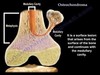
What kind of bone tumor is this?

Osteosarcoma (malignant)
Look for the Codman Triangle
- New sub-periosteal bone that raises the periosteum away from the bone
- This leads to a sunburst reaction

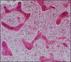
Which tissue is shown in this picture?

Bone marrow
Contains adipocytes and hematopoietic stem cells
What kind of bone tumor is this?
How do you know?

Multiple myeloma
- Lots of plasma cells undergoing clonal proliferation
- Kappa cells only; no lambda
Describe osteoid osteomas
- Benign or malignant:
- Typical patient:
- Common sites:
- Significance:
Osteoid osteoma
- Benign or malignant: Benign
- Typical patient: Young adult (<25), M>F
- Common sites: Cortex of long bones (esp. femur)
- Significance: Excellent prognosis after nidus removal

What bone tumor is this?
How do you know?

Fibrous dysplaisa (benign)
- Expansile, well-circumscribed lesion
- Variable internal density
- May have “ground glass” appearance
Describe the histologic findings of osteoid osteoma
Sclerotic bone surrounding a nidus
The nidus = anastomosing woven bone + osteoblastic rimming + vascular stroma

Describe an osteochondroma
- Benign or malignant:
- Typical patient:
- Common sites:
- Significance:
Osteochondroma
- Benign or malignant: Benign
- Typical patient: Males <25 y/o
- May arise after trauma or radiation
- Common sites: Distal femur, proximal tibia, arises from metaphysis
- Significance: Most common benign bone tumor
-
Good prognosis - rarely transforms to a chondrosarcoma
- Cap > 2 cm suggests malignant transformation
-
Good prognosis - rarely transforms to a chondrosarcoma
Describe the treatment of osteosarcoma
- Preoperative chemotherapy
- 2-6 cycles
- Tumor excision + histologic examination
- <90% tumor necrosis = consider changing chemo + local therapy if margins are positive
- >90% tumor necrosis = continue chemo + local therapy if margins are positive
- Fit for prosthesis
- Postoperative chemotherapy
Which bone tumors are most likely to arise in the epiphysis?
Giant cell tumors (benign)

What kind of bone tumor is this?

Multiple myeloma
- Multiple punched out lesions
- Caused by clonal proliferation of plasma cells
Which bone tumors are most likely to arise in the metaphysis?
- Malignant
- Osteosarcoma
- Chondrosarcoma
- Benign
- Endochondoma
- Osteochondroma

Which benign bone lesion?
“<2cm, appears in long bones, responds to NSAIDs”
Osteoid osteoma
Which tumors appear in bone marrow?
- Myeloma
- Malignant lymphoma
Both are malignant
“CRAB” findings of
HyperCalcemia, Renal insufficiency, Anemia, and lytic Bone lesions
are characteristic of which malignant bone tumor?
Multiple myeloma
All statements are true of osteochondroma except:
- Recurrences may occur if the cartilaginous cap is not completely excised.
- Radiologically and grossly, the tumor projects away from the joint space.
- The cartilaginous cap exhibits microscopic features similar to the cartilage composing the growth plate.
- Presence of a cartilaginous cap >2cm, especially in an adult, is worrisome for sarcomatous transformation.
- In Multiple Hereditary Exostoses (Osteochondromatosis), the incidence of malignant transformation of the cartilaginous cap is over 50%
E.
In Multiple Hereditary Exostoses (Osteochondromatosis), the incidence of malignant transformation of the cartilaginous cap is over 50%
Which benign bone lesion?
“Linked to developmental arrest of bone”
Fibrous dysplasia
Which bone tumor is shown in this image?

Chondroma (benign)
Expansile lytic lesion with calcified matrix
What kind of cartilaginous tumor is this?

Chondroma
- Nodules of mature cartilage within a fatty bone marrow
- Low cellularity, lack of pleomorphism (just mature cartilage)
What is the characteristic features of a bone tumor that has metastasized from another site?
Usually osteolytic (punched-out lesion)
Exception = prostate carcinoma: osteoblastic lesion
Which malignant bone tumor is caused by clonal proiferation of plasma cells?
Multiple myeloma

What kind of bone tumor is shown in this x-ray?

Osteochondroma
- Cartilagionous cap on a bony stalk continuous with the medulla of the bone
- Looks like an ice cream cone
What kind of bone tumor is this?

Giant cell tumor of the bone
- Many multinucleated giant cells
- May look like osteoclasts
- Giant cells have lots of nuclei, lots of cytoplasm
Which benign bone lesion?
“<2cm, appears in long bones, does not respond to NSAIDs”
Osteoblastoma
Which bone tumors are most likely to arise in the middle of the diaphysis of the bone?
- Benign
- Fibrous dysplasia
- Malignant
- Ewing sarcoma
- Myeloma

What bone tumor is this? How do you know?
Fibrous dysplasia
- All elements of bone are present, but they are not maturing
- Irregularly-shaped “C” and “S” shaped spicules of woven bone
- Without osteoblastic rimming
- Spindled and collagenous stroma
What kind of bone tumor is this?

Ewing Sarcoma (malignant)
- Diaphysis of long bones (esp. femur)
- Cortical destruction: “onion skin” pattern
- Some soft tissue extension
Describe Multiple Myeloma
- Benign or malignant:
- What:
- Typical Patient:
- Common sites:
- Prognosis:
- Benign or malignant: Malignant
- What: Clonal proliferation of plasma cells (IgG) -> “punched out” lesions
- Typical Patient: Males 65-70 y/o
- Common sites: Vertebra, ribs, skull, pelvic bones, femur
- Prognosis: Poor

What kind of bone tumor is this?

Osteoid osteoma
Which tissue is shown in this picture?
(The pink part)

Bone
What kind of tumor is this?

Chondrosarcoma (malignant)
- Cartilage matrix w/ stipplied and arc&ring calcifications
- Cortical destruction
- Zero or minimal periosteal reaction

What kind of tumor is this?

Giant cell tumor of the bone
- Soap-bubble appearance
- Lytic lesions in the epiphysis
Describe the radiographic findings of an osteoma
Same density as thte surrounding bone
Made from dense, compact bone

What kind of bone tumor is this?

Chondrosarcoma (malignant)
- Multiple chondrocytes in each lacunae
- More cellular than normal cartilage
- Higher-grade tumors will be very cellular; very little matrix
- Matrix that is there is poorly differentiated
All statements are true of osteosarcoma except:
- In the vast majority of cases, the radiologic appearance of conventional osteosarcoma mimics other benign osseous tumors.
- A relatively higher percentage of pelvic and craniofacial osteosarcomas occur in patients over the age of 30 years.
- By definition, an osteosarcoma is a tumor in which bone matrix is produced by malignant mesenchymal cells.
- Patients exhibiting at least a 90% histological response to chemotherapy have a much improved survival rate compared with patients who show less than 90% response.
- In children, osteosarcoma is a more commonly encountered primary bone tumor than chondrosarcoma
A
In the vast majority of cases, the radiologic appearance of conventional osteosarcoma mimics other benign osseous tumors.
This is false; osteosarcomas have a characteristic “Codman Triangle”

Which bone tumors are most likely to arise on the edge of the diaphysis of the bone?
Osteoid osteoma (benign)
Which bone tumor is characterized by a “bony stalk with a cartilaginous cap?”
Osteochondroma

Describe giant cell tumors of the bone
- Benign or malignant:
- Typical patinet:
- Common sites:
- Significance:
- Benign or malignant: Benign
- Typical patinet: 20-40 yo, F > M
- Common sites: Epiphysis of long bones, esp. knee
- Significance: Locally aggressive, benign. May recur

If a patient has Café-au-lait spots and a bone histology that shows irregularly contoured spicules of woven bone without osteoblastic rimming, which syndrome would you suspect?

McCune-Albright sydrome
Suspect in polyostic fibrous dysplasia;
Also look for endocrine dysfunction
What kind of bone tumor is characterized by
“nocturnal bone pain releived by aspirin”
Osteoid osteoma

Describe the radiographic findings of osteoid osteoma
Radiolucent central focus of woven bone with a vascular stroma.
Surrounding cortical bone is thickened

Which bone tumor causes cortical destruction in an “onion skin” pattern?
Ewing Sarcoma

Describe a Chondrosarcoma
- Benign or malignant:
- What:
- Typical patient:
- Common sites:
- Prognosis:
Chondrosarcoma
- Benign or malignant: Malignant
- What: Neoplasm of chondrocytes
- Typical patient: M>F, Decades 5-7
- Common sites: Central portion of the skeleton, within the medullary cavity
- Pelvis, shoulder, rib
- Prognosis: Depends on grade and surgical accuracy
- Metastases are rare and occur late

What kind of bone tumor is this?
Is it benign or malignant?

Osteochondroma - benign
- Arises from the metaphysis
- Usually in the distal femur or proximal tibia
- Bone + cartilaginous cap; continuous with the medullary cavity
Note; cartilaginous cap suggests malignant transformation
What kind of bone tumor is this?

Osteosarcoma (malignant)
- Malignant cells directly produce osteoid/woven bone
- Highly cellular - cells are highly pleomorphic

What bone tumor is this?
How do you know?

Ewing Sarcoma
- Diffuse proliferation of small, neoplastic cells
- May have focal necrosis
- Cells have rounded nuclei with very little cytoplasm
Describe the appearance of a chondroma on x-ray
Exapansile lytic lesion with a calcified matrix
