Haematology: Blood Coagulation, Haemostasis & Its Investigation Flashcards

1
Q

What is haemostasis?

A
  • Protective process designed to stop bleeding and maintain blood flow
2
Q

The circulatory system of the horseshoe crab contaisn haemolymph. What does haemolymph contain?

A
  • Contains amebocytes which contain:
    • Proteins of the coagulation system
    • Proteins & peptides of the immune system
3
Q

How does the fact that the horseshoe crab’s circulatory system is made up of haemolymph affect the way it fights infection?

A
  • It means that in order to fight infection coagulation of that haemolymph would also have to occur
4
Q

What are some of the functions of haemostasis?

A
  • Respond to tissue injury
  • Curtail blood loss
  • Restore vascular integrity & promote healing
  • Limit infection
5
Q

Name the 4 main components of haemostasis

A
  • Endothelium
  • Coagulation
  • Platelets
  • Fibrinolysis
6
Q

What are the components of a thrombus (blood clot)?

A
  • Fibrin mesh
  • Platelets
  • Red blood cells
7
Q

Name the 3 different stages of haemostatsis and state what occurs during each stage

A
  • Primary haemostasis
    • ​Vasoconstriction
    • Platelet adhesion
    • Platelet aggregation and contraction
  • Secondary haemostasis
    • ​Activation of coagulation factors
    • Formation of fibrin
  • Fibrinolysis
    • ​Activation of fibrinolysis
    • Lysis of the plug
8
Q

What are the different stages of blood clot formation?

A
  • Initiation
  • Amplification
  • Stable clot
  • Lysis
9
Q

Describe the process of primary haemostasis

A
  • Damage to endothelium exposes sub-endothelium to flowing blood
  • This results in sub-endothelial cells releasing Von willebrand factor (VWF)
  • Von willebrand factor binds to platelets and causes them to adhere to exposed collagen
    • Platelets also become activated as a result of this
  • Activated platelets release various pro-thrombotic substances, e.g. thromboxane A2, which help to clump (aggregate) platelets together
    • This forms a loose platelet plug
  • Loose platelet plug contracts to form a dense, adherent plug
  • Platelets in platelet plug form negatively charged phospholipid surface which is required for coagulation
    • Allows coagulation factors to interact much better with each other
10
Q

Why is the phospholipid surface formed by the platelet plug required for secondary haemostasis (coagulation)?

A
  • Allows coagulation factors to interact much better with each other
11
Q

When released by sub-endothelial cells von willebrand factor goes through a conformational change. Describe what this conformational change is and why it occurs

A
  • As a result of increased shear stress due to endothelium damage when released, von willebrand factor goes from a globular form to a filamentous form
  • This filamentous form von willebrand factor then begins to bind to platelets as well as exposed collagen
12
Q

Where are most of the coagulation factors produced?

A
  • In the liver
13
Q

Name some of the coagulation factors and state their functions in coagulation (secondary haemostasis)

A
  • Fibrinogen (I) - Forms clot (fibrin)
  • Prothrombin (II) - Active form (IIa) activates factors I, V, VII, XIII, protein C and platelets
  • Tissue factor (III) - Co-factor of VIIa
  • Factor V - Co-factor of X with which it forms the prothrombinase complex
  • Factor X - Activates prothrombin by forming prothrombinase complex with factor V
  • Von willebrand factor - Binds to VIII, mediates platelet adhesion
14
Q

Explain how the original waterfall hypothesis describes secondary haemostasis (coagulation)

A
  • States that there are 2 seperate pathways for coagulation - extrinsic and intrinsic pathway which convene to form common pathway
  • Extrinsic pathway
    • Tissue damage causes factor VII to activate and become VIIa
    • Activated factor VII (VIIa) binds to tissue factor (TF) to form a complex
    • This complex then activates factor X to form activated factor X (Xa)
  • Intrinsic pathway
    • Blood comes in contact with non-physiological surface
    • This causes kallikrein to activate factor XII
    • Activated factor XII (XIIa) forms complex with high-molecular weight kininogen (HMWK) which activates factor XI
    • Activated XI (XIa) activates factor IX
    • Activated factor IX (IXa) forms complex with activated factor VIII (VIIIa) which activates factor X
  • Common pathway
    • Activated factor X (Xa) causes factor II (prothrombin) to activate and become IIa (thrombin)
    • Thrombin then catalyses conversion of fibrinogen to Fibrin
15
Q

Deficiencies in particular clotting factors may or may not cause bleeding. Deficiencies in which factors cause bleeding?

A
  • Factor VIII deficiency (Hemophilia A)
  • Factor VII deficiency
16
Q

Deficiencies in which factors don’t cause bleeding?

A
  • Factor XII deficiency
17
Q

The thrombin produced by the extrinsic pathway isn’t enough for it to catalyse the production of fibrin. How is the extra thrombin needed to produce fibrin produced?

A
  • Extra thrombin produced via a thrombin burst
  • This thrombin burst is caused as a result of the thrombin formed via the extrinsic pathway activating factors IX and XI
18
Q

How does the revised waterfall hypothesis differ in the way it describes the process of secondary haemostasis (coagulation) comapred with the original waterfall hypothesis?

A
  • Revised waterfall hypothesis states that there aren’t seperate pathways that join together in clotting cascade but instead all the reactions are interlinked
  • Also states that each reaction needs:
    • Ca2+
    • Phospholipid surface
    • ± Specific co-factors
19
Q

Explain the cell-based model of coagulation

A
  • Initiation of coagulation occurs when sub-endothelial tissue is exposed to the circulation at a site of injury.
  • Sub-endothelial cells express tissue factor at their surface, which binds to endogenous activated Factor VII to form a complex
  • This complex binds small amounts of Factor X and Factor V to the exposed endothelial surface, which produce small quantities of thrombin
  • The thrombin activates platelets that are attracted to the site by the process, as well as other plasma-borne clotting factors
  • The activated factors (among them Factor VIII and Factor IX) enable the binding of activated Factor X and Factor V to the surface of activated platelets
  • The surface membranes of the activated platelets have gone through conformational changes which expose the ‘reaction sites’ necessary for continuation of the process
  • Activated platelets with Va and Xa bound foes on to cause prothrombin to be converted to thrombin which leads to the ‘thrombin burst’
  • This thrombin burst is necessary for the large-scale production of fibrin and so the development of an effective clot
20
Q

What are the main functions of fibrinolysis?

A
  • Limiting blood clots
  • Repair and healing
21
Q

Name the main components of fibrinolysis

A
  • Plasminogen
  • Tissue plasminogen activator (t-PA) & urokinase (u-PA)
  • Plasminogen activator inhibitor -1 and -2
  • α2-plasmin inhibitor
22
Q

Describe the process of fibrinolysis

A
  • Tissue plasminogen activator (tPA) catalyses the reaction that converts plasminogen into plasmin
  • Plasmin then degrades cross-linked fibrin of fibrin clot to produce D dimers
23
Q

Plasmin can also degrade non-cross linked fibrin or fibrinogen. What is formed as a result?

A
  • Fibrin degradation products (FDP)
24
Q

How can tPA be used therapuetically?

A
  • tPA and a bacterial activator, streptokinase, can be used in therapeutic thrombolysis for myocardial infarction
25
Q

What role do anticoagulant proteins play in haemostasis?

A
  • Anticoagulant proteins cause the inactivation of the coagulation factors
26
Q

What is thrombosis?

A
  • The formation of a blood clot inside a blood vessel resulting in the obstruction of blood flow
27
Q

Imbalance of factors that contribute to haemostasis can lead to thrombosis. What

A
  • Increased activation of coagulation factors/inability for coagulation factors to be deactivated
  • Increased no. of platelets
  • Decreased no. of fibrinolytic factors
  • Decreased no. of anticoagulant proteins
28
Q

What condition can thrombosis lead to?

A
  • Deep vein thrombosis
29
Q

What condition can deep vein thrombosis become if it worsens? What are the symptoms of this condition?

A
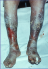
  • Chronic venous insufficiency, symptoms include:
    • Atrophic changes
    • Hyperpigmentation
    • Ulceration
    • Infection
30
Q

Imbalance of factors that contribute to haemostasis can also lead to bleeding. Describe the imbalances in these factors that lead to bleeding

A
  • Absence of clotting factors or platelets
  • Presence of too many fibrinolytic factors or anticoagulant factors
31
Q

What are the principles of clotting tests?

A
  • Incubate plasma with reagents necessary for coagulation
    • Phospholipid, co-factors
    • Trigger or activator
    • Calcium
  • Measure time taken to form fibrin clot
32
Q

What is prothrombin time (PT)?

A
  • It’s a blood test that evaluates the extrinsic pathway and to a lesser extent common pathway
  • It is Tissue Factor driven
33
Q

What is activated partial thromboplastin time (APTT)?

A
  • It’s a blood test that evaluates the intrinsic pathway and to a lesser extent common pathway
  • It is contact activated
34
Q

What is thrombin time (TM)?

A
  • It’s a blood test that evaluates how quickly fibrinogen can be converted into fibrin
35
Q

For each of the clotting tests state the average time it takes for them to be completed

A
  • Prothrombin time: 11-13 seconds
  • Partial activated thrmoboplastin time: 30-45 seconds
  • Thrombin time: 12-14 seconds
36
Q

What are the limitations of the clotting tests?

A
  • They only test the initiation stage of the process of blood clot formation rather than the entire process
37
Q

What anticoagulant is used when collecting blood for a clotting test?

A
  • Blood is anticoagulated with 3.2 % (0.109 M) Sodium citrate
38
Q

How much blood and how much anticoagulant is needed in order for the blood collected to be used in a clotting test?

A
  • Most tubes contain 0.3 mL anticoagulant and require 2.7 mLs of blood.
39
Q

Why is it important to fill the tube with the exact quantity of blood needed for a clotting test?

A
  • Under filling the tube yields grossly inaccurate results.
40
Q

What are some pre-analytical errors that can occur during a clotting test?

A
  • Problems with blue-top tube
    • Partial fill tubes
    • Vacuum leak and citrate evaporation
  • Problems with phlebotomy
    • Heparin contamination
    • Wrong label
    • Slow fill
    • Underfill
    • Vigorous shaking
  • Biological effects
    • Heamatocrit ≥55 or ≤15
    • Lipaemia, hyperbilirubinaemia, haemolysis
  • Laboratory errors
    • Delay in testing
    • Prolonged incubation at 37°C
    • Freeze/thaw deterioration
41
Q

What are the 2 reasons why a clotting test time would be prolonged?

A
  • Clotting factor deficiency
  • Something else interferring with clot formation
42
Q

How can you differentiate the reason for a prolonged clotting time?

A
  • You perform a mixing study
  • This involves mixing the prolonged clotting time plasma with control plasma at a 50/50 mix
43
Q

What does it mean if you perform a mixing study and the time for a clotting test is no longer prolonged?

A
  • It means the reason for the prolonged time of the clotting test was that there was a deficiency in a clotting factor/s
44
Q

What does it mean if you perform a mixing study and the time for a clotting test is still prolonged?

A
  • It means the reason for the prolonged time of the clotting test was the presence of an antibody or another thing interefering with clot formation
45
Q

What is D-dimer testing?

A
  • A test that measures levels of the D-dimer, a fibrin degradation product
46
Q

What conditions are associated with increased D-dimer levels?

A
  • Found elevated in the situation of enhanced fibrinolysis (Thrombosis, Disseminated intravascular coagulation)