MSK 2 - Upper Limbs Flashcards

1
Q

a

A

Lesser tubercle

2
Q

b

A

Coronoid fossa

3
Q

c

A

Trochlea

4
Q

d

A

Greater tubercle

5
Q

e

A

Intertubercular groove

6
Q

f

A

Deltoid tuberosity

7
Q

g

A

Radial fossa

8
Q

h

A

Capitulum

9
Q

i

A

Surgical neck

10
Q

j

A

Radial groove

11
Q

k

A

Head

12
Q

l

A

Anatomical neck

13
Q

m

A

Shaft/diaphysis

14
Q

n

A

Olecranon fossa

15
Q

o

A

Medial epicondyle

16
Q

what are muscle compartments?

A

An important concept when studying anatomy of the extremities is the arrangement of muscle groups within fascial compartments.

Fascial compartments contain groups of muscles and neurovascular structures enveloped by a layer of connective (fibrous) tissue.

Muscles in each compartment often (but not always) act similarly on a joint, sharing a common nerve and blood supply

17
Q

what is in the Anterior Compartment of the Arm?

A

The anterior compartment contains 3 important muscles and their accompanying neurovascular supply. The muscles act on the elbow joint and superior radio-ulnar joint

18
Q

1

A

Biceps brachii

19
Q

2

A

Brachialis

20
Q

3

A

Musculocutaneous nerve

21
Q

4

A

Coracobrachialis

22
Q

where is the biceps brachii located?

A

The biceps brachii is the most superficial muscle of the anterior compartment.
It arises from the scapula via. two heads. Both heads unite at the distal third of the upper arm forming a short tendon which inserts onto the radial tuberosity.
It also attaches through the bicipital aponeurosis to the deep fascia of the forearm
The muscle has a powerful action on the shoulder, elbow and radio-ulnar joints

23
Q

The short head of the biceps muscle originates from the ________ process of the scapula

A

coracoid

24
Q

The long head of the biceps muscle originates from the __________ tubercle of the scapula

Its tendon passes through the shoulder joint and runs down the _______ groove of the humerus

A

supraglenoid

bicipital

25
Q

what is the function of the biceps brachii?

A

It flexes both the shoulder and elbow joint (action)

At the superior radio-ulnar joint it is involved in supination (the action in which the radius rotates over the ulna)

26
Q

The coracobrachialis slender, rounded muscle passing from its origin at the tip of the _______ process and inserting to the ______ aspect of the mid-shaft of the humerus

A

coracoid

medial

27
Q

what is the action of the coracobrachialis?

A

Its action is to flex the arm at the shoulder joint

28
Q

The brachialis muscle arises from the _______ surface of the distal shaft of the humerus. It is a deep muscle of the anterior compartment, passing downwards and attaching to the _______ process of the ____

A

anterior

coronoid

ulna

29
Q

what is the fucntion of the brachialis?

A

Its main action is to flex the elbow joint

30
Q

where does the musculocutaneous nerve arise from?

A

This nerve arises as a terminal branch from the lateral cord of the brachial plexus

31
Q

what is the function of the musculocutaneous nerve?

A

It is the mainnerve of the anterior compartment of arm, supplying all the muscles within this compartment

32
Q

what is the location and route of the musculocutaneous nerve?

A

It enters the arm by perforating the coracobrachialis, descending distally between the biceps brachii and brachialis muscles. After crossing the elbow joint it gives off its terminal branch to supply the skin as the lateral cutaneous nerve of the forearm

33
Q

What is the segmental or root value of the musculocutaneous nerve?

A

C 5 to C 7

34
Q

Does the musculocutaneous nerve supply any muscles in the forearm or hand?

A

no

35
Q

1

A

Axillary artery

36
Q

2

A

Median nerve

37
Q

3

A

Ulnar nerve

38
Q

4

A

radial nerve

39
Q

The brachial artery is a direct continuation of the _______ artery

A

axillary

40
Q

what is the route of the brachial artery?

A

It begins at the lower border of teres major and ends in the cubital fossa by dividing into radial and ulnar arteries, close to the neck of the radius

The profunda brachii (or deep artery of the arm) is one of the main branches given off passing deep to the triceps muscle, along with the radial nerve

41
Q

Clinical:

Non-invasive blood pressure (NIBP) measurement can be taken from a number of sites in the body, but most commonly on the arm through compression of the brachial artery on the humerus using a _________________.

Using a stethoscope to auscultate the ________ artery at the cubital fossa, changes in the flow of blood are heard during inflation and deflation of the cuff. These changes in sound (_________ sounds) correspond to different phases of the blood ________ (systolic and diastolic) and allows measurement of blood pressure in millimetres of mercury (mmHg).

A

sphygmomanometer

brachial

Korotkoff’s

pressure

42
Q

Median nevre in the arm:

Arises in the axilla by two roots – one from the _______ cord and one from the ______ cord of the brachial plexus. It descends along the ______ side of axillary artery and upper part of brachial artery.

In the middle part of the arm the nerve crosses to the ______ aspect of the brachial artery and enters the cubital fossa. It gives off __ branches either in the axilla or in the upper arm

A

lateral

medial

lateral

medial

no

43
Q

Ulnar nevre in the arm:

The ulnar nerve arises from the ______ cord of the brachial plexus. It descends along the ______ side of brachial artery and then it enters the _______ (flexor) compartment of arm through the medial intermuscular septum. It runs along the medial head of triceps and to the lie behind the ______ epicondyle at the elbow

A

medial

medial

anterior

medial

44
Q

Are any muscles in the arm supplied by the median or ulnar nerve?

A

no

45
Q

what occupies all of the posterior compartment of the arm?

A

The triceps occupies the entire posterior compartment of the arm

46
Q

what does the triceps arise from and where does it insert?

A

It arises by a long head from the scapula and by two shorter heads - the lateral and medial heads - from the humerus. The common tendon of triceps is inserted into olecranon process of the ulna

47
Q

olecranon process is part of what bone?

A

ulna

48
Q

what is the ocmmon tendon of the triceps?

A

this large tendon is the convergence of the 3 heads of the triceps muscle

49
Q

Which head of the triceps attaches onto the infra-glenoid tubercle of the scapula?

A

long

50
Q

What is the main action of triceps on the elbow joint?

A

extension

51
Q

what is the anconeus?

A

the anconeus muscle is a small muscle at the elbow and aids the triceps muscle action at the elbow joint
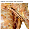
52
Q

What is the action of the triceps and anconeus muscle?

A

extension

53
Q

What is the nerve supply to triceps and anconeus?

A

radial nerve

54
Q

radial nerve:

Is the one of the terminal branches of the ________ cord of the brachial plexus and nerve to the ________ compartment of arm and forearm

A

posterior

posterior

55
Q

what does the radial nerve innervate?

A

It innervates all the extensor muscles of elbow and wrist joint

56
Q

The radial nerve enters the arm anterior to the ____ head of triceps and runs with the _______ ______ artery (deep artery of the arm), a main branch of the brachial artery

It descends down the around the midshaft region of the humerus in the _____ groove, then appears as one of the most lateral structures of the distal arm lying between the _________ and ____________ muscles

A

long

profunda brachia

radial

brachialis

brachioradialis

57
Q

What is the segmental (root value) value of the radial nerve?

A

C5 to T1

58
Q

What is the characteristic clinical sign of radial nerve injury?

A

wrist dorp

59
Q

Clinical: Nerve injuries in the arm

We’ve already looked at how the nerve supply to the upper limb can be affected through damage to the roots of the brachial plexus.

The terminal branches are also at risk with fractures of the humerus

Match the common site of humerus fracture to potential nerve injury:

  1. Radial Nerve
  2. Ulnar Nerve
  3. Axillary Nerve
    a. Fracture to surgical neck
    b. Fracture to medial epicondyle
    c. Fracture to spiral groove
A
  1. 3 - a
  2. 2 - b
  3. 1 - c
60
Q

what is the antecubital fossa?

A

The antecubital (or cubital) fossa is a triangular depression or hollow lying in front of the elbow. It corresponds to the popliteal fossa at the back of the knee.

It acts as a conduit for passage of major neurovascular structures from the arm down to the forearm, and ultimately the hand.

61
Q

what are the boundaries of the antecubital foosa?

A

superior: the line joining the medial and lateral humeral epicondyles (epicondylar line)
lateral: medial border of brachioradialis
medial: lateral border of pronator teres
apex: formed by the meeting of the lateral and medial boundaries (proximal intersection of the brachioradialis and pronator teres)

roof:

  • deep fascia of forearm reinforced medially by the bicipital aponeurosis
  • medial cutaneous nerve of forearm; lateral cutaneous nerve of forearm
  • basilic vein; cephalic vein; median cubital vein
    floor: brachialis muscle (medial), and supinator muscle (lateral)
62
Q

what is the contents of the antecubital fossa?

A

From medial to lateral:

median nerve

brachial artery and brachial veins (venae comitantes)

tendon of biceps

radial nerve

posterior interosseous branch of radial nerve

63
Q

a

A

Brachial artery

64
Q

b

A

Median nerve

65
Q

c

A

Biceps

66
Q

d

A

Bicipital aponeurosis

67
Q

e

A

brachioradialis

68
Q

f

A

Posterior interosseous nerve

69
Q

g

A

Extensor carpi radialis longus

70
Q

Which vein lies anterior to the roof of the antecubital fossa?

A

Median cubital vein

71
Q

At what level does the brachial artery bifurcate into the radial and ulnar artery?

A

neck of the radius in the cubital fossa

72
Q

The cephalic vein lies over the ______ aspect, while the basilic artery lies over the _____ aspect of the fossa

A

lateral

medial

73
Q

what type of joint is the elbow joint?

A

This is a synovial hinge type of joint, located 2- 3 cm below the humeral epicondyles

74
Q

a

A

Lateral epicondyle

75
Q

b

A

Coronoid fossa

76
Q

c

A

Radius head

77
Q

d

A

Radial notch on the ulnar bone

78
Q

e

A

Medial epicondyle

79
Q

f

A

Olecranon (ulnar)

80
Q

what are the ligaments of the elbow joint?

A

Support to the joint is provided by the thickening of the medial and lateral aspects of the capsule – this forming the medial and lateral collateral ligaments.

The capsule is loose on both the anterior and posterior aspect of the joint to allow for movement

81
Q

Which ligament holds the ulnar in place?

A

Ulnar collateral ligament

82
Q

Which ligament holds the head of the radius?

A

annular ligament

83
Q

Which muscles cause flexion of the elbow joint?

A
  1. biceps brachii
  2. brachialis
  3. brachioradialis (weakest out the free, mid point)
84
Q

Which muscles cause extension of the elbow joint?

A
  1. triceps
  2. Anconeus muscle
85
Q

Clinical: Eponymous Elbows

Students, Tennis, Golfers…

A number of structures can be injured in the elbow through overuse and injury to these structures comes with a number of eponymous titles

what are the different ones?

A

Students Elbow

The subcutaneous olecranon bursa is one of these structures and can be damaged through repeated excessive pressure and friction. This inflammation (commonly occurring when you’re leaning on them studying) is called olecranon bursitis

Tennis elbow

Repetitive overuse of the muscles in the forearm causes inflammation of the muscle tendons attached to the lateral epicondyle of the elbow – this is lateral epicondylitis

Golfers elbow

Repetitive overuse of the muscles in the forearm causes inflammation of the muscle tendons attached to the medial epicondyle of the elbow – this is medial epicondylitis

86
Q

The radius and ulnar are the bones found in the forarm, they articulate at _ points

Both of these joints (proximal and distal radio-ulnar joints) are ______ type synovial joints, allowing ________ and _______ of the forearm

The proximal radio-ulnar joint is supported by another ligament arising at the elbow – this keeps the head of the radius in place. What is the name of this ligament?

A

2

pivot

supination

pronation

Annular ligament

87
Q

Supination and pronation are the movements of the radio-ulnar joints

what is which?

A
88
Q

Movement at Radio-Ulnar Joint = supination

Muscles involved = ?

A
  1. supinator muscle
  2. biceps brachii muscle
89
Q

Movement at Radio-Ulnar Joint = Pronation

Muscles involved = ?

A
  1. pronator teres muscle
  2. pronator quadratus muscle
  3. brachioradialis