9.12 Vessels of the Lower Limb Flashcards
The origin, course and branches of the major arteries of the lower limb • The locations at which the femoral, popliteal, dorsalis pedis and posterior tibial pulses can be felt • The course of the principle veins of the lower limb • The role of the perforator veins and the function of the “muscle pump”. •The organisation of inguinal lymph nodes and their role in lymphatic drainage of the limb, trunk skin and perineum (38 cards)
The abdominal aorta splits at what level?
What does it split into and what are the branches of these?
What do the supply?
It splits at the level of L4 on posterior abdominal wall into the left and right common ileac arteries.
These further branch into the external ileac artery (lower limb) and the internal ileac artery (into the pelvis).

What happens to the external ileac artery as it descends down the foot?
- The arteries change their name as the progress down the length of the limb.
- They give off branches to supply adjacent structures as they descend
- They follow the nerves of the foot
Over what surface do the arteries generally course over?
What is the implication of this?
They generally course over flexor surfaces (anterior in the arm and posterior in the legs) and thus obtain protection as they pass over them.
What is the first major branch coming from the external ileac artery and where does it become this?
The external ileac artery courses over front of the head of femur and passing midway between ASIS and pubic crest under the inguinal ligament (midpoint) into the femoral triangle.
Before this, there are small branches that come off to supply the lower abdominal wall and crest of the ileum

How and when does the femoral artery make its way into the flexor compartment?
It starts anteromedially trying to get to flexor compartment and achieves this at the popliteal fossa protected here. (Thus it is vulnerable as it passing more proximally)
Describe the location of the artery in relation to other structures within the femoral triangle
It is the lateral most structure within the femoral sheath of the femoral triangle (but the femoral nerve lies lateral to it outside the sheath)
However as it courses down towards the apex of the triangle, the artery comes to lie anterior (superficial) to the femoral vein

Where can the femoral artery be palpated?
In the femoral triangle as it comes across the head of the femur

In the femoral triangle, the femoral artery divides into a superficial and deep part. What are each parts?
What separates them?
- In the femoral triangle, the (deep femoral artery) profunda femoris artery arises from the posterolateral aspect of the femoral artery.
- THIS SUPPLIES THE THIGH
- The superficial femoral artery continues down on the medial side and wraps around to the rest of the leg.
They lie superficial to and deep to the adductor longus
What are the important branches of the profunda femoral artery?
“Put My Leg Down Please”
Profunda gives off
- Medial Femoral Circumflex Artery (very important)
- Lateral Femoral Circumflex Atery
- Descending branch of the lateral circumflex femoral artery
These circumflex arteries encircle the neck of the femur and pass throgh the retinacular bundle of fibres to supply neck to the epipyseal line during growth from below.
- Perforating branches

Describe the perforating branches of the profunda femoris artery
A group of arteries that perforate the adductor magnus, contributing to the supply of the muscles and bone in the medial and posterior thigh.
- 2 branches above adductor longus
- One behind adductor longus
- The termination is underneath adductor longus

Both the superficial femoral artery and the profunda femoral artery are very large vessels.
What is the implication of this?
Injury or direct trauma to the area leads to significant blood loss - particularly anteromedial of the thigh
Describe the path of the superficial femoral artery down the leg?
It descends continues down the anterior surface of the thigh through the adductor canal suppling the anterior thigh muscles as it goes.
The adductor canal ends at an opening in the adductor magnus, called the adductor hiatus. The femoral artery moves through this opening, and enters the posterior compartment of the thigh, to behind the knee.
The femoral artery now known as the popliteal artery.
It has gives off no branches until it is at the knee joing

How do the perforating branches access the posterior thigh muscles?
Through spaces where the adductor magnus attaches to bone (along the medial lip of the linea aspera to the adductor tubercle)

Describe the path of the popliteal artery
It descends down the posterior thigh giving off genicular branches that supply the knee joint. It moves down through the popliteal fossa and exits it between gastrocnemius and popliteas muscle.
It then gives off the anterior and posterior tibial arteries at the lower border of the popliteus muscle.
The posterior tibial artery continues and divides again to give off the fibular artery (continues laterally down while the tibial goes medially)

Where can the popliteal muscle be palpated in the leg?
The posterior aspect of the knee joint
(medially)

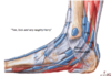
Describe the path of the anterior tibial artery
It goes across the front of the interosseus membrane and it continues across the dosum of the foot where it changes its name to dorsalis pedis artery (palpable on dorsum of the foot).
Here it divides again into a proximal and distal arcade and then digital branches to the digits.

Describe the path of the posterior tibial artery down the leg
It continues inferiorly on the medial side of the posterior leg, along the surface of the deep muscles.
It accompanies the tibial nerve in entering the sole of the foot via thetarsal tunnel.
Together they pass behind medial mallelous, here it divides into medial plantar artery to large toe and lateral plantar artery (more significant contribution to the foot itself)

Describe the path of the fibular artery down the leg
The fibular artery is given along the descent of the posterior tibial artery. It moves laterally down the leg and penetrates the lateral compartment of the leg. It thus supplies the muscles in the compartment: fibularis brevis and fibularis longus (and adjacent muscles in the posterior compartment)

What part of the tibia is most vulnerable to fracture and why?
The distal third of tibia is vulnerable to fracture due to potential of poor blood supply to the area (not as many anastomotic branches)
As the anterior artery continues down to the dorsum of the foot, what structures does it pass through?
It is bound down by the extensor retinaculum and by the fibular retinaculum with the deep fibular nerve in the space.

Where can the dosalis pedis artery be palpated?
The dorsalis pedis pulse is found by palpating on the dorsum of the foot, just lateral to extensor hallucis longus tendon.
Through where does the posterior tibial artery enter the foot?
What is the significance of this?
Posterior tibial artery comes down with the posterior tibial nerve and passes deep to the flexor retinaculum (and through the tarsal tunnel)
It can be impacted in tarsal tunnel syndrome (but this syndrome is most commonly due to nerve problems)
Where can the posterior tibial artery be palpated?
At the point just under medial mallelous

Where in the compartments of the leg do the vessels run through?
- Deep in anterior compartment resting against the immovable interosseus membrane is a bundle of neurovascular structures (vena comitante, anterior tibail vein and nerve)
- Lateral compartment has nerve
- The deep compartment of the posterior leg, contains neurovascular bundle to the posterior compartement and one for the fibular compartments (2)


