Anatomy Flashcards
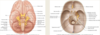
the SCALP
- Skin
- Connective tissue
- Aponeurotic tissue
- Loose Connective Tissue
- Pericranium
- extends over the neurocranium
- Innervation to the scalp is from the trigeminal nerve and spinal cutaneous nerves
Bones of the skull
- the Neurocranium and the viscerocranium = 22 bones
- Neurcroniaum bones
- Occipital
- 2 x temporal
- 2x parietal
- Sphneoid
- ethmoid
- frontal
- Viscercranium
- 2x nasal conchae
- 2x nasal bones
- 2x maxilla
- 2x palatine bones
- 2x zygomatic bones
- 2x lacrimal bones
- vomer
- mandible
Foramina of the cranium
Cribiform: plate CN 1
Optic Canal: CN 2, Ophthalmic A
Superior Orbital Fissure: CN 3,4,6, 5 V1 (LFTSNIA)
Rotundum: CN5 V2
Ovale: CN 5 V3, AMMA
Spinosum: Middle Meningeal Artery
Lacerum: *Carotid Artery
Internal Acoustic Meatus: CN 7 and 8
Jugular foramen: CN 9, 10,11, IJV
Hypoglossal Canal: CN12
Magnum: Spinal Cord
Dura Mater
- 2 layers of dura mater around the brain: create a sinus
- endosteal layer: outer layer stops at the foreman magnum, only lines the skull
- Meningeal layer around the brain and the skull
- falx cerebri: separates cerebral hemispheres
- tentorium cerebelli: separates the cerebellar hemisphere from the cerebral hemispheres
- falx cerebelli- separates cerebellar hemispheres
- supplied by CNX 5 (trigeminal), 10(vagus), C1-3 and sympathetic
- blood supply is the middle meningeal artery
Arachnoid Mater
- thin avascular layer between the pia and the dura
- loosely applied layer with projections
- all structures passing to/ from brain pass through the subarachnoid space
- Subarachnoid space contains cerebrospinal fluid produced by choroid plexus in brain ventricles
- the CSF provides buoyancy for the brains
- any excess CSG in the subarachnoid space goes to the arachnoid granulations which project into the superior sagittal sinus (the space between the two dura layers)
- these granulations affect the transfer of CFS into the venous system
Pia Mater
- very delicate vascular membrane: nourishes
- closely invests brain following gyri/sulci
- Cerebral arteries entering the brain carry a bit of the pia with it
Leptomengittis
- infection and inflammation in the arachnoid and the pia mater
- infection may enter the subarachnoid space and enter into the blood (septicemia)
Dural Sinuses
- Sinuses sit between the dural folds.
- Drained blood and CSF from the brain via cerebral veins.
- Communicate with the veins of the skull and scalp.
- Thick-walled endothelium. No Valves or smooth muscle.
- Drain into the internal jugular vein.
Blood supply to the brain

Stroke
Occurs when the supply of blood to the brain is reduced or blocked completely, which prevents brain tissue from getting oxygen and nutrients.
Describe the embryological stage of the face
- Development begins in week 4 and forms from 5 swellings.
- Frontonasal
- Maxillary X2
- Mandibular X2
- By week 5 two events;
- Maxillary prominences enlarge in medial direction
- Nasal placodes appear and form medial and lateral processes.
- The medial nasal processes merge towards each other and form intermaxillary segment
- The maxillary prominences fuse with the lateral and medial nasal processes to form the upper lip

What are the key muscles of the face and what are there roles
- Occipitofrontalis- elevates eyebrows
- Orbicularis oculi- closes eyelids
- Orbicularis oris- closes mouth
- Zygomaticus major- elevates labial commissure
- Buccinator- compresses cheek
- Platysma- depresses mandible against resistance, tenses

Innervation of the fascial muscles
- Cutaneous innervation by the Trigeminal nerve (Cranial nerve 5)
- All muscles of facial expression supplied by the Facial Nerve (Cranial nerve 7)
- Sensory, Taste and a general motor and visceral motor nerve
- Exits through:
- the internal acoustic meatus
- facial canal
- stylomastoid foramen
- Branches: Tempra facial and Vercofacial branch from the Posterior auricular nerve
- Motor-Posterior auricular, temporal, zygomatic, buccal, marginal mandibular, cervical
- Parasympathetic- branches: to pterygopalatine ganglion.
- Taste- via chorda tympani via lingual nerve from anterior two thirds of tongue.
- General sensory: skin over external auditory meatus.
`
The Parotid glands place in the face
- largest of the three salivary glands in the head and is superficial to the muscles in the face
- Parotid duct leaves gland at anterior edge and passes towards the corner of the mouth but turns deep through buccinator.
- The parotid duct opens into oral cavity at upper second molar tooth.
- The retromandibular vein and external carotid artery run through it.
- Facial nerve passes through the parotid gland.

Muscles responsible for mastication (chewing)
- Temporalis- elevation, retraction
- Masseter- elevation
- Medial Pterygoid- elevation, side to side
- Lateral Pterygoid- protrusion and depression
Innervation of the mastication muscles
- supplied by the motor nerve of V3
- the Mandibular branch of the trigeminal nerve
Cranial NErve 5
- the Trigeminal Nerve
- it’s a somatic and somatic motor to derivatives of the 1st pharyngeal arch
- It has three main divisions
- Ophthalmic (V1): exits through the Superior orbital fissure
- Maxillary (V2): exits through the Foramen Rotundum
- Mandibular (V3): exits through the Foramen Ovale

Explain the Ophthalmic Nerve V1
- Type: Sensory fibres skin, mucous membranes, conjunctiva, front of head and nose
- Path: Branches into lacrimal, nasociliary, frontal
- Exit: Superior Orbital Fissure

Explain the Maxillary Nerve V2
- Type: Sensory fibres dura, nasal, upper cheek, lip, teeth
- Path: Enters pterygopalatine fossa, gives off branches to the pterygopalatine ganglion, through the inferior orbital fissure.
- Exit: Foramen rotundum
- Branches: Infraorbital, zygomatic, superior alveolar.

Explain the Mandibular Nerve V3
- Type: Mixed, sensory, lower face, lip, teeth. Motor to muscles of mastication
- Exit: Foramen Ovale
- Branches:
- Sensory-auriculotemporal, buccal, lingual, inferior alveolar, mental
- Motor- temporalis, masseter, medial and lateral pterygoids, nerve to mylohyoid
- Parasympathetic (hitchhiking)- to salivary glands

The Temporal and infratemporal fossa
- The temporal fossa is a fan-shaped space that is located on the lateral surface of the skull.
- The temporal fossa contains: temporalis muscle, branches of V2
- The infratemporal fossa is inferior to the temporal fossa.
- The infratemporal fossa contains: medial and lateral pterygoids, maxillary artery, V3, branches of the facial nerve, glossopharyngeal nerve and pterygoid plexus of veins.

Describe the arterial supply to the face
arterial supply from the external carotid artery
- Lingual
- Facial
- Maxiallry
- Superficial temporal
in this order

Venous drainage of the face
- the facial vein drains the majority of the face, starting near the eye
- the facial vein passes inferiorly and drains into the internal jugular vein
- the superficial temporal vein drains into the external jugular vein

Draw out this image

Overview of the tongue
- The tongue is divided into an anterior two thirds (oral) and a posterior third (pharyngeal), demarked by a V shaped sulcus (terminal sulcus)
- Papillae cover the tongue and all except filiform have taste buds on their surface.
- Undersurface contains medial fold (frenulum) which internally separates the right and left sides of the tongue.

Muscles of the tongue
- Intrinsic muscle: creates precise movement for speech, eating and swallowing.
-
Extrinsic:
- Genioglossus: depresses and protrudes the tongue
- Hyoglossus: depresses tongue
- Styloglossus: retracts tongue
- Patalglossus: elevates back tongue and depresses soft palate
Give an overview of Cranial Nerve 12: Hypoglossal
- provides motor function to intrinsic and extrinsic muscles of the tongue (save palatoglossus)
- exits through the hypoglossal canal: this is just lateral of the Foramen Magnum

Give an overview of the framework of the larynx
- Provides a protective sphincter for the air passages and produces phonation, made up of cartilages:
- Thyroid, Cricoid, Arytenoid, Cuneiform, Corniculate and Epiglottis
- Contains ligaments, most important:
- Vestibular and the Vocal ligament.

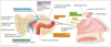
Give an overview of the Pharynx
- the most posterior part of the neck, situated behind the nasal cavity and the larynx.
- It’s a funnel-shaped tube with three sections:
- Nasopharynx
- Laryngopharynx
- Oropharynx
- Muscles consist of three constrictors: Superior, Middle and Inferior
- also the stylopharyngeus

Give an overview of the neck Fascia
- The deep fascia of the neck creates three important layers
- Pretracheal fascia- Pink
- Prevertebral fascia- Blue
- Investing fascia- Green
- Important as reduces the spread of infection
- Enables structures to move past each other in movement and swallowing etc
- Carotid sheath (Red) blends with the pretracheal and prevertebral fascia. It contains the
- Common and internal carotid arteries.
- Internal jugular vein.
- Vagus nerve (CN 10).
- Some deep cercal lymph nodes.
- Carotid sinus nerve

Identify the surface anatomy of the Neck
- the triangles
- The neck is divided into the anterior and lateral/posterior compartment by the presence of the sternocleidomastoid muscle.
- The anterior compartment is further subdivided into three paired triangles and one unpaired;
- the unpaired submental triangle,
- the paired submandibular, carotid and muscular triangle.
- The posterior compartment is bounded posteriorly by the trapezius muscle and is divided into two triangles by the presence of the posterior belly of omohyoid into:
- the large occipital triangle
- the smaller omoclavicular triangle.

Muscles and contents of the Anterior triangle
- Submandibular triangle bounded by: anterior and posterior bellies of digastric.
- Contains: the submandibular gland, facial artery and vein.
- Submental triangle bounded by: digastric.
- Contains lymph nodes.
- Muscular triangle bounded by: omohyoid, Supraclavicular Muscles SCM
- Contains supra and infrahyoid muscles
- Carotid triangle bounded by: omohyoid, stylohyoid, digastric, SCM.
- Contains: common carotid artery, IJV, hypoglossal nerve, vagus nerve, accessory nerve.

Give details of the muscles of the muscular triangle
Suprahyoid muscles
- Stylohyoid, digastric, mylohyoid and geniohyoid
- Innervation:
- Facial nerve for the stylohyoid and posterior belly of digastric.
- Mylohyoid by CN 5 and
- Geniohyoid by CN 12
Infrahyoid muscles
- Omohyoid, sternohyoid, thyrohyoid, and sternothyroid
- Innervation: C1-C3 of ansa cervicalis

Muscles and contents of the posterior triangle?
- Posterior triangle contains: Subclavian artery, EJV, Brachial plexus, CN 11, Cervical plexus
- Occipital triangle bounded by: SCM, trapezius and omohyoid.
- Supraclavicular triangle bounded by: clavicular head of SCM, clavicle and omohyoid

Describe arterial supply of the neck and throat
- Brachiocephalic ÷ Common carotid ÷ Internal and external carotids
- External carotid ÷ 6 branches to supply the neck and head
- Superior thyroid,
- Ascending pharyngeal,
- Lingual,
- Facial,
- Maxillary,
- Superficial temporal
- Subclavian arteries supplied by the inferior thyroid
- Internal carotid forms no branches until it’s inside skull

Give an Overview of Cranial Nerve 8: Vestibulaochlear Nerve
- Type: Sensory, special somatic afferent for hearing, equilibrium and motion.
- Path: Divides into Vestibular and Cochlear Nerves.
- Exit: Internal acoustic meatus.

Give an Overview of the Cervical Plexus
- Roots of cervical plexus C1-C4 lies anterior to Levator scapulae.
- The cervical plexus provides cutaneous branches and deep motor branches (ansa cervicalis and phrenic nerve)

Give an Overview of the Brachial Plexus
- Roots of the brachial plexus C5-T1 appear between the anterior and middle scalene muscles.
- Five rami unite to form the three trunks of the brachial plexus
- Supply cutaneous and motor to the upper limb.

What are the 12 Cranial Nerves?
- Olfactory: Smell.
- Optic: Sensory nerve of vision it is more correctly called a brain tract.
- Oculomotor: innervates four of the extrinsic eye muscles—muscles that move the eyeball in the orbit.
- Trochlear: innervates an extrinsic eye muscle that hooks through a pulley-shaped ligament in the orbit.
- Trigeminal: The trigeminal nerve provides general sensory innervation to the face and motor innervation to the chewing muscles.
- Abducens: innervates the muscle that abducts the eyeball (turns the eye laterally).
- Facial: This nerve innervates the muscles of facial expression as well as other structures.
- Vestibulocochlear: sensory nerve of hearing and equilibrium/ balance
- Glossopharyngeal: innervates structures in the tongue and pharynx,
- Vagus: this nerve “wanders” beyond the head into the thorax and abdomen.
- Accessory: carries motor innervation to the trapezius and sternocleidomastoid muscles.
- Hypoglossal: The name hypoglossal means “below the tongue.” This nerve runs inferior to the tongue and innervates the tongue muscles.

Give an overview of the structure of the Nose
- Bones: comprised of paired Nasal bones, Maxillae, Frontal bone and Septum.
- Cartilages: comprised of 3 main lateral nasal cartilage, major alar cartilage and septal cartilage• Septum divides the chamber into two cavities
- Internal surface: lateral wall has three projections; superior, middle and inferior conchae
- Nasal cavity opens into the nasopharynx

Give an Overview of Cranial Nerve 1: Olfactory
- Special sensory afferent nerve
- Exits through the Cribiform Plate
Explain Olfaction
- Olfaction is aided by the airflow through the nasal cavity
- Conchae help warm and moisten the air
- Olfaction receptors are in the epithelium lining the roof and walls,
- The epithelium secretes a mucus fluid, odoriferous gases are dissolved into the fluid and then detected by the olfactory nerves

Explain the purpose and organisation of the Paranasal sinuses

- Paranasal sinuses are air filled extensions of the nasal cavity
- Paired Frontal sinuses in frontal bone that drain through frontonasal duct.
- Ethmoidal cells (sinuses) are invaginations in the ethmoid bone and drain into middle meatus (if anterior, or middle) or superior meatus (if posterior).
- Sphenoidal sinuses in sphenoid bone that drain into spheno-ethmoidal recess.
- Paired Maxillary sinuses in maxilla drain into middle meatus.

7 of them
Name the bones that make up the Orbit

Frontal, Maxilla, Ethmoidal, Lacrimal, Sphenoidal, Temporal-zygomatic, Nasal

What are the Extraocular muscles that control eye movement?
- and how does the movement occur?
- Levator Palpebrae, Superior Oblique, Superior Rectus, Medial Rectus, Lateral Rectus, Inferior Rectus, Inferior Oblique
- movement occurs around 3 axes: vertical, transverse and anterioposterior

Give an Overview of the Cranial Nerve 3: Oculomotor
- Somatic Motor (general somatic efferent) to extraocular muscles and general visceral efferent-parasympathetic via short ciliary nerves to the ciliary body and sphincter pupillae.
- doesn’t provide motor supply to the superior oblique and lateral rectus
- exits through the Superior orbital fissure

Give an Overview of Cranial Nerve 4: Trochlear
- Somatic Motor (general somatic efferent) to Superior Oblique
- Found on the posterior view of the pons
- Exits through Superior orbital fissure

Give an Overview of Cranial Nerve 6: Abducent
- Somatic Motor (general somatic efferent) to Lateral Rectus
- Found in the pons at the floor of the fourth ventricles, at the same level as the facial colliculus
- Exit: Superior orbital fissure

Describe how crying is able to take place
- lacrimal apparatus
- innervation
- Lacrimal apparatus consists of lacrimal glands
- Orbital
- Palpebral
- secretes lacrimal fluid which passes across the eye and into the lacrimal papilla, lacrimal sac and to the inferior meatus via the nasolacrimal duct.
- Innervation
- Parasympathetic: Secretomotor (via facial nerve CN7)
- Sympathetic: Vasoconstrictive (superior cervical ganglion, via internal carotid)

Describe the parasympathetic supply of the lacrimal gland
Give an Overview of Cranial Nerve 5: Trigeminal
- Somatic (general sensory) and somatic motor to derivatives of 1st pharyngeal arch.
- Three divisions-
- Ophthalmic- V1
- Maxillary- V2
- Mandibular- V3
- Exits through the
- V1: Superior orbital fissure
- V2: Foramen Rotundum
- V3: Foramen Ovale

Give an Overview of V1- the Ophthalmic branch
- Sensory fibres skin, mucous membranes, conjunctiva, front of head and nose
- Branches into the lacrimal, nasociliary, frontal
- Exits through the Superior Orbital Fissure
