Lesson 12B: Brain Flashcards
What are the membranes called that cover and protect the CNS?
Meninges
what are the three layers (types) of Meninges?
- Dura Mater: tough outer layer
- Arachnoid Mater: Has space filled with CSF below (ie is the roof to the space filled with CSF); impenetrable to fluid
- Pia Mater: Floor of space filled with CSF; intimate contact with brain cells themselves
The space below the arachnoid mater that is filled with CSF is called the:
Subarachnoid space
Label the layers of the brain:


Label the brain membranes seen


The arachnoid is an ______ layer
epithelium
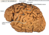
The hills of the brain are called ______ (outer cortical tissue)
The spaces between the hills (valleys) are called _______
The hills of the brain are called Gyri (outer cortical tissue)
The spaces between the hills (valleys) are called sulci

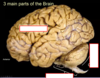
Label the three main parts of the brain:

What are the five main functions of the CEREBRUM?
- Higher cognitive functions
- thought, intellect, planning, creativity
- Language and speech
- Somatic motor function
- regulate skeletal muscle activity
- regulate and coordinate movement (basal ganglia)
- Somatic sensory function
- Interpret stimuli from environment
- Regulates emotional aspects of behaviour
The space separating the left and right hemispheres of the cerebrum is called the:

Longitudinal Fissure
The space between the Frontal Lobe and the Temporal lobe is called the:
Lateral (sylvian) fissure

The space between the Frontal lobe and the parietal lobe is called the:
Central Sulcus
The central sulcus is the ________ (starts every signal to move muscle)
Primary motor cortex
Which lobe is associated with
- thinking, planning, personality
- motor planning
- has primary motor cortex
Frontal Lobe
The Parietal lobe contains what important cortex?
Primary Somatosensory Cortex
Which lobe of the brain is associated with:
- perception of self in space (proprioception)
- Houses the primary somatosensory cortex
Parietal Lobe
Which lobe is associated with vision?
Occipital lobe
Which lobe is associated with Learning and memory and hearing?
Parietal Lobe
The neurons reside in the ______ of the brain
Cerebral Cortex
(grey matter on outside of the brain)
What is the map of the primary motor cortex called?
Motor Homunculus
(the dark yellow border is the grey matter of the cortex)

The brain map of the somatosensory cortex is called:
Somatosensory Homunculus

White matter is caused by:

White matter is caused by: Myelinated axons (myelinated by oligodendrocytes cns))

Neuronal cell bodies of the brain reside in the _____ (_____ matter)
Neuronal cell bodies of the brain reside in the cortex (grey matter)

Axons of the brains neurons extend from the cortex into the ______ (____matter)
Axons of the brains neurons extend from the cortex into the medulla (white matter)

What type of fibre tract are between gyri within the same hemisphere? (ie intrahemispheric connections; short or long)
Association fibres
What type of fibre connects one hemisphere to another?
(ie interhemispheric connections)
Commissural fibres

What type of fibre tract travels to and from the cortex of the brain?
Projection fibres

Projection fibres can be ______ (toward midline - to cortex) or ______ (motor - away from midline)
Projection fibres can be afferent (toward midline) or efferent (away from midline)
Red line = efferent
Blue line = afferent

What aer the three components of the brainstem?
- Midbrain
- Pons
- Medulla

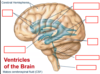
What happens in the ventricles of the brain?
Cerebral spinal fluid is produced (released into subarachnoid space)

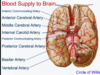
Which artery comes from the neck and supplies the brainstem, and the posterior part of the brain
Vertebral Artery

Label the:
- Anterior communicating artery
- Middle cerebral artery
- Internal carotid artery
- Posterior communicating artery
- Posterior Cerebral Artery
- Basilary Artery
- Vertebral Artery

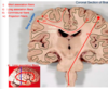
Label the:
- Anterior Horns
- Fourth Ventricle
- Inferior Horns
- Posterior Horns
- Central Canal
- Third Ventricle
- Lateral Ventricles

What makes up the circle of willis?
Anterior, middle and posterior cerebral
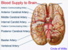
The internal carotid artery branches to the _________
Middle cerebral artery

The middle cerebral artery supplies the:
lateral surface of the brain

The anterior cerebral artery supplies the:
2 cerebral hemispheres from midline view
