Retinal Image Processing and Receptive Fields Flashcards
What are photoreceptor visual fields determined by?
Light sensitivity of their visual pigment and position of their Outer Segment in the retina (i.e., where it ‘looks’)
What is the receptive field of all retinal neurones except photoreceptors determined by?
Synaptic inputs received from photoreceptors &/or other retinal cells in the vertical or lateral pathways
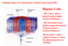
What are the difference in Receptive Fields (RF) of Photoreceptors vs that of other retinal neurones (i.e. Bipolar and ganglion cells)?
Photoreceptors:
- RF is Tiny (as they only correspond to region of space they are looking out towards which is <0.01 degrees),
- Shape is circular & uniform
- RF has Luminance(Brightness)- & wavelength-dependent responses
- Response to light is a Graded change in membrane potential
Bipolar & Ganglion Cells:
- Larger than photoreceptor RFs (Larger because of ) Convergence & spatial summation of synaptic inputs they receive
- Circular but non-uniform (they have two different parts to their RF)
- RFs of bipolar and retinal ganglion cells are more complex as they have two different parts – they have a central region and a surrounding region.
- RF is Concentric (basically means circle within a circle- have a look at the diagrammatic picture) and Antagonistic – light shining in one area e.g. the centre antagonizes light shining in the surroundings – this converts bipolar ang ganglion cells into contrast detecting cells.
- RFs have Luminance difference (Contrast)-dependent responses (explained in point above but basically these RFs aren’t interested in brightness they are interested in brightness of stimulus that falls in centre region vs that which falls into surrounding region in order to detect contrast)
- Bipolar cells respond via graded changes in their membrane potential but ganglion cells fire action potentials.

In receptive fields of bipolar cells which part is receiving input from which pathway?
When are photoreceptors depolarised and hyperpolarised?
Photoreceptors: depolarized in the dark & hyperpolarize in response to light.
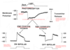
What are the two types of bipolar cells?
On bipolar cells and off bipolar cells.

In terms of a bipolar cell what is an ‘OFF’ response and what is an ‘ON’ response?
OFF’ response = cell is depolarized = excited by light OFF or ‘darkness’ in its RF (= like photoreceptors)
‘ON’ response = cell is depolarized = excited by light ON or ‘brightness’ in its RF (= opposite to photoreceptors)
Describe Briefly:
How can different Cone Bipolar Cells respond in opposite ways to input from the same cone?
Neurons (the cone photoreceptor in this case) increase neurotransmitter release when they are depolarized/excited & stop releasing neurotransmitter when they are hyperpolarized/inhibited
The effect of Neurotransmitter release depends on the type of receptor it activates in the post-synaptic cell
‘OFF’ & ‘ON’ bipolar cells have different receptors in their dendrites to the transmitter (glutamate) released at cone synapses
Describe the morphology of the two types of bipolar cells and what happens when they are stimulated.
Invaginating or non-invaginating refers to whether the dendrites slot into the grooves of the cone pedicel or not.
Receptors on dendrites for glutamate which is released by the cone are different.
Know the names of the receptors and what they do.

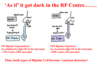
How does the concept of an antagonistic surround in the bipolar receptive field work?
And how does the concept of lateral inhibition work?
Light ‘ON’ in the Receptive Fields of Surrounding (red) cones (a.k.a imaging light is hitting the surround region of the receptive field so where the circle (or half circle in this case) of red cones is), Means:
- Horizontal Cell (HC) hyperpolarizes
- (Because these cones are directly connected to it via conventional, sign-conserving synapses).
- Horizontal Cells normally release an inhibitory neurotransmitter back onto cones in the dark = gamma-amino-butyric acid (GABA): But this is now inhibited (as its hyper polarised).
- This ‘disinhibition’ of the HC (inhibition of inhibition) is equivalent to an excitatory effect
- So all the cones connected to it, including the Central (blue) ones in the dark, depolarize
- For the Central cones connected to the Bipolar Cell, it’s as if it just got ‘darker’ in the RF centre and they start to depolarise and release more neurotransmitter.

How do both types of bipolar cells respond to it being as if the receptive field centre got darker ( i.e. light detected by surrounding field)?
Basically at rest its dark current and since bipolar cell receptve fields are anatgonistic if light is detected in surrounding field than it must be evevn darker in centre.
Do the signals from the two types of bipolar cells mix as they travel to retinal ganglion cells- expand on this.
And how are these signals carried forward?
Both types of bipolar cells make excitatory connections onto ganglion cells. The two physiological/morphological types do not mix they stay separate. So within the Inner Plexiform layer:
ON-Centre/OFF-Surround Bipolar Cell inputs generate ON-Centre/OFF-Surround Ganglion Cells, via their synapses in the inner zone of the IPL
OFF-centre/ON-surround Bipolar Cell inputs generate OFF-centre/ON-surround Ganglion Cells, via synapses in the outer zone of the IPL (i.e., these bipolar cells have shorter dendrites & axons than the ON bipolar cell class).
[2 Main Classes of Retinal Ganglion Cell = Midget & Parasol
•Both classes have different sub-types with either ON/OFF or OFF/ON Receptive Field organizations……]
What are the two classes of retinal ganglion cells- describe how their subtypes work?
Midget and Parasol ganglion cells.
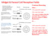
•Both classes have different sub-types with either ON/OFF or OFF/ON Receptive Field organizations……
When do Retinal ganglion cells ( so our midget and parasol cells) respond maximally and minimally?

What is a major advantage of Ganglion cell Contrast Enhancement?
Perpectual constancy
Ganglion cells enhance the contrast that you process things in ur brain with so even though in th edifferent conditions the photoreceptors pick up the image to look like the bottom two (in the slide) your brain sees it as the top image.

What is a disadvantage of increased contrast due to retinal ganglion cells?
Perceptual Illusion - fancy way of saying optical illusions.
