Lung Path 1 - Embryo Histo Malformation Atelactasis Edema ARDS (Singh) Flashcards
Requirements for normal fetal lung development?
Space in the thoracic cavity for growth (lungs are soft and cannot compete for additiona room, if it is not provided)
Enough room for the chest wall to be able to move (again, due to paranchymal softness)
Sufficient amount of amniotic fluid for inhalation to occur
What are the rigidly open structures of the airway and what is these sections primarily role?
Trachea
Main bronchus (primary)
Lobar bronchus (secondary)
Segmental bronchus (tertiary)
Their fxn is to conduct air to the terminal acinar units
What structures have thin walls and high vascularity?
What is their function?
Bronchioles
Terminal Bronchioles
Respiratory Bronchioles
Fxn of resp bronchioes: Gas Exchange
Except for the vocal cords, the entire respiratory tree is lined by what type of epithelium?
Pseudostratified, tall, columnar, ciliated epithelium

How will respiratory tissue appear histologically?
Mucosal epithalium is inner most layer (cillia, goblet cells, submucosal glands)
Muscularis mucose - smooth muscles, provide peristalsis
Bronchial ring - provides rigidity

Numerous mucus-secreting goblet cells and submucosal glands are dispersed throughout the walls of which parts of the respiratory tree?
- Trachea
- Bronchi
- NOT the bronchioles

Bronchial mucosa contains population of neuroendocrine cells with neurosecretory granules containing which factors?
- Serotonin
- Calcitonin
- Gastrin-releasing peptide (bombesin)
What are 2 functions of the Type 2 pneumocytes of the alveolar epithelium?
Type 2 pneumocytes are puliprotent
- Produce surfactant
- Repair of alveolar epithelium by giving rise to type 1 pneumocytes

What are the components of the Interstitium?
Alveolar epithelium
Fused basal laminae of the alveolar epithelium and the capillary endothelium
Capillary endothelium

What is the significance of alveolar pores?
What are they called?
Alveolar pores may also be referred to as “Pores of Kohn”
They conduct air (somehwat redundant) between alveoli –> good
Also allow the spread of bacteria and cancer cells to be exchanged between alveolar cells –> not so great
What plays a particularly important role in the synthesis of surfactant?
Glucocorticoids
Analysis of what in the amniotic fluid provides a good estimate of the level of surfactant in the alveolar lining?
Phospholipids
Pulmonary hypoplasia occurs in utero and what are 2 major causes?
- Congenital diaphragmatic hernia
- Inability to inhale
- Oligohydramnios (d/2 renal agenesis) Must know Potter Sequence!
- Airway malformation (stenosis)
- Chest wall motion disorders
–> high mortality rate

Foregut cysts are most often located where in the lungs and which classification/type is most common?
Complications?
Treatment?
Hilum or middle mediastinum
Bronchogenic cysts of the respiratory lining are most common
but can be esophageal or gastroenteric
Complications: rupture, infection, or pushing/compressing airway
Treatment: Excision curative!

What are some histologic findings on an excised bronchogenic cyst?
Typical respiratory lining filled with humorous substance

Congenital pulmonary adenomatoid malformations (CPAM/CCAM) are caused by what?
“Arrested development” of pulmonary tissue –> formation of intrapulmonary cystic masses WITH connection to tracheobronchial airways and pulmonary vasculature
No differentiation! Cell division is stuck in one development stage, and keeps reproducing the same cell line.

Congenital pulmonary adenomatoid malformations can be deadly due to what complications?
What is unique about this developmental disorder if discovered early in-utero?
- Hydrops or pulmonary hypoplasia
- Can get infected later in life
Can be removed in-utero –> corrects course of development by removing the “irregular, lumpy tumor” and making space in the thoracic cavity
Pulmonary sequestration refers to a discrete area of lung with what 2 features?
Non-functioning lung tissue forming abberant “lung bud” that
- Lacks any connection to the airway system
- Has abnormal blood supply arising from aorta or its branches

When do intralobal pulmonary sequestrations typically present and are often due to what?
Older children/adults
Due to recurrent localized infection or bronchiectasis due to “stasis” caused lack of airway
Extralobar pulmonary sequestrations most commonly come to attention in infants how?
As mass lesions in the chest or abdomen
Usually associated w/ other congenital anomalies
May have independent airways

Via which imaging modality can congenital pulmonary adenomatoid malformations be detected?
Fetal ultrasound

What are the differences between CPAM and Pulmonary sequesteration?
CPAM
always inrapulmonary
connection to airways and pum vasculature
Sequesteration
intra- or extrapulmonary
NO connection to bronchial tree of pulm vasculature
What are the 3 main types of acquired atelectasis and what is each caused by?
- Resorption due to obstruction of airway (mucus plugs) which gradually reduces lung expansion
- Compression due to accumulation of material or air/material within pleural cavity (i.e., transudate/exudate/blood or pneumothorax) that compresses the parenchyma
- Contraction due to fibrosis or restrictive processes in pleura preventing the lungs from filling completely

What are the histological findings of pulmonary edema due to HF?
dilated capillaries full of blood.
“Microhemorrhage” in alveolar spaces
scattered hemosiderin- laden macrophages accumulate within alveoli –> “heart failure cells”

What is the histological appearance of of the alveolar capillaries in hemodynamic pulmonary edema?
Engorged, and an intra-alveolar transudate appears as finely granular pale PINK material

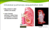
Acute lung injury (aka noncardiogenic pulmonary edema) is characterized by the abrupt onset of significant _________ and __________ in the absence of __________.
Acute lung injury (aka noncardiogenic pulmonary edema) is characterized by the abrupt onset of significant hypoxemia and bilateral pulmonary infiltrates in the absence of cardiac failure.

What is the term used to describe widespread ALI of unknown etiology associated with a rapidly progressive clinical course?
Acute interstitial pneumonia (aka idiopathic ALI-DAD)

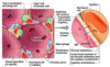
What is an important early event in the pathogenesis of ALI/ARDS?
Endothelial activation

Which factor is released inside of alveoli during ALI/ARDS that acts to sustain the ongoing pro-inflammatory response leading to more endothelial injury and local thrombosis?
Macrophage migration inhibitory factor (MIF)

During the acute stage of ALI/ARDS what is seen morphologically in the lungs?
- Lungs are heavy, firm, red, and boggy
- Exhibit congestion, interstitial and intra-alveolar edema, inflammation, fibrin deposition, and DAD
- Alveolar walls become lined with waxy hyaline membranes

The thickened protein-rich edema fluid + debris from dead alveolar cells accumulate in ALI/ARDS, and lead to the formation of what?
HYALINE membranes

The histologic manifestation of both ALI and ARDS is what?
Diffuse alveolar damage (DAD)

Following the fibroproliferative phase in ARDS, what 2 pathways may ensue and the result of each?
- Resolution –> restoration of normal cellular structure and function
- Fibrosis –> destruction and distortion of normal cellular structure –> IRREVERSIBLE
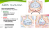
If there is resolution of the injury in ARDS/ALI, what factors are released from macrophages which stimulate fibroblast growth and collagen deposition leading to fibrosis of alveolar walls?
- TGF-β
- PDGF

What are the various stages of ARDS?
EXUDATIVE
Edema, hyaline membranes, neutrophils
PROLIFERATIVE
Fibroblast proliferation, organizing pneumonia, early fibrosis
FIBROTIC
Extensive fibrosis, loss of normal alveolar architecture
