S4) The Thigh Flashcards
What is the femur?
The femur is the only bone in the thigh and acts to transmit forces from the tibia to the hip joint
Identify the structures visible in the proximal area of the femur

Describe the structure and function of the head in the proximal area of the femur

- Structure: smooth surface with a depression on the medial aspect (ligament of head of femur attaches)
- Function: articulates with the acetabulum of the pelvis to form the hip joint

Describe the structure and function of the neck in the proximal area of the femur

- Structure: cylindrical, projecting in a superior and medial direction
- Function: connects the head of the femur with the shaft

Describe the structure and function of the greater trochanter in the proximal area of the femur

- Structure: bony projection angled superiorly and posteriorly, lateral to the neck
- Function: attachment site for many of the muscles in the gluteal region

Describe the structure and function of the lesser trochanter in the proximal area of the femur

- Structure: smaller bony projection, on the posteromedial side of the femur, inferior to the neck-shaft junction
- Function: attachment site for the psoas major and iliacus muscles

Describe the structure and function of the intertrochanteric line in the proximal area of the femur

- Structure: a ridge of bone running inferomedially on the anterior surface of the femur, connecting the two trochanters
- Function: attachment site for the iliofemoral ligament

Describe the structure and function of the intertrochanteric crest in the proximal area of the femur

- Structure: ridge of bone that connects the two trochanters on the posterior surface of the femur
- Function: attachment site for quadratus femoris (quadrate tubercle)

What is the pectineal line?

The pectineal line is the line formed when the intertrochanteric line passes the lesser trochanter on the posterior surface of the proximal femur

What are linea aspera?
Linea aspera are roughened ridges of bone found on the posterior surface of the femoral shaft

Describe the features of the linea aspera in the proximal region of the posterior femur
- Pectineal line → medial border of the linea aspera
- Gluteal tuberosity → lateral border of the linea aspera

Describe the features of the linea aspera in the distal region of the posterior femur

- Linea aspera widens and forms the floor of the popliteal fossa
- The medial and lateral borders form the medial and lateral supracondylar lines

Identify the structures visible on the anterior surface of the distal femur

Identify the structures visible on the posterior surface of the distal femur

Describe the articulations of the medial and lateral condyles of the distal femur
- Posterior & inferior surfaces articulate with the tibia and menisci
- Anterior surface articulates with the patella

Describe the structure and function of the intercondylar fossa in the distal femur
- Structure: a depression on the posterior surface of the femur, between the two condyles
- Function: contains two facets for attachment of internal knee ligaments

Where do the posterior and anterior cruciate ligaments of the knee attach to on the distal femur?
- Facet for attachment of the posterior cruciate ligament – found on the medial wall of the intercondylar fossa (large, rounded, flat)
- Facet for attachment of anterior cruciate ligament – found on the lateral wall of the intercondylar fossa (smaller)
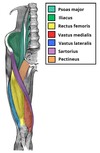
Identify the 4 muscles found in the anterior thigh
- Pectineus
- Sartorius
- Quadriceps femoris muscles
- Iliopsoas
Which two individual muscles compose the illiopsoas muscle?
- Psoas major
- Illiacus

Describe the structure, function and innervation of the psoas major muscle

- Structure: insert as the same tendon with the illiacus
- Function: hip flexion, lateral rotation (both muscles)
- Innervation: anterior rami of L1-3

State the origin and attachment of the psoas major
- Origin: lumbar vertebrae
- Attachment: lesser trochanter of the femur (with illiacus)

Describe the structure, function and innervation of the illiacus muscle

- Structure: insert as the same tendon with the psoas major
- Function: hip flexion, lateral rotation (both muscles)
- Innervation: femoral nerve

State the origin and attachment of the illiacus muscle

- Origin: iliac fossa of the pelvis
- Attachment: lesser trochanter of the femur (with psoas major)

Which 4 muscles compose the quadriceps femoris?
- Rectus femoris
- Vastus medialis
- Vastus intermedius
- Vastus lateralis
State the function and innervation of the vastus lateralis muscle

- Function: knee extension and stabilises the patella
- Innervation: femoral nerve

State the origin and attachment of the vastus lateralis muscle

- Origin: greater trochanter and the lateral lip of linea aspera
- Attachment: base of patella
State the function and innervation of the vastus intermedius muscle

- Function: knee extension and stabilises the patella
- Innervation: femoral nerve

State the origin and attachment of the vastus intermedius muscle

- Origin: anterior and lateral surfaces of the femoral shaft
- Attachment: base of the patella

State the function and innervation of the vastus medialis muscle

- Function: knee extension and stabilises the patella
- Innervation: femoral nerve

State the origin and attachment of the vastus medialis muscle

- Origin: anterior and lateral surfaces of the femoral shaft
- Attachment: base of patella

State the function and innervation of the rectus femoris muscle

- Function: hip flexion, knee extension
- Innervation: femoral nerve

State the origin and attachment of the rectus femoris muscle

- Origin: ilium (superior to the acetabulum)
- Attachment: base of patella

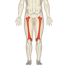
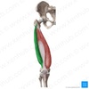
Describe the structure, function and innervation of the sartorius muscle

- Structure: superficial, long and thin, runs in an inferomedial direction
- Function: hip flexion, knee flexion, abduction, lateral rotation
- Innervation: femoral nerve

State the origin and attachment of the sartorius muscle

- Origin: anterior superior iliac spine (ASIS)
- Attachment: superomedial surface of the tibia

Describe the structure, function and innervation of the pectineus muscle

- Structure: flat muscle, forms the base of the femoral triangle (transitional muscle between anterior thigh and medial thigh)
- Actions: hip flexion, adduction
- Innervation: femoral nerve (& obturator nerve)

State the origin and attachment of the pectineus muscle

- Origin: pectineal line on the anterior surface of the pelvis
- Attachment: pectineal line on the posterior side of the femur (inferior to the lesser trochanter)

Describe the structure, function and innervation of the adductor magnus muscle

- Structure: lies posteriorly, consists of an adductor part and a hamstring part
- Function: adduction (both), flexion (adductor part) extension (hamstring part)
- Innervation: adductor part – obturator nerve, hamstring part – tibial nerve

State the origin and attachment of the adductor magnus muscle

- Adductor part
I. Origin: inferior rami of pubis & ramis of ischium
II. Attachment: linea aspera of femur
- Hamstring part
I. Origin: ischial tuberosity
II. Attacment: adductor tubercle & medial supracondylar line of femur

Describe the structure and function of the adductor longus muscle

- Structure: large and flat, partially covers adductor brevis and magnus, forms the medial border of the femoral triangle
- Function: adduction, medial rotation

State the origin and attachment of the adductor longus muscle

- Origin: pubis
- Attachment: linea aspera of the femur (broadly)

Describe the structure and function of the adductor brevis muscle

- Structure: short muscle, lying underneath the adductor longus and between the anterior and posterior divisions of the obturator nerve
- Function: adduction

State the origin and attachment of the adductor brevis muscle

- Origin: body of pubis and inferior pubic rami
- Attachment: linea aspera of the femur (proximal to the adductor longus)

Describe the structure and function of the obturator externus muscle

- Structure: small muscle, located most superiorly
- Function: lateral rotation

State the origin and attachment of the obturator externus muscle

- Origin: membrane of the obturator foramen, and adjacent bone
- Attachment: posterior aspect of the greater trochanter (passes under the neck of femur)

Describe the structure and function of the gracilis muscle

- Structure: most superficial and medial muscle
- Function: adduction, knee flexion

State the origin and attachment of the gracilis muscle
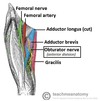
- Origin: inferior rami and body of the pubis
- Attachment: superomedial surface of the tibia (between sartorius and semitendinosus tendons)

What is the femoral triangle?
The femoral triangle is a hollow area in the anterior thigh in which many large neurovascular structures pass through

Identify and describe the superior, lateral and medial borders of the femoral triangle

- Superior border: inguinal ligament (ASIS → pubis tubercle)
- Lateral border: medial border of the sartorius muscle
- Medial border: medial border of the adductor longus muscle

Identify and describe the anterior and posterior borders of the femoral triangle

- Roof: fascia lata
- Base: pectineus, iliopsoas and adductor longus muscles
Describe the role of the inguinal ligament
The inguinal ligament acts as a flexor retinaculum, supporting the contents of the femoral triangle during flexion at the hip

What are the contents of the femoral triangle?

- Femoral nerve
- Femoral artery
- Femoral vein
- Femoral canal (deep lymph nodes and vessels)
Femoral artery, vein and canal are contained in a femoral sheath

The femoral canal is located in the anterior thigh, within the femoral triangle.
Describe and identify its borders
- Medial border: lacunar ligament
- Lateral border: femoral vein
- Anterior border: inguinal ligament
- Posterior border: pectineal ligament, superior ramus of the pubic bone, pectineus muscle

Describe the opening of the femoral canal

The femoral ring is the opening to the femoral canal, located at its superior border and enclosed by a connective tissue layer (femoral septum)

What are the contents of the femoral canal?

- Deep lymph node (lacunar node)
- Empty space
- Lymphatic vessels (draining the deep inguinal lymph nodes)
- Loose connective tissue

Identify and describe the borders of the adductor canal
- Anterior: sartorius
- Lateral: vastus medialis
- Posterior: adductor longus and adductor magnus

Describe the contents of the adductor canal

- Femoral artery
- Femoral vein
- Nerve to the vastus medialis
- Saphenous nerve

What happens to the femoral artery and vein once they exit the adductor canal?
As the femoral artery and vein exit the canal, they become the popliteal artery and vein respectively

What is the adductor hiatus?
The adductor hiatus is a gap between the adductor and hamstring attachments of the adductor magnus

Describe the structure, function and innervation of the biceps femoris muscle

- Structure: most lateral muscle in posterior thigh, long head and a short head
- Function: knee flexion, hip extension, lateral rotation
- Innervation:
I. Long head – tibial part of sciatic nerve
II. Short head – common fibular part of sciatic nerve

State the origin and attachment of the biceps femoris muscle

- Origin:
I. Long head – ischial tuberosity of pelvis
II. Short head – linea aspera of femur
- Attachment: head of the fibula

Describe the structure, function and innervation of the semitendinosus muscle

- Structure: largely tendinous muscle, lies medially to the biceps femoris, and covers most of semimembranosus
- Function: knee flexion, hip extension, medial rotation
- Innervation: tibial part of the sciatic nerve

State the origin and attachment of the semitendinosus muscle

- Origin: ischial tuberosity of the pelvis
- Attachment: superomedial surface of the tibia

Describe the structure, function and innervation of the semimembranosus muscle

- Structure: flattened and broad, located underneath the semitendinosus
- Function: knee flexion, hip extension, medial rotation
- Innervation: tibial part of the sciatic nerve

State the origin and attachment of the semimembranosus muscle

- Origin: ischial tuberosity (superior to semitendinosus and biceps femoris)
- Attachment: medial tibial condyle

The presence of a femoral pulse means that blood is reaching the lower extremity.
Where can this pulse be palpated?
- In the femoral canal, inferior to where the femoral artery crosses the inguinal ligament
- It crosses exactly midway between the pubis symphysis and anterior superior iliac spine

How does one test the femoral nerve?
- The quadriceps femoris muscles are used to test for femoral nerve palsies
- The patient is positioned supine with the knee slightly flexed and is asked to extend their leg (at the knee) against resistance

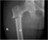
What is a fracture of the femoral shaft?
- Femoral shaft fractures are spiral fractures which occur due to a lot of force on the shaft of the femur
- They are relatively uncommon and cause leg shortening due to bony fragments overriding and the proximal pull of the associated muscles

What is a proximal intracapsular femur fracture?

- A proximal intracapsular femur fracture is a fracture within the capsule of the hip joint which commonly occurs in the elderly, especially women
- It can damage the medial femoral circumflex artery, carries a risk of avascular necrosis and presents with the distal femur fragment being pulled upwards and laterally rotated (shortening)

What is a proximal extracapsular femur fracture?

- A proximal extracapsular femur fracture is a fracture outside the hip joint capsule, which occurs more commonly in young and middle aged people
- The femoral circumflex artery is intact, there is no risk of avascular necrosis and it presents with leg shortening as well as the lateral rotation of the thigh
What is a femoral hernia?
- A femoral hernia is a condition where part of the bowel is displaced and protrudes into the femoral canal, underneath the inguinal ligament
- It presents as a lump or bulge in the area of the femoral triangle
