Histology of Accessory GI Organs (Dennis) Flashcards
What are the functions and 3 main types of digestive glands?
- functions: lubrication, digestion, and absorption mediated by secretory products
- 3 types:
1) major salivary glands: a/w oral cavity through independent excretory ducts
2) exocrine pancreas: secretes its alkaline aqueous and enzymatic product into the duodenum
3) liver: endocrine and exocrine gland w/ extensive access to blood circulation
What is the histological structure of salivary glands?
- CT capsule w/ septa, dividing gland into lobes and smaller lobules (route for neurovasculature)
- secretory cells organized in an acinus produce saliva via ANS control
- myoepithelial cells aid in release of secretions
- released via ducts: acinus > intercalated duct > striated duct > excretory duct

What is the pathway for saliva flow?
acinus
↓
intercalated duct
(low cuboidal epithelium)
↓
striated duct
(simple cuboidal-to-columnar epithelium)
↓
excretory duct
(simple cuboidal > pseudostratified columnar or stratified cuboidal)

- largest salivary gland, adipocytes may be present
- acini consist of serous secretory cells (pyramidal cells w/ basally located nucleus, prominent RER in basal region, secretory granules visible in apical region)
parotid (serous) gland

- branched tubule-alveolar gland w/ both serous and mucous cells
- mixed gland, but predominantly mucous
- lacks defined capsule, but is divided by CT into small lobes
- intercalated and striated ducts are poorly developed
sublingual gland

- serous cells are predominant in this gland, but mucous cells are also present
- mucous cell: contains acini and are capped by serous demilunes (‘bonnet’)
- intercalated ducts are shorter and striated ducts are longer than those in the parotid gland
submandibular gland

Identify what gland in the body this image was obtained and associated structures:


Identify what gland in the body this image was obtained and associated structures:


Identify what gland in the body this image was obtained and associated structures:


- large gland w/ endocrine and exocrine functions, bulk of gland is exocrine
- thin layer of loose CT forms capsule, divides gland into ill-defined lobules (neurovasculature and ducts extend within septa)
- exocrine component: synthesizes/secretes enzymes that are essential for digestion in the intestines
- endocrine component: synthesizes/secretes hormones (insulin/glucagon) into the blood > regulate glucose, lipid, and protein metabolism
pancreas

Describe the histological structure of the exocrine pancreas:
- serous acinus: functional unit of exocrine pancreas, structurally unique, contains pancreatic acinar cells
- intercalated duct begins within acinus centroacinar cells (duct cells inside the acinus)
- centroacinar cells: continuous w/ low cuboidal epithelium of intercalated duct; secrete HCO3-, Na+, and H20 which alkalinize secretions
- hallmark: acinar cells stain intensely, centroacinar cells stain lightly, pancreas often confused w/ parotid

Describe the histological structure of pancreatic acinar cells:
- characterized by: well-developed RER, prominent golgi apparatus, and apical domain of zymogen granules
- granules contain ~20 different pancreatic proenzymes

What are the functions of the pancreatic acinar cell products?
granules contain ~20 different pancreatic proenzymes:
- trypsinogen, chymotrypsinogen > digest proteins
- amylolytic enzymes (α-amylase) > digest carbs
- lipases > digest lipids
- deoxyribonuclease, ribonuclease > digest nucleic acids
functions:
- increase synthesis of proteases w/ protein-rich diet
- increase amylases and decrease in proteases w/ carb rich diet
Identify where in the body this image was obtained and associated structures:

acinar cells of the pancreas

Describe the structure and secretory products of endocrine pancreas:
- spherical masses of endocrine cells, surrounded by thin reticular capsule
- most islets contain several hundred cells, pancreas has more than 1 million islets
- islets arise from endodermal epithelial outgrowths
- α cells = glucagon
- β cells = insulin (most numerous)
- δ cells = somatostatin (least abundant)
- PP cells = pancreatic polypeptide

Identify where in the body the following image was obtained and identify associated structures:

endocrine pancreas

Describe the histological structure of the liver:
- enclosed in thin CT capsule lined w/ mesothelium of visceral peritoneum which is absent where liver directly adheres to diaphragm/other organs
- hepatocytes: function in metabolism, storage, and bile prod (exocrine); arranged in cellular ‘cords’
- liver structure varies depending on the functional unit: hepatic lobule, portal loble, and liver acinus

Describe the histological structure of hepatocytes:
- large, polygonal cells w/ eosinophilic cytoplasm and microvilli
- large, spherical nuclei, many cells are binucleate, most are tetraploid
- numerous peroxisomes and lysosomes, extensive sER, large golgi

- mixture of H20, bile salts, pigments, phospholipids, and electrolytes secreted by by hepatocytes
- functions in fat absorption, and excretion of cholesterol, bilirubin, iron, and copper
- drains into bile canaliculus, spaces located between adjacent hepatocytes
bile

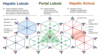
What is the structure/function relationships of the different parts of the liver?
- hepatic lobule: drains blood from portal vein and hepatic artery to hepatic or central vein; emphasizes endocrine function of hepatocytes as blood flows toward central vein
- portal lobule: drains bile from hepatocytes to the bile duct; emphasizes hepatocyte exocrine function and flow of bile from classic lobules toward bile duct in portal triad (area drained by each bile duct is triangular)
- hepatic acinus: supplies oxygenated blood to hepatocytes; emphasizes different oxygen and nutrient contents of blood at different distances along the sinusoids (blood from each portal area supplies 2+ classic lobules)
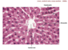
What is the histological structure of hepatic lobules?
- liver parenchyma is organized into hepatic lobules: hepatocytes form irregular plates radiating from a central vein, plates are supported by a stroma of reticular fibers, plates separated by sinusoids
- peripheral angles of each lobule contains a portal triad in fibrous CT: venule branch of portal vein (increases nutrients, decreases O2); arteriole branch of the hepatic artery (supplies O2); bile ductules (1-2) branches of the bile conducting system
- blood and bile flow in opposite directions

Identify where in the body this image was obtained and associated structures:

hepatic lobule
C: central venule
H: hepatocytes
S: sinusoids

Identify where in the body this image was obtained:

bile canaliculi
(brown-staining ‘stick’ like structures located in between hepatocytes)

Identify where in the body this image was obtained and associated structures:

portal triad
BD: bile ductules
HA: branches of hepatic A.
PV: branches of portal V.

Where does bile collect in the liver?
- bile collects in bile canaliculi, interconnected channels formed by hepatocyte plates and tight junctions
- bile canaliculi drain into canals of Hering, composed of cuboidal epithelial cells called cholangiocytes

- consists of hepatic, cystic, and common bile ducts
- lined w/ a mucous membrane having a simple columnar epithelium of cholangiocytes
- cystic duct has some areas w/ mucous glands
- all ducts have a thin lamina propria and submucosa, surrounded by a thin muscularis
- muscularis layer becomes thicker near the duodenum and forms a sphincter that regulates bile flow into the duodenum
biliary tree

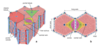
- anastomosing capillaries that perfuse hepatocytes w/ portal and arterial blood
- Kupffer cells are located within surrounding endothelium and hepatic stellate cells are located within space of Disse
hepatic sinusoids

- stellate macrophages within endothelium of sinusoids
- larger than endothelial cells
- detect and phagocytose effete erythrocytes
- distinguishes hepatic sinusoids
Kupffer cells

- cells w/ small lipids droplets that store vit A and other fat-soluble vitamins
- found in the perisinusoidal space of the liver, also known as the space of Disse
hepatic stellate cells (Ito cells)

- located between hepatocytes and sinusoidal endothelium
- facilitates uptake/release of nutrients, proteins, and potential toxins
- creates a potential space for exchange of materials between blood and hepatocytes: microvilli project into this space, plasma fills the space and directly bathes microvilli, increase surface area available for material exchange
perisinusoidal space (of Disse)

- where excess fluid from perisinusoidal space is collected and then drained by lymphatic vessels
- located at edges of canals between stromal CT and hepatocytes
- also houses Ito cells that store fat/vit A
periportal space

- central axis is the bile duct (portal triad)
- ID portal triad > draw imaginary lines between 3 central veins > ____ _______
- triangular block of tissue, outlines bile drainage pathway from adjacent lobules into the same bile duct
- provides a description comparable to that of other exocrine glands
portal lobule

- diamond shaped structure that occupies parts of adjacent classic hepatic lobules
- hepatocytes are ‘arranged’ in concentric zones around short axis based on [O2] gradient along sinusoids of adjacent lobules
- flow of arterial blood creates gradient of O2/nutrients
- cells within each zone have different metabolic functions and distribution of hepatic enzymes
- this explains distribution of liver damage resulting from ischemia and/or exposure to toxic substances (damage occurs from zone 3 > 1)
hepatic acinus
What are the hepatic acinus zones, how do they differ in terms of oxygen/nutrient content, and what implications does this have in liver damage?
- hepatocytes are arranged in concentric zones around short axis based on [O2] gradient along sinusoids of adjacent lobules
- the activity of each hepatocyte is determined by location along oxygen/nutrient gradient
- periportal cells of zone I: first to receive blood, high in oxygen/nutrients
- zone II: second to receive blood, moderate amount of oxygen/nutrients
- pericentral hepatocytes of zone III: last to receive blood, lowest amnt of oxygen/nutrients
- liver damage resulting from ischemia and/or exposure to toxic substances occurs first in zone III due to the low oxygen content and then works it way to zone II then zone I

Describe the histological structure of the gallbladder:
- sac-like structure that stores/concentrates bile, releases it into duodenum after meals
- highly folded mucosa (arrows), w/ a simple columnar epithelium (w/ microvilli) overlying a lamina propria (LP)
- no muscularis mucosae or submucosa
- muscularis (M) w/ bundles of muscle fibers oriented in all directions to facilitate emptying of organ
- external adventitia (A) where it is against the liver, but a serosa where it is exposed to peritoneal activity

What are Rokitansky-Aschoff sinuses and what are their clinical relevance?
- Rokitansky-Aschoff sinuses: deep diverticula of the gallbladder mucosa that may extend through muscularis externa
- develop as the result of hyperplasia and herniation of epithelial cells through the muscularis externa
- bacteria may accumulate, causing chronic inflammation and increased risk for gallstones

Identify where in the body this image was obtained and associated structures:


Identify where in the body this image was obtained and associated structures:


Identify where in the body this image was obtained and associated structures:


Identify where in the body this image was obtained and associated structures:


Identify where in the body this image was obtained and associated structures:


Identify where in the body this image was obtained and associated structures:


Identify where in the body this image was obtained and associated structures:


Identify where in the body this image was obtained and associated structures:


Identify where in the body this image was obtained and associated structures:


Identify where in the body this image was obtained and associated structures:


Identify where in the body this image was obtained and associated structures:


Identify where in the body this image was obtained and associated structures:


Identify where in the body this image was obtained and associated structures:


Identify where in the body this image was obtained and associated structures:


Identify where in the body this image was obtained and associated structures:


Identify where in the body this image was obtained and associated structures:


Identify where in the body this image was obtained and associated structures: