Aesthetic Surgery Flashcards
(141 cards)
Describe the development of the breast.
Embryology
- The breast is ectodermally derived.
- In week 6 the milk ridge develops, extending from the axilla to the groin. From week 7 to birth, mammary anlage develops into an pithelial bud with 15-20 ducts and nipple develops into circular smooth muscle fibers.
- Normal breast development in anterolateral pectoral region at the level of the 4th intercostal space.
Development
- Puberty begins at age 10-12 and anterior pituitary releases follicle stimulating hormone (FSH) and luteinizing hormone (LH).
- FSH stimulates ovarian follicles to secrete estrogen which stimulate longitudinal growth of breast ductal epithelium.
- The corpus luteum releases progresterone, which along with estrogran leads to complete mammary development.
List 5 arteries providing blood flow to the breast.
- Perforating branches of internal mammary artery.
- Lateral thoracic artery.
- Thoracodorsal artery.
- Intercostal perforators.
- Thoracoacromial artery.
Describe the innervation of the breast.
- Anterolateral and anteromedial branches of the thoracic intercostal nerves T3-T5.
- Supraclavicular nerves from lower fibers of cervical plexus contribute.
- Nipple-areolar sensation deried from T4 intercostal nerve.
- Intercostal brachial nerve courses across axilla to supply upper medial arm and lateral breast.

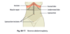
Describe Würinger septum.
- Würinger septum:
- Horizontal septum that originates from the pectoralis fascia along the 5th rib - which merged with lateral and medial vertical ligaments and ran anteriorly towards the NAC.
- Breast prenchyma bipartitioned as the septum ran anteriorly towards the NAC.
- Cranial aspect carried branches of thoracoacromial and lateral thoracic arterial branches and the caudal aspect carried branches of the 4th-6th intercostal arteries.
- Main contributorynerve to the nipple (lateral cutaneous branch of the intercostal nerve) was always found within the septum.
Describe the functional anatomy of the breast, including the lobule, lactiferous ducts, morgani tubercles, montgomery glands.
- Lobule is the functional unit of the breast and are located in a radial distribution.
- Lobules are composed of acini.
- Each acinus has secretory potential.
- Acinus are connected to lactiferous ducts by interlobular ducts.
- Lactiferous duct dilates as it approaches the nipple forming lactiferous sinus or central collecting duct. The nipple then contains orifices to drain each lactiferous duct.
- Morgagni tubercles are elevations formed by the opening of the ducts of the Montgomery glands located at the periphery of the areola.
- Montgomery glands are large sebaceous glands capable of secreting milk.
Describe the fascia, supporting ligaments of the breast, and the IMF.
- Breast is supported by layers of superficial fascia:
-
Superficial layer of superficial fascia:
- Located near the dermis and challenging to distinguish unless the patient is thin.
-
Deep layer of superficial fascia:
- On the deep surface of the breast.
- A loose areolar plane exists between this layer and the deep fascial layer that overlies the musculature.
-
Superficial layer of superficial fascia:
- Cooper ligaments penetrae deep layer of superficial fascia into parenchyma breast to dermis; ptosis results in attenuation of these attachments.
- The inframammary fold (IMF) is the lower border of the breast and is a disctint anatomic structure. It represents fusion of the deep and superficial fascia with the dermis.
Describe ideal ideal breast measurements.
- Less full above areola (upper pole) and fuller below (lower pole).
- NAC: 19-21cm from sternal notch, 9-11 from midline, and 7-8cm from IMF.

Describe the generations of silicone gel-filled breast implants.

List contraindications to breast augmentation.
- Significant breast disease (severe fibrocystic disease, ductal hyperplasia, breast cancer).
- Collagen vascular disease.
- Body dysmorphic disorder.
- Psychological instability.
- Social instability (e.g. divorce or separation, searching for relationship)
- Patient responding to pressure from friends, family, partner.
- Patient < 18 years of age.
Key elements on medical history for breast augmentation consult.
- Personal or family history of breast disease or breast cancer.
- Pregnancy history and plans for future pregnancy.
- Breast size before, during, and after pregnancy.
- Mammography history (recommended for patients >35 years of age and those with significant breast cancer risk).
- Patient without significant history should have a mammogram every 2 years starting at age 40 and every year begining at age 50.
- Previous surgeries or procedures on breasts.
- Previous cosmetic procedures.
- Tobacco or nictoine replacement use.
- Anticoagulation use.
- Current breast size.
- Desired breast size.
Breast measurements to take in a breast augmentation consult.
- Intermammary distance.
- Sternal notch to nipple distance.
- Nipple to IMF during stretch.
- Base width.
- Breast height.
- Parenchymal coverage (pinch test)
- Superior pole
- Inferior pole
- Anterior-pull skin stretch (cm of anterior stretch with pull at edge of areola)
- Parenchymal fill (% of skin envelope filled by parenchyma)
Describe advantages, disadvantages, and technique for subglandular breast agumentation plane.
- Subglandular: implant rests under the breast gland.
- Technique:
- Dissection on top of pec major, below gland
- If pinch test > 2cm, implant can safely be placed in the subglandular plane
- Advantages:
- Avoid implant distortion with pec activity.
- More anatomic.
- Better projection.
- Disadvantages:
- Higher capsular contracture rate.
- Visible rippling.
- Implant edges may be palpable.
- Interference with mammography.
Describe advantages, disadvantages, and technique for subpectoral breast agumentation plane.
- Technique:
- Implant placed completely under pec major
- Rarely performed in cosmetic surgery
- Advantages:
- Lowest capsular contracture rate (<10%).
- Good preservation of nipple sensation.
- Disadvantages:
- Animation with pec activity.
- Implant malposition.
- Difficult to control upper pole fill.
Describe advantages, disadvantages, and technique for dual plane breast agumentation plane.
- Technqiue:
- The origin of the pectoralis major is completely divided from its origin at the level of the IMF, stopping at the medial aspect of the IMF.
- Upper pole of the implant placed under the pectoralis; lower pole subglandular.
- Attachments of the pectoralis to the breast parenchyma are selectively divided (amount of dissection differential the type of dual plane: type I, II, III)
- Advantages:
- Decreases implant displacement caused by pectorlis contraction.
- Provides thick upper pole soft tissue coverage with subpectoral placemnet.
- Lower capsular contracture rates than with subglandular placement.
- Increased control of IMF position compared with submuscular.
- Breast parenchyma and the pectoralis can be dissected part to adjust for different types of breasts.
- Increases implant-parenchymal interface, which expands lower pole and prevent double-bubble deformity.
- Disadvantages:
- Restricted to IMF incision whenperformed dual plane II and III
- Contradinication:
- IMF pinch test < 0.4cm
Describe the relationship between the implant volume and the nipple to IMF distance.
N-IMF should correspond to implant volume; increasing volume needed for increasing N-IMF.

Pharmacotherapy options for capsular contracture.
- Leukotriene inhibitors
- Papaverine hydrochloride
- Oral vitamin E
- Intraluminal steroids: reduces contracture, but higher rate of implant rupture, skin erosion, atrophy, ptosis
- Cyclosporine
Rate of reoperation, rupture/deflation, and capsular contraction (III/IV) for saline and silicone implants.
2011 FDA Update Data
- Reoperation rate (~5%)
- 6.5% silicone
- 4.5% saline
- Rupture / deflation (~0-2.5%)
- 0.5% silicone (rupture)
- 2.5% saline (deflation)
- Capsular contracture
- 5% silicone
- 2.8% saline
Describe the impact of breast implants on cancer screening.
- Implants cause interference in mammogram imaging.
- Eklund mammogram views displace breast and implant to increase parenchymal imaging after breast imaging.
- With appropriate imaging:
- No increased risk for cancer is found.
- Diagnosis is not later.
- No difference in survival or recurrence.
At what cutoff of N-IMF do patients typically need a mastopexy in consideration of primary augmentation.
- Mild ptosis is improved with augmentation.
- Patients with N-IMF >9.5cm should undergo mastopexy.
What cut off in the pinch test is subglandular breast augmentation no longer recommended?
- Subglandular augmentation is not recommended with thing upper pole coverage (superior pole pinch test < 2cm).
Indications and contradinications to mastopexy.
-
Indications
- Women who desire change in breast contour witout a change in volume.
- Women who seek more lifted, ‘perky’, youthful breast, aim to correct upper pole deflation, ptosis of the areolar complex and breast tissue, and laxity of skin evelope.
-
Contraindications
- Active smoking.
- Women who desire volume change.
Describe two Periareolar mastopexy techniques.
-
Simple periareolar deepithelialization and closure
- Breast parenchyma is not repositioned
- Only useful for mild ptosis
- Permits nipple repositioning
- Limited elliptical techniques can elevate the NAC ~ 1-2cm.
-
Benelli technique
- Periareolar technique that can be applied to patients with larger degrees of breast ptosis
- Allows parenchymal repositioning
- Areola marked as well as a larger ellipse to resect redundant skin around the NAC
- Undermining to separate the breast from overlying skin
- Breast parenchyma is incised leaving the NAC on a superior pedicle.
- Medial and lateral parenchymal flap are mobilized inferiorly and are crossed in the midline to narrow the breast width and cone the breast shape
- Periareolar incision closed in a purse string fashion

Describe three vertical mastopexy techniques.
-
Vertical mastopexy without undermining (Lassus)
- Skin incision
- Inferior wedge of ptotic skin/fat/gland excised en bloc
- Nipple transposed superiorly without undermining
- Medial and lateral breast pillar are closed.
- Skin closed.
-
Vertical mastopexy with undermining and liposuction (Lejour)
- Skin incision
- Liposuction in large breasts to reduce parenchymal volume and facilitate mobilition of dermal-parenchymal pedicle
- Inferior wedge of ptotic skin/fat/gland excised en bloc
- Wide undermining is performed and medial and lateral breast pillars are closed inferiorly.
- Skin closed.
-
Short-scar periareolar inferior pedicle reduction mammaplasty (Hammond)
- Skin incision.
- NAC is transposed to desired location based on inferior pedicle.
- Nipple supported with parenchymal suspension sutures.
- Inferior skin tailor-tacked to create desired contour and closed in a vertical pattern.

Describe the technique for a inverted T / wise pattern mastopexy.
- Several variations of the skin incision.
- Parenchymal resection is indicated in hypertrophic breasts.
- Parenchymal support obtained with inferior clsure of medial and lateral breast pillars.
- Inferior parenchyma can be repositioned superiorly to restore superior pole fullness:
- Tunneled under a pectoralis sling
- Folded under a superior pedicle and secured to the pectoralis fascia.