all Flashcards
(777 cards)
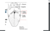
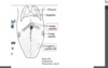
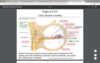
Sensory from a portion of external ear
General Somatic Afferent (GSA) Facial Nerve
Supposed existence carrying information from sublingual and submandibular glands
General Visceral Afferent (GVA)
Inf. Vagal Ganglion
SA and GVA Vagus
Supposed existence carrying information from palatine, pharyngeal
General Visceral Afferent (GVA from CN 7
Taste from the anterior 2/3 of the tongue and palate
SA CN7
Taste from the posterior 1/3 of the tongue
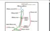
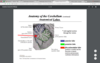
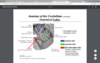
Cranial Nerve IX Glossopharyngeal Nerve
Sensory from a portion of external ear
GSA CN9(GP)
Taste from the epiglottis
SA CN10
GSA and SA fibrs
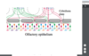
solitary tract nucleus
facial motor nucleus
CN 7 SVE: motor to muscle of facial expression
SVE: motor to stylopharngeus
CN 9
from nucleus ambiguous
SVE: motor to muscles of palate, pharynx
CN 10
Inf. GP gnaglion
SA and GVA CN9
SVE: motor to muscles of larynx, upper esophagus
CN 10
TF SVE fibers of CN9,10 start from
nucleus ambiguous
spinal tri nucleus
GSA fibers of CN 7 anf CN 9, CN 10 from ext ear
postganglionic parasympathetic fibers innervate thoracic and abdominal viscera
CN 10 GVE
Parasympathetic ganglion cell in the wall of the target organ
GVE of CN 10
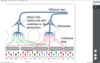
Superior Salivatory Nucleus
starts GVE fibers of CN7
Dorsal motor nuc
starts GVE fibers of CN 10
postganglionic parasympathetic fibers to sublingual and submandibular glands
from CN 7 GVE
goes to submandibular gang
postganglionic parasympathetic fibers to lacrimal, nasal palatine and upper pharynx glands
from CN 7 GVE
from Pterygopalatine ganglion
Otic gaglion
GVE fibers of CN 9
parasympathetic fibers(pre and post ganglionic neurons)