An Introduction to the Structure of Cells Flashcards

(148 cards)

1
Q

What do all cells have in common?

A

DNA

Cytoplasm

Plasma membrane surrounding the cell

2
Q

Where is DNA stored in prokaryotic cells

A

Nucleoid

3
Q

Where is DNA stored in eukaryotic cells

A

Nucleus

4
Q

Cytoplasm

A

Semi-fluid matrix

Contains sugars, amino acids, proteins etc In a cytosol

5
Q

How big are prokaryotes

A

1-10µm

6
Q

How big are eukaryotes

A

10-100µm

7
Q

Why aren’t cells bigger?

A

Would make diffusion less efficient

Surface area : volume ratio would be worse

8
Q

Why is the surface area : volume ratio important in cells

A

Communication and interaction with internal environment happens through the surface of the cell

If the volume in the ratio was too large it wouldn’t be able to keep up

9
Q

2 types of cell

A

Prokaryotes

Eurkaryotes

10
Q

Types of prokaryotes

A

Bacteria

Archaea

11
Q

Archaea

A

Ancient prokaryotes

Often adapted to extreme conditions

12
Q

Extreme conditions arachaea

A

Methanogens

Extreme halophiles

Extreme thermophiles

13
Q

Methanogens

A

Archaea

Metabolic activities produce methane

Poisoned by oxygen

14
Q

Extreme halophiles

A

Archaea

Salt lovers

15
Q

Extreme thermophiles

A

Archaea

Heat lovers

16
Q

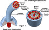
Structure of a prokaryotic cell

A

Nucleoid

Ribosomes

Cytoplasm

Pili

Plasma membrane

Cell wall

Flagellum (sometimes)

17
Q

Prokaryote cell wall

A

Outside plasma membrane

Quite porous

18
Q

Function of prokaryote cell wall

A

Protection

Maintains shape

Helps prevent excessive water uptake

19
Q

Gram positive bacteria

A

Thick, single layered cell wall

Retains dye

20
Q

Gram negative bacteria

A

More complex then gram positive

Many layers

Doesn’t retain dye

21
Q

How do antibiotics often work

A

By disrupting cell walls

22
Q

Bacteria

What is the cell wall often covered by What does it do

A

Capsule Is slimy, prevents it drying out and helps attachment

23
Q

Ways prokaryotic cells move about

A

Flagellum

Pili

24
Q

Interior organisation of prokaryotes

A

Simple

No internal compartmentalisation

No membrane bound organelles

No nucleus Cytoplasm with internal support structure

25
Types of Eukaryotes
Protists Fungi Plants Animals
26
Structure of eukaryote cell
Compartmentalised Plasma membrane Cytoplasm Organelles Nuclear envelope Nucleus
27
Which eukaryote cells have a cell wall
Plants Fungi
28
What does the cell wall do
Provides mechanical support Protects against infection
29
Eukaryote meaning
True nucleus
30
Prokaryote meaning
Before nucleus
31
Plant cell wall
Composed of fibres of cellulose embedded in polysaccharides and proteins Interconnected by plasmadesmata
32
Plasmadesmata
Physical passageway in cell wall Allows communication and movement of molecules between cells that the cell wall would stop
33
Central vacuole
Large membrane bound sac in plant cells Stores proteins, pigments and waste Presses against cell wall giving tugor pressure
34
How much of the cell does the central vacuole take up
Up to 80%
35
Contractile vacuole
Found in some protist cells Stores water coming in by osmosis then expels the water Stops the cell bursting from excess water
36
Types of vacuoles
Central (plant) Contractile (protists) Phagocytic Food
37
Cytosol
Region of eukaryotic cells that is inside the plasma membrane and outside the organeles
38
Cytoplasm =
Cytosol + Organelles
39
Cytoplasm is the site of...
Metabolism Catabalism
40
Metabolism
Utilisation of energy for synthesis of materials
41
Catabalism
Breakdown of materials to utilize energy and generate building blocks for construction macromolecules Each step catalysed by an enzyme
42
Nucleus
Contains DNA Spherical Surrounded by nuclear envelope
43
What amount of cell volume is the nucleus
10-20%
44
Nucleolus
Inside nucleus Site of intense ribosomal RNA synthesis
45
Nuclear envelope
Double membrane Has nuclear pores to allow passage of substance Interior filled with fluid containing chromosomes
46
What is eukaryotic DNA divided into?
Chromosomes
47
Chromosomal territory
Distinct Non-overlapping Where each chromosome is located within the cell nucleus of eurkaryotic cells
48
Chromosome
Unit of genetic material Composed of DNA and proteins
49
Types of histone proteins
Central histone Spacer histone
50
Nucleosomes
Formed by DNA coiled around clusters of histones Looks like beads on a string
51
Chromotins around surrounded by...
Nucleoplasm
52
Nuclear lamina
Made of intermediate filaments Lines the inner nuclear membrane Attaches to chromatins which helps maintain nuclear shape and keep chromosomes in their territories
53
Ribosomes
Site of protein synthesis
54
Structure of ribosomes
Ribosomal Ribonuceic acid (rRNA) bound with several dozen types of protein 2 subunits, 1 large and 1 small
55
When are ribosomes functional and why
Only when attached to messenger RNA (mRNA) mRNA contains the code required to make the protein
56
Free ribosomes
Make proteins that function in the cytoplasm
57
Proteins that function in membranes or for transport are made by...
Ribosomes on the rough endoplasmic reticulum
58
RER stands for
Rough endoplasmic reticulum
59
Why do cells in the same organism, with the same DNA, look and function differently
They share the same genome But the proteome is different
60
Genome
Complete genetic composition of a species
61
Proteome
The proteins that a cell can make
62
Different cells may produce...
Different proteins Different proportions of the same protein Slightly different variants of proteins Modifications of proteins
63
Endoplasmic reticulum
Smooth and rough Folded inner membranes (cisternae) give it an enormous surface area
64
Rough endoplasmic reticulum
Site of protein synthesis Covered with ribosomes Proteins may be chemically modified there
65
In what ways are proteins chemically modified in the RER
Glycosylated to become glycoproteins Have "address label" signal sequences added
66
Glycoprotein
A protein that has carbohydrate attached to it
67
Glycosylation
The attachment of carbohydrate to a protein or lipid Produces a glycoprotein or glycolipid
68
Proteins leaving the RER may be....
Exported Sent to the lysosomes or vacuoles Become part of the plasma membrane
69
When leaving the RER, what determines a proteins destination
The signal sequences attached
70
Steps of protein synthesis in the RER
Free ribosomes produced in cytosol If mRNA contains an ER signal sequence the ribosome attaches to the ER ER signal sequence recognised by signal recognition particle (SRP) SRP docks ribosome over channel protein in ER Channel protein opens Translation resumes and the growing polypeptide is threaded through the channel The ER signal sequence is removed Protein either released into ER lumen or embeds in ER membrane
71
Why is the rough endoplasmic reticulum rough
Has ribsomes attached
72
Contraslational sorting
Occurs during protein synthesis First step of the sorting process happens during translation Ribosome attaches to ER if mRNA contains an RE signal sequence
73
Smooth ER (SER)
Diverse functions Few ribsosomes
74
Functions of the SER
Chemically modifies small molecules like drugs and alcohol Hydrolyses glycogen Synthesises and modifies lipids and steroids Accumulates calcium ions
75
What happens to proteins leaving the RER
Cargo (maybe protein molecules) binds to receptors on ER membrane Cargo loaded into forming vesicle Vesicle pinches off the membrane and is released Coat proteins are shed V-snares bind to T-snares on target membrane Vesicle fuses with target membrane to deliver cargo
76
Coat proteins
Facilitate the formation of vesicles in transporting cargo from the ER
77
Golgi apparatus function
Receives proteins from ER and modifies them Concentrates, packages and sorts proteins Sends them on to other destinations in secretory vesicles Synthesises polysaccharides in plant cells
78
Amount of golgi apparatus in: Simple cells Animal cells Plant cells
Simple - 1 or few Animal - About 20 Plant - 100s
79
How proteins are delivered to the nucleus, peroxisomes, mitochondria and chloroplasts using mitochondria as the example
Chaperone keeps protein unfolded Sorting signal sequence binds to receptor on mitochonrion membrane Chaperone released, protein transferred into channel New chaperones bind to protein as it enters matrix Sorting signal sequence cleaved off Protein completely enters matrix Chaperone released and protein folds into final 3D structure
80
Why are chaperones needed in delivery of protons to the necleus, peroxsomes, mitochondria and chloroplasts
To prevent the protein from folding early and not fitting through the protein channels
81
What are the chaperones
Proteins
82
Lysosomes are features of
Animal cells
83
How man lysosomes do you have
Maybe dozens per cell
84
How are lysosomes formed
By budding from the endomembrane system
85
Functions of lysosomes
Digestion of molecules taken up from outside the cell by endocytosis Recycling cellular molecules
86
Lysosomes internal pH and how maintained
pH4.8 Maintained by H+ ion pumps
87
What stops a massive issue if a lysosome mebrane was damaged
Has a very acidic pH Because of this the enzymes inside work optimally at a pH of around 4.8 The pH of the cytosol is 7 (neutral) Therefor the enzymes would not be able to work very effectively outside the lysosme
88
How does the lysosome break things down if the enzymes stay inside it
Engulfs the thing it digests
89
Acid hydrolases
Used by lysosomes Hydrolitic enzymes Use a molecule of water to break a covalent bond Work at an acidic pH
90
What stage of cellular respiration happens in mitochondria In what part of the mitochodria
Oxidative phosphorylation On the inner membrane
91
What cells may require more mitochondria than usual
Liver Heart Muscle
92
What about mitochondria's shape makes it effecient
Much folded inner membrane to form cristae Gives it a large surface area
93
Role of mitochondria
Converts energy stored in organic molecules into ATP Synthesis, modification and breakdown of other molecules like hormones
94
What does the internal matrix of mitochondria contain
Enzymes Ribosomes
95
Describe mitochondria's membranes
Outer - Smooth and permeable Inner - Much folded, selectively permeable, embedded with protein complexs
96
Site of photosynthesis in plant cells
Chloroplasts
97
Semi-autonomous organelles
Have their own DNA and can stimulate it's own replication But depend on other parts of the cell for internal components
98
What organelles are semi-automonous
Mitochondria Chloroplasts
99
Internal compartment of chloroplasts contains...
Hundreds of granum surrounded by stroma
100
Grana
Inside chloroplasts Stacked Membranous Contains several compartments called thylakoids
101
Stroma
Fluid inside chloroplasts Contains enzymes, ribosomes and DNA
102
Thylakoids
Light capturing pigments on surface Often connected to make a complex membranous system
103
Peroxisomes
Hundreds per cell, especially in the liver Collect toxic hydrogen peroxide and breaks it down to harmless water and oxygen using catalase
104
Catalase
Enzyme Used by peroxides to break hydrogen peroxide into water and oxygen
105
Microbodies
In plant, animal, fungi and simple eukaryotic cells Formed by budding from the ER Grow by incorporating proteins and lipids and then divide
106
Endosymbiosis
Describes a symbiotic relationship where the smaller species lives inside the larger one
107
What makes mitochondria and chloroplasts weird organelles
They have their own DNA
108
How mitochondria are believed to have come around
Descended from bacteria that existed inside eukaryotic cells in endosybiotic relationship Over time (about 2 billion years) adapted to living in the cell and changed to mitochondria we see today
109
Why would endosybiosis with purple bacterium have been benificial
Eukaryotic cell able to synthesise greater amounts of ATP Purple cell may have benefited from stable environment and nutrients provided by cytosol (we don't really know)
110
What do we think mitochondria used to be?
Purple bacterium
111
Evidence for endosymbiosis of mitochondria
Double layered outer membrane Same size as prokaryotes Some prokaryotes have folded membranes resembling cristae Prokaryotes have similar sized and structured ribosomes Have circular DNA molecules like prokaryotes Share similar genes to prokayrotes Divide by binary fission Have own DNA
112
Why does mitochondria having a double membrane support endosymbioisis
Inner membrane from original engulfed cell Outer membrane from invaginated plasma membrane of host cell
113
Mitochondria DNA
Have their own that codes for proteins needed for some functions Some DNA moved to nucleus during evolution to help cell control mitochondria
114
3 types of structural components in cytoskeleton
Intermediate filament Microtubule Actin filament (microfilament)
115
Cytoskeleton
Network of 3 different types of protein filaments Found primarily in the cytosol and in the nucleus along the inner nuclear membrane
116
Actin filaments
Two protein strands made of actin subunits twirled together Formed spontaneously Have + and - ends
117
Which end does actin filaments grow at
The positive (+) end
118
Where do you often find actin filaments and why
Lie near plasma membrane aSupports and allows quick changes in cell shape
119
Actin filaments are responsible for....
Cell movement Crawling Pinching in cell division Formation of cellular extensions Supporting microvilli in intestine
120
Intermediate filaments
Most durable cytoskelatal element Stable, don't break down Like cables on suspension bridge Intermediate in size between actin and microtubules
121
Intermediate filaments roles
Stop cells stretching out of shape Bears tension
122
Structure of intermediate filaments
Tough, fibrous proteins twined in overlapping arrangement
123
Microtubules
Hollow cylinders Ring of 13 protofilaments + end faces away from nucleation centre, - towards Rapidly polymerise and depolymerise to rapidly change in length
124
Functions of micotubules
Rigid framework in cells Involved in organising cell wall (plant cells) Distributes chromosomes during cell division (mitotic spindle) Move materials within the cell Anchor organelles in place
125
Structure of microtubules
Have a + and -end Hollow cylinders Ring of 13 protofilaments Globular proteins made of dimers of alpah- and beta- tubulin polymerise to form microtubule
126
Where do microtubules form
From centrosome or microtubule organising centre Radiates out
127
Motor proteins
Consists of a head, tail and hinge ATP hydrolysed at head Hinge bends from hydrolysis of ATP Tail region attached to other components
128
How crawling cells move
At leading edge actin filaments polymerise pulling it forwards Microtubules polymerise to stabilise this region Myosin motors along actin filaments contract pulling cell towards newly extended front edge
129
How do crawling cells know which way to go
Receptors on cell surface respond to chemical signals
130
What do swimming cells use to move
Flagella or cilia
131
Are flagella the same in prokayrotes and uekaryotes
No Very different
132
Structure of flagella and cilia
Same but cilia shorter and more numerous Main part made up of axoneme Anchored to basal body that sits just within the cell boundry Linker protein nexin joins pairs of mircotubules
133
Flagella/cilia axoneme
2 central microtubules Surrounded by 9 microtubule doublets Each of 9 pairs joined by the linker protein nexin
134
Flagella/cilia basal body
Sits just within cell boundry Has 9 triplet microtubules
135
How does the flagella/cilia move
Dynein is activated to walk towards the - end of microtubules Linking proteins hold dynein and microtubules in place This tops dynein just walking up microtubules Instead causes it to exert a force that bends the microtubules This causes movement to start at the base of the flagella/cilia and progress with the same sequence towards the tip
136
Prokaryotic cells flagellum
Made of flagellin helix and rotated by a protein motor secured in the plasma One or more may be present Rotated like motar
137
Prokaryotic cells pili
Shorter but similar in structure to flagelli Project from cell surface Also allow attachment and exchange of genetic material
138
Structure of flagellum/cilia diagram
139
Structure of motor protein - diagram
140
Movement of motor protein along cytoskeletal filament
141
Structural components of cytoskeleton - diagram
142
Rough endoplasmic reticulum protein synthesis diagram
143
Ribosomes image
144
DNA diagram
145
Protein transport diagram
146
Nucleus image
147
Bacteria cell image
148
Mitochondria image