ANAT - Kidney and Urinary System Flashcards
(15 cards)
1
Q
INTRA VS RETROPERITONEAL
A
2
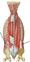
Q
POSTERIOR APPROACH TO KIDNEY
A
- Need to avoid the peritoneal cavity by a posterior retroperitoneal approach
- Risk of fibrous tissue/scarring of the mesentery from an anterior approach that can constrict portions of the GI tract
-
Latissimus dorsi
- Need to split the fibers of this muscle to reach kidney right below 12th rib
- Attached to spinus process by a broad flat tendon called the thoracolumbar aponeurosis that has anterior and posterior leaflets
- Has extensive aponeuroses which give rise to other abdominal muscles
- Maintains integrity of vertebral column and abdominal wall musculature
- Will also encounter muscles with posterior origins and insertions such as the external and internal abdominal obliques
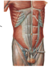
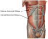
3
Q
ABDOMINAL WALL MUSCLES
A
-
External abdominal oblique
- Originates from ribs
- Attaches to rectus sheath
- Fibers run in direction of hands into pockets
-
Internal abdominal oblique
- Fibers orthogonal to external oblique
- Origin from the thoracolumbar aponeurosis
-
Transversus abdominus
- Below the 12th rib
- Fibers run in transverse plane
- Deepest of the three muscles
- Origin from the posterior side of the thoracolumbar aponeurosis
-
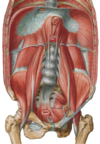
Quadratus lumborum
- Arises from tips of 12th ribs and transverse process of the upper lumbar vertebrae
- Pretty deep muscle
- Helps in postural adjustment and gait
- Maintains horizontal position of the pelvis
- Serves as a bed for kidneys
4
Q
KIDNEY POSITION
A
- Extends just slightly out from underneath 12th rib = not generally palpable
- R kidney is 2-3 cm lower because of the liver
- Kidneys are surrounded by peri/para renal fat in which they move vertically 2.5 cm during respiration
- During biopsy, you know you’re in the kidney when the needle is moving with respirations
- Surrounded by a renal fascia that contains the kidney BVs
5
Q
ASCENT OF THE KIDNEY
A
- Develops in the pelvis first and then ascends to adult position forming new and releasing old blood supplies from the aorta along the way
- Sometimes the old vessels persist or secondary vessels are made that the kidney moves into = polar arteries
- These will enter kidney at a pole, normal vasculature enters at hilus
- Since kidney develops in pelvis, parasympathetic innervation arises from the pelvic splanchnics
- Function of parasympathetic stimulation on kidneys…unknown
- Sympathetic stimulation = vasoconstriction of renal vascular bed
- Fused kidneys can become trapped under the inferior mesenteric artery → one larger horseshoe kidney
6
Q
RENAL BLOOD SUPPLY
A
- posterior branches before the hilum
- Each branch supplies an area of the kidney that is resectable in the event of disease/necrosis
- Left renal vein/right renal artery are longer that their compliments because of the positioning of the IVC and aorta
- Left renal vein crosses under the SMA which is dangerous because blood back up in the SMA can compress the renal vein (SMA syndrome) and affect the blood supply of the kidney and duodenum
- Both renal arteries give off branches to the ureters and adrenal glands
- Adrenal glands get their blood supply from three places:
- Directly from aorta
- Branches from R and L renal arteries
- Branches off inferior phrenic arteries
- Adrenal glands get their blood supply from three places:
7
Q
RENAL VEINS
A
- Renal veins have tributaries to adrenal glands, ureter, and ascending lumbar veins (which connect with the azygous system)
- L renal vein accommodates the L gonadal vein and has a connection to the azygous system which helps prevent backup of blood from the L testicle
- Right gonads drain directly to the VC
- Why connect to renal vein? Better for smaller veins to connect to a closer, larger vein than cross other structures to reach IVC
-
Azygous system serves as the venous drainage of the thoracic wall since the SVC and IVC are above and below the heart, but do not exist in the chest
- Allows collateral flow in the event of renal vein blockage
8
Q
KIDNEY STRUCTURE
A
- Outer fibrous capsule
- Hilus = point where renal pelvis meets the ureter and exits kidney
- Also point where blood vessels enter and leave
- Bean shaped area
- Open fat-filled space between renal pelvis and medulla = renal sinus
- Cortex/medulla (renal pyramid → collecting tubules merge → papilla) → merge into minor calyx → major calyx → renal pelvis → ureter
- Can visualize kidney outflow (and any points of blockage) with retrograde dye injection from ureter opening into bladder and a KUB X-ray
9
Q
URETER
A
- Outflow from kidneys → forces urine into bladder
- Extensive blood supply from renal, gonadal, and vesicular arteries
- Long course into pelvis = many points of referred pain from kidney stone movement through different dermatome levels
- Points where kidney stones can lodge:
- Narrowing at transition from renal pelvis to ureter
- Where the ureter crosses the bifurcation of the common iliac
- Before entering the bladder
- Enters bladder at an oblique angle
- Distended bladder → compression of ureter → prevents backflow from a full bladder
- Acts as a functional sphincter
- Ureter has pulsatile contractions that can force boluses or urine into a distended bladder
10
Q
INNERVATION OF KIDNEYS
A
-
Sympathetic
- From lesser (T10-11) and least (T12) thoracic splanchnics and first lumbar splanchnic
- Synapse in the aorticorenal plexus and at ganglia near renal artery
- Afferent fibers can create localized pain signals
- Area referred to as the superior mesenteric plexus
-
Parasympathetic
- From vagus nerve and pelvic splanchnics (S2-4)
- Vagus has no association with sympathetic fibers and goes directly to brain stem, so there can be some localization of pain
- Mostly create sensation of general pain/malaise and referred pain
- Kidney referred pain usually ends up in dermatomes T10-L2
11
Q
SUPRARENAL GLANDS
A
- Adrenal glands
- A type of endocrine gland
- Developmentally distinct from kidney
- Can have kidney agenesis (failure to develop during embryogenesis) without any effect on the adrenal glands
- Separated from kidney by a thin layer of renal fascia
12
Q
A
13
Q

A

14
Q
A

15
Q
A
QUADRATUS LABORUM

