Anatomy 4 - Space occupying lesions Flashcards
(30 cards)
What is the problem with space occupying lesions and define what a SOL is ?
Space occupying lesions (SOL) = abnormal tissue taking up space in the skull
The problem with SOL’s is that the base of skull foraminae do not readily permit ‘escape’ of contents ==> Acute or sub acute (expanding) intracranial pathologies can result in increased ICP, in turn the increased ICP can result in herniation

What are the 5 layers of the scalp and how do you remember them ?
SCALP:
- Skin
- Connective tissue
- Aponeurosis
- Loose connective tissue
- Pericranium
What does layer 2 of the scalp contain and why is this important ?
Contains the named arteries of the scalp
The scalp arteries form a rich anastomotic network just deep to the skin – scalp lacerations & incisions can bleed excessively

What are the 4 main sutures of the skull and what is their function ?
- Coronal
- Sagittal
- Lamboid
- Squamous - joins the parietal and temporal bones
Function is to stop fractures spreading (they are fibrous joints)

What are the main bones of the skull ?

What are the main bones of the skull (face)
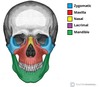
What are the main bones of the base of the skull

Go over some of the features of the skull

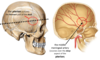
What is the pterion and why is it significant ?
It is the thinnest part of the skull formed between the Frontal, parietal, temporal and sphenoid bones
The middle meningeal artery courses deep to it ==> it is at risk to damage
State the following meningeal layers shown


What is the intracranial dura mater mainly innervated by ?
CN V
What is the dura mater adherent to ?
All of the bones of the skull
What is the tentorium cerebelli and the diaphragm sellae ?
Tentorium cereblli = a tough sheet of dura mater ‘tenting’ over the cerebellum
Diaphragm sellae = a tough sheet of dura mater forming a roof over the pituitary fossa

What is the falx cerebelli ?
A midline structure of dura mater splitting the brain into the left and right cerebral hemispheres

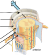
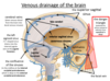
Label the venous drainage of the brain

Main veins to remember:
- Superior sagittal sinus
- Cerebral veins
- Left sigmoid sinus
- Opthalamic veins
- Internal jugular vein
- Facial vein
The dural venous sinuses lie between the periosteal and meningeal layers of the dura mater. They are best thought of as collecting pools of blood, which drain the central nervous system, the face, and the scalp. All the dural venous sinuses ultimately drain into the internal jugular vein

Label the arteries indicated

Note that the external carotid arteries remain external to the cranium and supply the face, scalp and neck

What does the internal carotid and what does the vertebral arteries pass through to enter the cranial cavity ?
- Internal jugular arteries pass through the carotid canal
- Vertebral arteries pass through the foramen magnum
Label the arteries of the circle of willis


What do each of the following arteries supply:
- Anterior cerebral
- Middle cerebral
- Posterior cerebral
- Anterior cerebral - supplies the anteromedial portion of the cerebrum
- Middle cerebral - supply the majority of the lateral part of the brain.
- Posterior cerebral - supply both the medial and lateral parts of the posterior cerebrum.

Describe the clinical relevance of the danger triangle of the face
- The danger triangle of the face consists of the area from the corners of the mouth to the bridge of the nose, including the nose and maxilla.
- Due to the special nature of the blood supply to the human nose and surrounding area, it is possible, albeit extremely unlikely, for retrograde infections from the nasal area to spread to the brain, causing cavernous sinus thrombosis, meningitis or brain abscess.
- This is possible because of venous communication (via the ophthalmic veins) between the facial vein and the cavernous sinus.
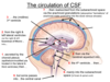
Describe the anatomy of the subarachnoid space in terms of:
- What is surrounds
- Where is it located
- What is contains
- Its function
Found between arachnoid mater and pia mater:
- completely surrounds both brain & spinal cord
- continuous
- cushions and protects the brain and spinal cord
Contains circulating cerebrospinal fluid (CSF)

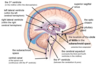
Where is CSF produced and how is it reabsorbed ?
- Produced in the choroid plexus of the ventricles
- Reabsorbed into the dural venous sinuses via arachnoid granulations
Where is the circle of willis located ?
Inferior to the midbrain, closely related to the pituitary stalk & the optic chiasm, within the subarachnoid space
Label the indicated structures