Anatomy Flashcards
(28 cards)
What nerve supplies sensory innervation of the facial skin and the anterior surface of the eyes?
CN V (the trigeminal nerve)
- all 3 divisions
Which parts of the facial skin/anterior surface of the eye does CN V1 (the opthalmic nerve) supply?
- upper eyelid
- cornea
- conjunctiva
- skin of the root/bridge/tip of the nose

Which parts of the facial skin/anterior surface of the eye are supplied by CN V2 (the maxillary nerve)?
- skin of the lower eyelid
- skin of the maxilla
- skin of the ala of the nose
- skin/mucosa of the upper lip

Which parts of the facial skin/anterior surface of the eye are supplied by CN V3 (the mandibular nerve) ?
- skin over the mandible and temperormandibular joint
- apart from the angle of the mandible- supplied by C2, 3 spinal nerve

Describe the steps of the ‘blink (corneal) reflex’
- Sensory (afferent) Limb
- Action potentials conducted from cornea via CN V1 (the opthalmic) branches
- Reaches trigeminal ganglion
- Goes along CN V
- Reaches pons
Central CNS connections between CN V and CN VII
- Motor (efferent) Limb
- Action potentials conducted via CN VII
- To eyelid part of orbicularis oculi

Why is the blink reflex important for corneal health?
Blinking is part of the immune system protection
Describe the anatomy of the oculomotor nerve (CN III) and be able to identify the nerve in illustrations/prosections/models
- connects with CNS at junction between midline and pons
- passes through cavernous sinus
- exits via orbital fissure
Which parts of the orbit/its surrounding structures recieve somatic motor innervation from CN III?
- Somatic motor to…
- superior rectus
- medial rectus
- inferior rectus
- inferior oblique
- levator palpebrae superioris (LPS)
What’s the connection between CN III (Oculomotor nerve) and ciliary ganglions?
CN III (Oculomotor) has presynaptic parasympathetic axons to the ciliary ganglion
What do the superior division and inferior division of the oculomotor nerve (CN III) supply?
- Superior Division
- Superior rectus
- Levator palpebrae
- Inferior Division
- Medial rectus
- Inferior rectus
- Inferior oblique
- Ciliary ganglion
Describe the anatomy of the ciliary ganglion.
Ciliary nerves supply autonomic axons to control diameter of iris** & **refractive shape of lens
- Long ciliary nerves:
- sympathetic
- somatic sensory
- form first part of the blink reflex
- Short ciliary nerves:
- sympathetic
- parasympathetic

List the autonomic reflexes of the eye
- Maximal eyelid elevation/wide eye opening of fight/flight
- Pupillary dilation/constriction adjusting light entry: (pupillary light reflex)
- Focussing lens far & near vision: accomodation reflex
- Lacrimation relfex tear production
- Vestibulo-ocular reflex:
- turns the eyes in the opposite direction to a head movement
- stablises gaze on an object during head movement
- CNS connections between CN VIII & CNs III, IV & VI
- Oculocardiac reflex
- reflex bradycardia in response to tension on extraocular muscles or pressure on eye
- CNS connections between CN V1 and CN X
Baby Elephants Play Lacrosse Too Much
What are the sympathetic functions of the eyelid?
- open eyes wider :. gets more light into eyes
- pupil dilates :. get more light into eyes
- focus on far objects (lens lengthens)
- emotional lacrimation
What are the parasympathetic (rest and digest) functions of the eye its surrounding structures?
- get less light into eye
- to protect retina from bright light or when asleep
- focus on near objects
- reflex lacrimation
- to wash away the stimulant foreign body and clean the cornea
- allow orbicularis oculi to work
- BUT this is more about somatic motor but closing the eye when resting and digesting could be explained by parasympathetic
How do we open our eyes wider?
- levator palpebrae superioris contains skeletal AND smooth muscle
- postsynaptic sympathetic fibres reach levator palpebrae superioris via:
- superior cervical sympathetic ganglion
- internal carotid nerve
- internal carotid plexus
- axons carried on the opthalmic artery…
- and on its branches to the orbital structures
Describe pupillary dilation
sympathetics dilate the pupil!
in dim light; fight/flight in the ‘sick’ patient
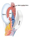
Dilator pupillae fibres are radially arranged
- orginate around the external circumference of iris- fixed
- insert around the internal circumference of iris-mobile

What is a non-physiologically enlarged pupil called?
a non-physiologically enlarged pupil is a MYDRIATIC PUPIL
mydriatic drugs induce dilation of the pupil
Describe pupillary constriction
parasympathetics constrict the pupil
in bright light and ‘rest & digest’
Sphincter pupillae fibres encircle pupil around internal circumference of iris
What is a non-physiologically constricted pupil known as?
a non-physiologically constricted pupil is known as a miotic pupil
e.g. a component of Horner’s syndrome
Briefly discuss fixed “pin-point” pupil and a “fixed-dilated” (“blown”) pupil
- Fixed “pin-point” pupil* is often a serious pathological sign
- opiate drugs
A “fixed-dilated” (“blown”) pupil is often a serious pathological sign
- CN III pathology
Describe the anatomical relation of the lens and the ciliary body
suspensory ligament of lens connects the circumferences of the lens and the ciliary body
- the ciliary body is:
- muscular and vascular
- smooth muscle like a sphincter around circumference
Describe what happens in “far vision”
- the ciliary muscle relaxes in “far vision”
- no parasympathetics
- ligament tightens & lens flattens to focus on an object in the distance
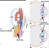
Describe what happens in “near vision”
- the ciliary muscle contracts in “near vision”
- parasympathetic
- ligament relaxes & lens becomes spherical to focus on near objects

What are the three different types of tears?
Basal tears
Reflex tears
Emotional tears
