Anatomy Flashcards

(78 cards)

1
Q

Coronal Plain

A

vertical plane that divides body ventral and dorsal parts
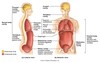
2
Q

cranial

A

superior ( nearer to head)

3
Q

caudal

A

inferior ( nearer to feet)

4
Q

proximal vs distal

A

proximal is closer to point of origin distal is farther from point origin. example shouder is more proximal than hand

5
Q

Ipsilateral

A

occuring on same side of body.

the right arm and the right lung are ipsilateral but not bilateral

not equilent to unilateral

unilateral= unpaired structure on one side

the liver is unilateral

bilateral= paired structure on L and R side of body

the lungs are bilateral

6
Q

contralateral

A

occuring on the opposite side of body

7
Q

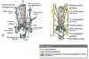
Flexion vs extension

A
8
Q

Adduction vs abduction

A
9
Q

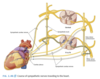
Dorsal Body Cavities

A
10
Q

Ventral Body Cavities

A
11
Q

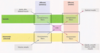
What are the tissue types

A
  1. Epethilias
  2. connective Tissue
  3. Muscle Tissue
  4. Nervous Tissue
12
Q

Epithelial tissue

function?

What are 3 examples of epithelial tissue in the body?

describe what it looks like

A

1)Epithelial tissue

  • Tightly packed cells that form a sheet
  • Functions: cover and line, seperate different environments

ex)

  • Cover body surface (epidermis)
  • Line internal body cavities (thoracic & abdominopelvic – serous membrane made of epiethlium( the pleura of the lungs are a serous membrane) )
  • Line tubes (GI tract & respiratory tract – mucosa- epiethelial tissue)
  • Internal lining of blood vessels (endothelium) and heart (endocardium)
  • Helps to form glands and ducts
  • Apical surface (interacts with lumen or the inside of whatever it is lining, has microvili) and basal surface (located on basement, or connective tissue side)
  • Avascular. Capillaries in underlying CT.
13
Q

Integumentary System

describe its structure

A

Integumentary system:

Skin protects the body and underlying tissues, prevents dehydration, participates in thermoregulation, is involved in sensation, and synthesizes/stores vitamin D

  1. Epidermis: epithelium (avascular)
  2. Dermis: dense connective tissue, tougher structure
  • Collagen and elastic fibers in ECM provide strength and skin tone
  • Hair follicles, arrector pili muscles (smooth), sebaceous (sweat) glands
  1. Subcutaneous tissue (superficial fascia; hypodermis)
  • Loose connective tissue
  • Most of body’s fat storage
  • Blood vessels, lymphatics, deep parts of sweat glands, cutaneous nerves
  1. Deep fascia (white layer)
  • Dense connective tissue
  • Surrounds body deep to skin and superficial fascia

skin = epidermis+dermis

14
Q

Deep fascia: function?

deep fascia vs investing fascia

A
  1. Extensions of deep fascia invests neurovascular bundles and muscles, divides groups of muscles, surrounds bone and lies between the body wall and membranes that line body cavities

  1. Deep fascia and investing fascia are synonymous

Deep fascia is like seran wrap holding everything together. It will surround a neurovascular bundle, artery nerve and vein that run together

Deep fascia surrounds bone, will lie between bone and body wall

15
Q

Muscle Tissue

What are the different types?

What are thier fuctions ?

tendon vs aponeurosis

A

Muscle tissue

  • Allows for movement and control
  • 3 types:

1) Skeletal muscle (Somatic; Voluntary)

  • Made up of striated muscle on one end and tendon (round) or aponeurosis (flat) on the other end
  • Attached to bone, cartilage, ligaments or deep fascia (sometimes skin)
  • Contracts to move or stabilize body, manipulate environment, produce facial expressions
  • Striated or banded appearance

2) Smooth muscle (Visceral; Involuntary)

  • Found in walls of hollow organs; arrector pili muscle in dermis
  • Contracts to propel substances through hollow tube to reduce diameter of organ
  • Not striated

3) Cardiac muscle (Visceral; Involuntary)

  • Found in wall of heart
  • Contracts and propels blood through circulatory system
  • Striated, branching
16
Q

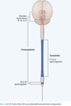
What is nervous tisssue?

What are the cells components?

A

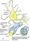
Neuron = functional unit of nervous system

Neuron consists of:

  • Cell body
  • Processes extending from cell body
  • Dendrites: carry impulses towards cell body
  • Axon: carry impulses away from cell body
  • Point of communication between 2 neurons = synapse (via neurotransmitters)

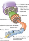
Other type of nervous tissue - neuroglia (glial cells or glia)

  • Support, nourish and insulate neurons
  • Schwann cells provide myelin sheath in PNS and enhance conductivity
  • Nerves may be myelinated (white) or unmyelinated (gray)
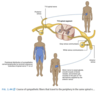
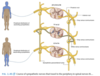
17
Q

Multipolar neuron

A

•motor neuron

Most abundant

  • Many dendrites and single axon
  • Dendrites receive stimulus
  • Axon terminals release neurotransmitter to effector

( 2 types of neurons)

18
Q

Unipolar neuron

A
  • Mainly in peripheral nervous system (cell bodies located in dorsal root ganglia)
  • No dendrites and one axon (peripheral and central process)
  • Peripheral process has receptive endings
  • Central process releases neurotransmitter
  • sensory neuron

2 types of neurons that we discuss in this anatomy class

19
Q

What is the function of the skeletal system?

What does the skeletal system consist of?

axial vs appendicular skeleton

A

•Divided into:

1) Axial skeleton – cranium, vertebrae, ribs and sternum
2) Appendicular skeleton – bones of upper and lower limbs

•Made up of bone and cartilage

•Bone is vascular (has a blood supply) and innervated

20
Q

What are the three types of cartilage and thier different functions?

A
  • Cartilage is avascular and is not innervated
  • Hyaline cartilage – most common (articular surfaces, synovial joints )
  • Elastic cartilage – recoil property (external ear)
  • Fibrocartilage – tensile strength (IV discs or areas that bear a lot of weight from upper body )
21
Q

What are the two types of joints?

Describe thier differences

A

Joints

•Where two skeletal elements come together (articulate)

•Are either classified as: Synovial or Solid joints

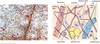
  • Synovial joints: highly moveable
  • Hyaline cartilage covering articular surfaces
  • Joint capsule with inner synovial membrane(pink) (produces synovial fluid) and outer fibrous membrane (dense CT)
  • Articular cavity with synovial fluid that reduces friction and noursishes cartiledge

  • Solid joints: some or little movement
  • Skeletal elements united by fibrocartilage
  • Moments at solid joints are more limited than those at synovial joints
22
Q

What is the muscular system?

origin vs insertion

prime mover vs synergist

agonist vs antagonist

fixator

A
  • Skeletal muscle that attaches directly or indirectly to bone
  • Origin: proximal/fixed end
  • Insertion: distal/movable end

•Prime mover (agonist): main muscle responsible for a specific movement

•Synergist: complements action of prime mover

•Antagonist: opposes action of prime mover

•Fixator: steady skeletal elements during certain movements

23
Q

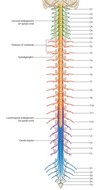
How are neurons organized?

A

Nerves are bundles of fascicles (and fascicles are bundles of long axons that are called nerve fibers).

•Each nerve has a connective tissue framework and blood vessels (vasa nervorum)

Nerves in the PNS are either spinal nerves (31 pairs) or cranial nerves (12 pairs)

A collection of cell bodies in the PNS is called a ganglion

24
Q

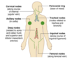
What is the lymphatic system? What does it do?

A

Recycles excess tissue fluid back to cardiovascular system

  • Interstitial fluid
  • Pathogens
  • Cell products (e.g. hormones)
  • Cell debris

Substance known as lymph

  • Carried from lymphatic capillaries to lymph nodes by way of lymph vessels
  • Lymph nodes act as body’s defense against foreign antigens
  • Filter, detect and phagocytose
25
What are lymph nodes and where are they located?
Clusters of lymph nodes found throughout the body * Node can undergo physical changes when draining regions that have an infection or are diseased * Clinically important Lymph nodes that drain infected or diseased regions can become hard or tender or larger. Clinicians can utilize these changes to track the spread of disease. Tumor cells that metastasize can enter lymphatic vessels and grow secondary tumors in lymph nodes.
26
Where do lymph vessels drain?
Lymph vessels form larger trunks and ducts These larger structures drain lymph into large veins * Lymph from the right upper limb, right thorax, right side of head and neck will drain into vein on right side of neck * Lymph from everywhere else will drain into vein on left side of neck Internal jugular vein- coming down from the neck Vein over from the upper limb= subclavian Where they meet=left venus angle and they form the brachiosyphalic vein At two veinous angle is where lymph is entering
27
Describe the structure of the vertebrae? numbers function
Spinal cord is surrounded by the vertebrae •Vertebral cavity 7- cervical 12- thoracic- this is where the ribs connect to the vertebrae 5-lumbar 5 -sacrall 3-4- coccyx There is an opening in each vertebrae where the nerves enter and exit
28
Describe the spinal nerves: number arrangement
Spinal nerves will communicate with the spinal cord at a specific level. ( There are cranial nerves in PNS as well, but we didnt discuss them in class so PNS has 12 cranial and 31 Spinal, jst so you know) Spinal cord (CNS) is surrounded by the vertebrae •Vertebral cavity Spinal nerves (PNS) are named according to their relationship with certain vertebrae 31 pairs of spinal nerves: *•**8** cervical* * 12 thoracic * 5 lumbar * 5 sacral * ***1** coccygeal*
29
Connective Tissue Function? Components? Types?
* Cells are more spread out compared to epithelial tissue * Cells are surrounded by extracellular matrix (ECM) * ECM is composed of ground substance ( jelly like tissue) and fibers (collagen, elastin, reticular) fibers help provide structural properties * Properties of CT depends on composition of ECM * Connective tissue proper * Loose connective tissue (e.g. superficial fascia): abundant ground substance and collagen fibers that are spread out and thin * Dense connective tissue (e.g. dermis): abundant in fibers Specialized CT: bone, cartilage, hematopoietic tissue, adipose and lymphatic tissue Fascia, tendons and ligaments are all CT
30
How is the body segmentally arranged
Each *spina*l nerve innervates tissue that is derived from a somite on the corresponding level as the nerve somites develop into skin and muscle innervated by nerve
31
32
Provide an overview of the Peripheral nervous system ( origin of S, Effector for E, how many neurons? GSA vs GVA GSE vs GVE
At the center of this diagram is the CNS and all input/output belongs to the PNS Impulses to CNS travel to C nervous system by one sensory neuron – In torso region sensory neuron (GSA AND GVA) has cell body in dorsal root ganglion With GSE, the a single neuron is going to deliver message from CNS to effector. the cell body is in CNS With VE, two neurons are going to deliver to effector. The first neuron cell body in CNS and the second neuron cell body in ganglion In torso region sensory neuron (GSA AND GVA) has cell body in dorsal root ganglion *note that GVE is not considered part of autonomic nervous system*
33
GVE of autonomic
Travel to viscera by a chain of two motor neurons that communicate with oneanother: * The first neuron is called presynaptic because it is the motor neuron before the synapse occurs or preganglionic and the second is postsynaptic or postganglionic * Synapse between the two motor neurons takes place at a ganglion (collection of neuron cell bodies located outside the CNS) *•The presynaptic neuron is myelinated because it is located in CNS and the postsynaptic neuron is unmyelinated*
34
Effect of sympathetic nervous system vs parasympathetic
Sympathetic prepares the body for exertion: raise blood pressure (how?) increases heart rate and strength of contraction dilate airway and pupils inhibits digestion reduces secretion,which allows for max airi exchange, vasoconstricts largepulmonary arteries to help support walls during an increase in blood pressure Parasympathetic nervous system(rest and digest ( brings the body to a resting state) : Lowers blood pressure decreases heart rate and strenght constriction in heart arteries constricts bronchi increases sectrion constricts airway and pupils constricts smooth mucle in GI tract neccessary for digestioni stimulates glandular secretion
35
Autonomic innervation of body walls or limbs sudomotion pilomotion
sympathetic arrector pili muscle (effector): causes hair to stand on end( pliamotion) Peripheral blood vessels(effector): causes blood vessels in skin to constrict and vasodilation in skeletal muscle sweat gland(effector): secretion sweating (sudomotion) Parasympathetic arrector pili muscle, sudiferous glands, and arrector pili= no effect does not innervate This is why all spinal nerves branching wil have post synaptic sympathetic fibers but not parasympathetic fibers
36
sympathetic vs parasympathetic neruons Where do impulses originate
Sympathetic division → Fight or flight CNS Impulses originate from thoracic & lumbar spinal cord (T1-L2) Thoracolumbar outflow Relatively long postganglionic axons Parasympathetic division → Rest & digest Impulses originates from brainstem and sacral spinal cord ((cranial) C3, C7, C9, C10) , (S2-S4) Craniosacral outflow Relatively short postganglionic axons O------------- Another difference between these two divisions is the type of neurotransmitter released by the postsynaptic neuron. * All pre-synaptic have axons and release achetycholine * Postsynaptic sympathetics: norepinephrine * Postsynaptic parasympathetics: acetylcholine O-------------------------------------
37
38
Parasympathetic Pathway ONly have to be familiar with vagus nerve which is?
Presynaptic parasympathetics: * Cranial nerves III, VII, IX, X * Pelvic splanchnic nerves Vagus nerve is cranial nerve 10: it innervates the lungs, the liver, the pancreas, the stomach, the heart , and the gall bladder, the small intestine, and the tranverse colon, ascending colon, and cecum (all thoracic viscera and most of abdominal viscera) •Pelvic splanchnic nerves supply visceral motor to the distal 1/3 of large intestine and pelvic viscera Preganglionic axons (very long) will synapse with postganglionic (axons very short) cell bodies at a ganglion located on or near the effector
39
Sympathetic Pathways ( In general what are they?)
Sympathetic trunk (Bilateral) Paravertebral (chain) ganglia are linked together to form the sympathetic trunk on both right and left sides of the vertebral bodies 3 Cervical ganglia in neck Ganglion impar unites trunks on either side at coccyx The inferior cervical ganglion is sometimes joined with the first thoracic ganglion and together they are called the cervicothoracic or stellate ganglion. Note in image how some sympathetic fibers are headed towards the viscera and other fibers are using somatic nerves (aka spinal nerves) to reach body periphery. *Sympathetic* outflow is limited to T1-L2, so other vertebrae have output as well just not sympathetic Sympathetic trunk provides a route for sympathetic impulses to reach all spinal nerves Note communication of thoracic spinal nerve ventral rami (i.e. intercostal nerves) with paravertebral ganglia. There are 6 pathways to learn:
40
41
Sympathetic Pathways to blood vessels and structures in skin (T1-L2 nerves)
Remember that sympathetic output only originates in the spinal cord at T1-L2 levels. Cell bodies of presynaptic sympathetic neurons are located in the lateral horn of gray matter. How sympathetic impulses reach spinal nerves T1-L2: 1. Presynaptic sympathetic fibers travel on the ventral root to the spinal nerve proper and ventral ramus. 2. Fibers reach sympathetic trunk via the white ramus communicans (myelinated) 3. Synapse with postsynaptic sympathetic neuron with cell body in a paravertebral ganglion 4. Postsynaptic fiber uses gray ramus communicans (unmyelinated) to reach spinal nerve and make it’s way to effector via spinal nerve branches ● Destination: Blood vessels, structures in skin take note: gray and white ramus communicans on ramus not nerve
42
Sympathetic pathway to blood vessels and structures in the skin (C1-C8, L3-Cox0)
Sympathetic impulses to spinal nerves C1 – C8 and L3 – C0: 1. Presynaptic neuron travels on ventral root, spinal nerve proper and ventral ramus 2. Reaches sympathetic trunk via white ramus communicans and then travels up or down the sympathetic trunk before synapsing with second motor neuron to reach destined level 3. Synapses with second motor neuron in a paravertebral ganglion at new level 4. Leaves sympathetic trunk via gray ramus communicans and travels to effector via spinal nerve Destination: Blood vessels, structures in skin
43
44
Sympathetic Pathway to head Which plexus is used? when?
1. Presynaptic neuron travels on ventral root, spinal nerve proper and ventral ramus 2. Reaches sympathetic trunk via white ramus communicans and then travels up or down the sympathetic trunk before synapsing with second motor neuron to reach destined level 3. Synapses with second motor neuron at superior cervical ganglion 4. post synaptic fibers use plexuses surrounding arteries (periarterial plexus) to reach effectors Periarterial plexuses are autonomic plexuses that surround arteries Dont worry about this too much
45
Sympathetic Pathway to Heart and lungs
1. Origin from T1 – T4 2. Synapse between the first and second motor neuron occurs in paravertebral ganglion (either at same level or above) 3. Postsynaptic neuron travels via upper thoracic splanchnic nerve to reach cardiac plexus or pulmonary plexus 4. Travels through the plexus to reach effector
46
Autonomic Plexus
All autonomic plexuses will typically have: * Visceral sensory fibers * Presynaptic parasympathetic fibers * Presynaptic or postsynaptic sympathetic fibers (depending on the sympathetic pathway) • Review innervation of the trachiobronchial tree: * Sympathetic stimulation: inhibit contraction of smooth muscle of the bronchial tree thereby dilating the airways, inhibits glandular secretion, and vasoconstricts large pulmonary arteries * Parasympathetic stimulation: constricts smooth muscle of bronchial tree and narrows airways, stimulates glandular secretion, and vasodilates smaller pulmonary arteries Review autonomic innervation of the heart: * Sympathetic stimulation: increases heart rate and force of heart contractions * Parasympathetic stimulation: decreases or slows heart rate and reduces the force of contraction; also vasoconstricts coronary arteries
47
Sympathetic pathway to the abdominal and pelvic viscera
1. Originate from T5-L2 2. The first motor neuron travels through the sympathetic trunk without synapsing 3. Presynaptic sympathetic neuron utilizes a splanchnic nerve to reach prevertebral (preaortic) plexus near aorta (lower thoracic and lumbar splanchnic nerves). 4. Synapse between first and second motor neuron occurs in a prevertebral (preaortic) ganglion (except sacral splanchnic nerves) 5. Postsynaptic sympathetic neuron utilizes a periarterial plexus to reach effector Greater splanchnic nerve (T5-T9): Foregut & Midgut; Spleen Lesser splanchnic nerve (T10-T11): Midgut Least splanchnic nerve (T12): Kidneys & Gonads Lumbar splanchnic nerve (L1-L2): Hindgut & pelvic organs Sacral splanchnic nerve (L1-L2): Pelvic organs
48
49
Sympathetic Pathway to the suprarenal gland(medulla) what is the difference between this pathway and the abdominopelvic pathway? skin pathway? cardiac pathway?
To suprarenal gland (medulla): 1. First motor neuron (T8-T12) travels onto sympathetic trunk, does not synapse there, and continues onto a splanchnic nerve (Greater, lesser and least splanchnic nerves) 2. Once reaching the prevertebral plexus the presynaptic sympathetic fibers branch off to reach effector 3. Synapse occurs in Medulla and cells in medulla release neurotransmitter directly into bloodstream different type of pathway because they pre and post synapse is at the effector( the medulla) . This structure kind of looks more parasympathetic becasue preganlionic fibers are typically short but here it looks long and it reaches it postganlionic neuron close to effector so post ganglionic is short and close to effector
50
Visceral sensory sympathetic vs parasypathetic
Travel along with visceral motor (GVE) fibers * Those with sympathetics detect chemical changes, irritation, temperature etc. * Those with parasympathetics detect normal physiological processes (monitor blood pressure and oxygen levels) and are involved in visceral reflexes (detect stretching/distention; influence peristalsis, urination, and defecation)
51
Body wall
branches of spinal nerves
52
somatic pain vs visceral sensation
Somatic pain is intense sensation carried by GSA (general somatic afferent) neurons in the body wall (branches of spinal nerves) •Parietal serosa is innervated by GSA nerves in the body wall and pain is well localized Visceral sensation is mostly dull sensation carried by GVA (general visceral afferent) neurons from the abdominal viscera •Visceral serosa is innervated by GVA neurons that detect stretch and chemical irritation; pain is poorly localized Referred pain or sensation originates in one location and is misinterpreted as somatic pain from a dermatome associated with the same spinal cord segment
53
What are the divisions of the mediastinum?
T2 marks the end of th superior mediastinum T6 marks the bottom of the mediastinum From T1 to sternum is the clavicle
54
What are the contents of the middle portion of the mediastinum
55
What are the contents of the superior mediastinum
Aortic Arch, Pulmonary Trunk, Left & Right Brachiocephalic Veins, Trachea, Upper Esophagus, Thoracic Duct
56
What are the contents of the middle mediastinum?
The Heart! Pericardium, Phrenic nerves & Pericardiacophrenic vessels on sides of Pericardium
57
What does the anterior mediastinum contain?
the thymus if you are under 25 years old
58
what does the posterior medistinum contain ?
Descending Aorta, Sympathetic Trunk, Vagus nerves, Lower Esophagus, Azygos Venous System, Thoracic Duct esophagus is still going through here but his
59
What is this structure?
left atrium
60
What is this structure?
left ventricle
61
what is this structure?
Apex of the heart
62
What is this structure? What is the specific name for this part of the strucutre?
Right Atrium What is shown in this picture and the picture of the left atrium is only part of the atrium called the auricle. The auricles are remnants of early embryonic atria ,
63
What is this structure?
64
The cardiovascular system consists of two _______ pathways which form a _______ loop connected by the \_\_\_\_\_\_\_. Arteries carry blood ______ from the heart. Veins carry blood ______ the heart.
The cardiovascular system consists of two circulatory pathways which form a closed loop connected by the heart. Arteries carry blood away from the heart. Veins carry blood toward the heart.
65
How many outflows does the heart have? Name the strucure
aorta The adult heart hass two outflows: pulmonary trumk and the aorta note that the that the aorta is red because oxygenated blood is leaving the heart to go to the body but tthe pulmonary trunk is blue because deoxygenated blood is leaving the heart to go to the lungs and be oxygenated
66
Name this structure
Pulmonary trunk
67
How many inflow systems does the adult heart have? What are they? fill in the labels of the dorsal side of the heart
there are 2 inflow system deoxygenated blood flowing into the pulmonary trunk (left and right pulmonary veins) and deoxygenated blood flowing into superior and inferior vena cava Note that the inferior and superior vena cava are blue because the incoming blood is deoxygenated but the pulmonary veins are red because the incoming blood is oxygenated
68
What is the direction of blood flow?
blood goes through inferior and superior vena cava to enter the right atrium of the heart then it goes into the right ventricle which carries it to the rpulmonary artery( pulmonary trunk) from there goes to lungs exchanges co2 for o2 and comes back through pulmonary vein. from pulmonary vein flows into left atrium from left atrium to left ventricle and then to aorta which pumps blood to the body
69
the heart in situ Name these structures realize there is an error with the subclavian and internal jugular
70
Identify the cardiac valves Where should a physican listen for heart sounds
body of the sternum covers heart and its difficult for sound to travel through bone so hard to hear. So doctors listen at specific places around the bone
71
What is the image shown and what are the sounds produced?
72
ventral view ( there will be a lateral view) Identify the structures of the heart in the xray
73
Name these structures in a lateral xray of the heart
74
75
Identify the strucutes of the thorax
76
What dermatome corresponds to the jugular notch? the sternal angle? the xiphoid process? the umbilicus? the pubic symphysis?
Sternum Jugular Notch, Dermatome C4, T2 vertebra Sternal Angle, Dermatome T2, T4 vertebra Xiphoid- Dermatome T6, T9 vertebra Umbilicus- Dermatome T10 Pubic Symphysis- T12-L1
77
Identify the structures which nerves innervate the diaphram what type of PNS fibers innervate the diaphram?
C3, C4, and C5 prhenic nerves innervate the diaphram the phrenic nerves carry both GSE and GSA fibers (voluntary muscle) Reffered pain from the diaphram is felt in the shoulder the descening aorta, inferior vena cava, and esophagus travel through openings in the dorsal side of the diaphram
78