Anatomy Flashcards
(38 cards)
What are the layers of the body wall? Which parts are derived from ectoderm? Mesoderm?
Layers in order:
Superficial Body Wall:
1) Epidermis (keratinized squamous stratified epithelium)
2) Dermis of skin (dense irregular connective tissue)
3) Superficial Fascia (fat + loose connective tissue)
Deep Body Wall:
4) Deep investing fascia
5) Skeletal Muscle/Bone
6) Parietal layer of celom lining (simple squamous mesothelium)
Epidermis = derived from ectoderm
Everything else: mesoderm
What are the parts of the sternum?
Manubrium -> sternal angle -> sternal body -> xiphoid process
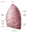
What are the key structures of the female breast?
Nipple, areola, lactciferous duct, mammary gland (lobule shaped when resting), fat

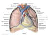
Describe pectoralis major and pectoralis minor
These muscles help contorl the movement of the arm. They grow over the body wall during development. Pectoralis major is superficial to pectoralis minor and attaches to the humerus and sternum. Pectoralis minor, which is superficial to the thoracic skeletan, attaches to the ribs and scapula.

Describe serratus anterior
Located on the lateral side of the chest. Used in pushing.
Attached to scapula
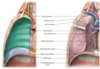
What are the “true” body wall muscles? Which direction are the striations in each of the muscles?
External oblique (45 degree angle down from rib down)
Internal Oblique (90 degree rotation from the external oblique)
Transverse Thoracis (parallel with ground if person sitting)
These muscles are also called external intercostal, internal intercostal, and innermost intercostal if they are located between the ribs.
What are the functions of the intercostal muscle?
1) Protect viscera
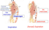
2) Respiration:
External intercostal -> pulls down on ribs -> expands thoracic cavity -> inspiration
Internal Intercostal -> pushes ribs in -> contracts thoracic cavity -> expiration
What is segmentation? Why do neurovascular bundles appear in series? Where do neurovascular bundles run?
Seegmentation -> wall of abdoment/thorax organized like stack of similarly stacked circles (tires/pancakes) Neurovascular bundles are present on each of these stacked circles, thus giving them a “in series” look. Neurovascular bundles run between the middle and innermost muscle layers.
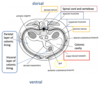
Describe the circulation of the typical body segment? What is function of aorta? What is function of internal thoracic artery?
Aorta-> posterior intercostal artery
Subclavian -> internal thoracic artery -> anterior intercostal artery
What does the autonomic nervous system innervate? What is unique about the autonomic nervous system? What are the divisions of the autonomic nervous system?
Cardiac muscle, smooth muscle, glands
In autonomic nervous system, neurons are arranged so that there are always 2 neurons in sequence from brain to target tissue.
Parasympathetic, sympathetic
What is the role of the sympathetic nervous system? Parasympathetic?
Sympathetic: increase HR, Increased BP, dilate airways, dilate pupils
Parasympathetic: Decrease HR, Decrease BP, constrict airway, constrict pupils, increase gland constriction (saliva/stomach acid), peristalsis
What is difference between efferent and afferent neurons?
Afferent = adding = sensory neurons (brings input into the CNS)
Efferent = exiting = motor neurons from CNS to organs/muscles
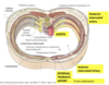
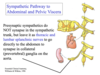
What is the anatomy of sympathetic neurons? What is the siginificance of the sympathetic trunk? What is thoracolumbar flow?
Presynaptic cell body located in the lateral horn of the spinal chord. The axons of the presynaptic neuron leave via the entral root and enter the sympathetic trunk. In the sympathetic trunk, they innervate the post synaptic neuron which goes on to act on the target tissue (primarily controlling blood flow).
Sympathetic trunk is the location where the presynaptic neuron innervates the post synaptic neuron. Additionally, since sympathetic presynaptic neurons only exit from T1 - L2 (thoracolumbar flow), the sympathetic trunk allows presynaptic neurons to innervate postsynaptic neurons on other levels of the spinal chord.

What is are white rami communicans? What are grey rami communicans?
White rami communicans = pre-synaptic neuron
Gray rami communicans = post-synaptic neuron
** every spinal nerve recieves postsynaptic sympathetics from the sympatethic trunk
white = myelinated
gray = non-myelinated
What is the pathway for sympathetic neurons?
Lateral horn T1-L2 -> ventral root T1-L2 -> white ramus communicans (presynaptic communicating ramus) -> synapse in the sympathetic trunk -> grey ramus communicans (post synaptic communicating ramus) -> spinal nerve -> ventral/dorsal ramus
Which is more lateral? Presynaptic communicating ramus or postsynpatic communicating ramus?
Presynaptic
What are splanchnic nerves?
Sympathetic nerves also innervate organs (i.e. heart, trachea). Splanchnic nerves are sympathetic nerves which travel from the sympathetic trunk directly to innervate the organ (i.e. heart).
Splanchnic nerves always innervate inferiorly to the location where they branched off the sympathetic trunk.

What is unique about sympathetic pathway for abdominal/pelvic splanchnic nerves?
The presynaptic neurons do NOT synapse in the sympathetic trunk for abdominal splanchnic nerves. Instead, they synpase on collateral ganglia (next to organ). Post synaptic neurones then follow arteries to their target organs.
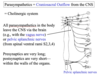
Where do parasympathetic nerves exit? What does vagus nerve do? What is the main mechanism of parasympathetics? Where do parasympathetic presynaptic neurons innervate post synpatic neurons?
Craniosacral outflow
Vagus Nerve (Cranial Nerve 10): Innervates smooth muscle in GI tract, glands, basically everything in gut
Mainly uses acetylcholine
Synapses usually occur in the wall of organ. Presynpatic neurons -> very long. Post synaptic -> very short.
What is difference between visceral and general sensory?
General sensory -> provide innervation for the body wall
Visceral -> follow splanchnic nerves to provide sensation in organs (usually provide dull sensation)
** only exception is that they will provide sharp pain from stretching/inflamation (gall stones, appendicities, lesions etc)
What is referred sensation?
Pain/signal originates from visceral location and is sensed by the same spinal segment in the body wall. (i.e. heart pain interpretted by brain as coming from the arm at T1)
What is the coelimic cavity? What happens to it during development?
Coelimic cavity is a fluid filled body cavity which gives rise to the pleural cavity, pericardial cavity, peritoneal cavity
What does the phrenic nerve do?
The phrenic nerves originate from C3-C5 and they innervate the diaphragm. Therefore, they are crucial for breathing.
How does the parasympathetic nervous system work on the heart?
The post synaptic neuron releases Acetylcholine onto the muscarinic GPCR in the heart. The beta and gamma subunits of the activated G-proteins then bind to K+ channels on the membrane and HYPERpolarize the muscles, therefore decreasing the propagation of action potentials and slowing the heart.