Anterior triangle and root of the neck week 1 Flashcards
What are the boundaries of the neck? (superior and inferior borders)
General Information on Neck
- Extends from base of skull and mandible to thoracic inlet
- Superior boundary is the mandible and posterior occiput
- Inferior boundary is sternum, clavicles, acromium and spinous process of C7
- Contains segments of digestive (pharynx and esophagus) and respiratory (larynx and trachea) systems

List the skeletal and cartilaginous components of the neck.
7 cervical vertebrae
Hyoid bone
Thyroid cartilage
Cricoid cartilage
Tracheal rings
Generally, what suspends the hyoid bone within the neck? What bony structures is it suspended from?
The hyoid bone is a interface between the ____ ____ superiorly, the ____ inferiorly, and the ____ posteriorly.
What activity is the hyoid bone important for?
Hyoid bone
- small U shaped bone at angle between floor of mouth and superior limit of neck
- only bone that does not articulate with another bone
- suspended from skull and mandible by ligaments and muscles
- moveable platform/anchor for muscles and soft tissue structures
- interface between oral cavity superiorly, larynx inferiorly and pharynx posteriorly
- body, greater and lesser horns
- important for swallowing. elevates the larynx during swallowing

What structure does the thyroid cartilage attach to? How is it connected to this structure?
What is the cricoid cartilage connected to and what connects it to this structure?
What structure are the thyroid and cricoid cartilages a part of?
Thyroid cartilage: attached to hyoid bone via thyrohyoid membrane. Part of larynx
Cricoid cartilage: attached to thyroid cartilage via cricothyroid ligament. Part of larynx

Define the following terms and state in what situations they are performed:
cricothyrotomy
tracheotomy/tracheostomy
Cricothyrotomy- easiest/ fastest access to airway when upper airway is blocked (by swelling, foreign object, etc); placement of endotracheal tube through Cricothyroid ligament; only small vessels located superficially
Tracheostomy- must be done surgically through anterior tracheal wall; for long term respiratory ventilation; more complicated due to thyroid gland and larger vessels

Where is the mastoid process located? What bone is it a part of?
What are the 4 parts of the mandible?
What are the 2 prominent cartilages of the larynx? Where is the larynx located in relation to the hyoid?
Surface Anatomy and Palpable Landmarks:
- mastoid process of the temporal bone behind and below the auricle of the ear
- ramus, angle and body of mandible; mental protuberance
- hyoid bone -midline of the neck below the mandible
- larynx below the hyoid: 2 of its prominent cartilages:
– thyroid cartilage: laryngeal prominence (“Adam’s apple”) created by union of the two lamina anteriorly; angle is more acute in men
– cricoid cartilage.

What structures do the superificial fascia of the neck extend from?
What muscle is located within this fascia?
Superficial fascia
- extends from the mandible to the superficial fascia of the thorax
- outermost layer surrounding the neck
- the platysma muscle located within it
- platysma is a muscle of facial expression-clenching neck

List the 4 layers of the deep cervical fascia.
- Investing
- Prevertebral
- Pretracheal/visceral
- Carotid sheath
What structures does the investing layer of the deep cervical fascia extend from?
What structures does the investing layer surround?
Investing: tubular investment surrounding entire neck; from the mandible, zygomatic arch, mastoid process and base of the occiput to the scapula, clavicles and manubrium
- splits to surround the trapezius, sternocleidomastoid and infrahyoid ms.
- encloses parotid and submandibular glands
- forms roof of anterior and posterior triangles of neck

What structures does the prevertebral layer of the deep cervical fascia envelop?
Prevertebral
- cylinder of fascia that surrounds the vertebral column, prevertebral, scalene and deep neck/back muscles.
- extends into the posterior mediastinum
- splits into 2 layers as it descends

What strucutres does the pretracheal/visceral layer of the deep cervical fascia invest?
What is the buccopharyngeal fascia?
Pretracheal (visceral)
- invests the trachea, pharynx, esophagus and thyroid/parathyroid glands. thyroid moves with swallowing due to this layer of fascia
- anteriorly, it extends from the hyoid bone, thyroid cartilage to the upper thoracic cavity
- Buccopharyngeal fascia
- posterior part of the visceral fascia covering posterior walls of pharynx and esophagus
- extends from the base of the skull into the middle mediastinum
• Pretracheal / buccopharyngeal fascias also collectively called the visceral fascia

What structures does the carotid sheat envelop?
Carotid sheath
- located laterally
- receives contributions from the other fascias
- surrounds the internal jugular vein, common carotid artery and vagus nerve
- extends from the base of the skull to the superior thoracic aperture

List the 3 fascial spaces? Why are they clinically significant?

- Pretracheal space
- Retropharyngeal space
- Within prevertebral layer (remember this fascia splits in 2 as it descends)
Fascial planes and spaces are clinically significant because infections tend to spread within the planes or travel in the fascial spaces from the head and neck into the mediastinum. Can close trachea and cause difficulty breathing.
What fascial layers is the pretracheal space between? What structures does it extend from?
What fascial layers is the retropharyngeal space between? What structures does it extend from? What movements does this space accommodate?
What strucutres does the space within the prevertebral layer extend from?
Which space is the most clinically significant?
- Pretracheal space - between the investing fascia and pretracheal fascia (anterior part) and extends from the neck to the superior mediastinum. Limited by attachments of fascia to thyroid cartilages superiorly. Infection can spread into thorax anterior to pericardium
- Retropharyngeal space - between the buccopharyngeal (posterior surface of the pharynx and esophagus) and the prevertebral fascia; extends from base of the skull to upper posterior mediastinum; accommodates the movements of the pharynx, larynx, trachea, and esophagus during swallowing. ** Largest and most significant space in neck
- Space within the prevertebral layer: where the 2 lamina create a space between the base of the skull through the posterior mediastinum to the diaphragm.
The retropharyngeal space is the most clincally significant. If one has an infection in their oral cavity, it can go through the buccopharyngeal fascia and into the mediastinum through this space.

What are the boundaries of the anterior cervical triangle? (anterior, posterior, superior, roof, floor)
Boundaries:
Anterior – Midline of the neck
Posterior – Anterior border of Sternocleidomastoid ms.
Superior – Inferior border of the body of the mandible
Roof- Investing layer of deep cervical fascia
Floor- Larynx and pharynx

Contents of the anterior triangle of the neck:
What muscles are contained within the anterior triangle of the neck?
What nerves are contained within the anterior triangle of the neck?
What arteries/veins are located here?
What glands/organs are located here?
Muscles
- Platysma
- Suprahyoid /Infrahyoid group
Nerves
- Laryngeal/Pharyngeal Nerves
- Cranial nerves or branches of (V, VII, IX, X, XII)
- Spinal nerves – Cervical plexus
- Autonomic – Cervical sympathetic trunk and parasympathetics from Vagus
Arteries and Veins
• Carotid and Jugular systems
Glands/Organs
- Submandibular gland
- Thyroid/Parathyroid gland
- Larynx/Trachea • Esophagus
Lymphatics

Name the suprahyoid muscles.
What are the functions of the suprahyoid muscles?
digastric muscle (anterior and posterior belly)
stylohyoid
mylohyoid
geniohyoid
The suprahyoid muscles:
- suspend hyoid from mandible or base of skull
- raise hyoid during swallowing
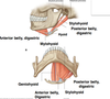
Name the suprahyoid muscles in this picture.

Note: There are 2 geniohyoid muscles (left and right). They are deep to the mylohyoid muscle.
Notice that the stylohyoid muscle splits as it descends to surround the intermediate tendon of the digastric muscle.
The anterior belly of the digastric attaches to the mandible while the posterior belly attaches to the mastoid process.

State what nerves the suprahyoid muscles are innervated by.
anterior belly of digastric muscle: Mandibular division (V3) of the Trigeminal Nerve (CN V)
posterior belly of digastric: Facial Nerve (VII)
Stylohyoid: Facial Nerve (VII)
Mylohyoid: Mandibular division (V3) of the Trigeminal Nerve (V)
Geniohyoid: ventral ramus of C1 (which travels with the hypoglossal nerve, CNXII). note this is the only suprahyoid muscle that is not innervated by a cranial nerve.
Identify the infrahyoid muscles in the attached picture.


What nerves innervate the infrahyoid muscles?
- Sternohyoid, omohyoid and Sternothyroid innervated by anterior rami of C1 to C3 via the Ansa Cervicalis
- Thyrohyoid innervated by its own nerve from C1 (Nerve to Thyrohyoid)

What muscle does the submandibular gland lie between?
What is the relationship of the submandibular gland to the facial artery and vein?
- The submandibular gland lies in btwn the anterior and posterior bellies of the digastric muscle.
- The facial vein passes superificial to the submandibular gland while the facial artery passes deep to it.

Explain the course of the Hypoglossal nerve (CN XII). What is its relationship to the posterior belly of digastic, hyoglossus, and mylohyoid muscles along its course?
What is the relationship of the muscular branch of the occipital aa to the Hypoglossal nerve?
What muscles does the Hypoglossal nerve innervate?
- enters neck by coursing deep to posterior belly of digastric ms., then passes superficial (lateral) to hyoglossus ms. and deep to mylohyoid ms. to enter oral cavity.
- innervates intrinsic muscles of tongue, hyoglossus and genioglossus (i.e. Motor for all muscles of the tongue EXCEPT the palatoglossus (innervated by CN X). (innervates muscles ending in glossus except for palatoglossus)

Fibers from the ventral ramus of C1 travel with what nerve?
They then leave this nerve to innervate what muscles? What are named branches of these nerves?
Fibers from ventral ramus of C1 travel with the Hypoglossal N. and then leave to provide Nerve to geniohyoid, Nerve to thyrohyoid and Superior root of ansa cervicalis which innervate the remaining infrahyoid muscles (sternohyoid, sternothyroid, omohyoid).

What is the relationship of the ansa cervicalis to the carotid sheath?
What nerve fibers contribute to the inferior root of the ansa cervicalis? What muscles do the inferior root of the ansa cervicalis innervate?
The Ansa Cervicalis
- Loop of nerve fibers from cervical plexus
- branches innervates strap (infrahyoid) ms
- lies on the anterior surface of the carotid sheath
- Fibers from ventral ramus of C1 run briefly with the hypoglossal nerve (CN XII); some descend to form the superior root of the ansa cervicalis and innervate the upper portions of strap ms
- Inferior root of the ansa cervicalis originates from the ventral rami of C2 and 3, passes around the carotid sheath and innervates lower strap muscles
- Separate nerves leave hypoglossal as N. to thyrohyoid and N. to geniohyoid (originate from C1).

What is the relationship of the sympathetic trunk to the carotid sheath?
The Carotid Sheath
- receives contributions from the deep cervical fascias
- surrounds the internal jugular vein, common carotid artery, internal carotid artery and vagus nerve
- extends from the base of the skull to the superior thoracic aperture
- Medial- Common carotid artery
- Lateral- Internal Jugular vein
- Posterior and in between- Vagus nerve (CN X)
- Note: The sympathetic trunk lies posterior to the medial portion of the carotid sheath.

Identify the branches of the external carotid artery.

Notes: ascending pharyngeal aa is btwn the external and internal branches of the common carotid aa
lingual aa dives deep to the oral cavity
facial aa is deep to the submandibular gland and posterior belly of digastric muscle
occipital aa: superior to hypoglossal nerve

Where is the carotid sinus located? What is contained within it?
Where is the carotid body located? What is contained within in?
What nerve innervates the carotid body and carotid sinus? What kind of nerve fibers are contained within it?
Carotid Sinus- dilation at origin of distal end of common Carotid A./beginning of Internal carotid A.; contains baroreceptors that monitor blood pressure
Carotid body- tissue at carotid bifurcation; Chemoreceptors monitor O2 and CO2 levels
The Carotid sinus and body innervated by the Glossopharyngeal (CN IX) nerve (GVA fibers): afferent limb of Baroreceptor Reflex.

What is the common place within the carotid aa where plaques form?
What are 2 treatment options for plaque formation?
- Atherosclerotic plaque in carotid artery: common at bifurcation. Presence is noted via US
- Can cause stroke
- Treatment:
(1) Carotid angioplasty with stent placement; balloon pushes plaque aside and stent placed to prevent re-occurrence
(2) Endarterectomy: Surgical removal of plaque

What foramen does the vagus nerve (X) exit through from the skull?
What are the branches of the vagus nerve in the neck? What do the branches innervate?
- Exits skull via jugular foramen in temporal bone
- Descends in the neck inside the carotid sheath between and posterior to the vessels
Branches in neck
- Pharyngeal- to most of the muscles of pharynx and soft palate
- Superior Laryngeal Nerve
- internal branch
- external branch - Recurrent laryngeal nerve/inferior laryngeal: in tracheoesophageal groove
- Cardiac
numbers correspond to numbers in picture

What structure does the internal branch of the superior laryngeal nerve pierce?
What does the internal branch of the superior laryngeal nerve innervate? What kind of innervation does it provide? (afferent or efferent)
What vessels does the internal branch of the superior laryngeal nerve travel with?
What does the external branch of the superior laryngeal nerve innervate? What kind of innervation does it provide? (afferent or efferent)
The Internal branch of the Superior laryngeal Nerve (also called the Internal laryngeal N.) pierces the thyrohyoid membrane to provide sensory innervation to the laryngeal mucosa as far inferiorly as the vocal cords; travels with the Superior laryngeal artery, a branch of the Superior thyroid Artery.
The External branch of the Superior laryngeal Nerve (also called the External laryngeal N.) travels inferiorly to provide motor innervation to the cricothyroid and inferior pharyngeal constrictor ms.

What structure does the right recurrent laryngeal nerve loop around? What structure does the left recurrent laryngeal nerve loop around?
What do these nerves innervate? What kind of innervation (sensory, motoer) do they provide?
The Recurrent Laryngeal Nerves
- The Right loops around the subclavian artery in root of neck
- The Left loops around the arch of aorta in the superior mediastinum
- Ascend in trachea-esophageal groove; are then also known as inferior laryngeal nerves
- Sensory innervation to laryngeal mucosa below vocal folds
- Motor to intrinsic muscle of larynx

What is the relationship of the thyroid gland to the thyroid cartilage? Cricoid cartilage? Trachea?
What are the lobes of the thyroid? What connects the lobes?
What is the relationship of the thyroid to the infrahyoid muscles?
What fascia surrounds the thyroid?
What lymph nodes drain the thryoid gland?
Thyroid Gland
- Anterior in neck, lateral and inferior to the thyroid cartilage
- 2 lateral lobes cover lower part of thyroid cartilage, cricoid cartilage and antero-lateral trachea
- Isthmus in midline connecting 2 lobes; H shaped
- In visceral compartment, deep to strap muscles.
- Surrounded by pretracheal/ visceral fascia holding gland onto larynx and trachea (therefore moves with swallowing)
- A pyramidal lobe may be present extending from isthmus superiorly
- Drained by deep cervical lymph nodes

Explain the arterial supply and venous drainage of the thyroid gland. State what arteries the thyroid arteries branch from/what veins thyroid veins drain into.
What thyroid artery is present in up to ten percent of people? What aa(s) does it arise from? What portion of the thyroid gland does it supply?
Thyroid Arteries
- Superior thyroid artery from the External carotid artery.
- Inferior thyroid artery from the Thyrocervical trunk.
Thyroid Veins
- Superior and middle thyroid veins drain into the Internal jugular vein
- Inferior thyroid veins typically drain into the Brachiocephalic veins
Thyroid Ima Artery in up to 10 % of people: arises from brachiocephalic trunk or aortic arch to supply isthmus (Clinically significant!!)

Where are the parathyroid glands located? What is their vasculature and lympatic drainage like?
- usually 2 pairs on deep surface of thyroid lobes
- variable in number and location
- Vasculature and lymphatic drainage same as thyroid gland

The two main arteries supplying the thyroid gland are accompanied by nerves that can be damaged by a thyroidectomy.
What nerves travel with the arteries that supply the thyroid? What are the consequences if they are damaged during surgery?
The two main arteries supplying the thyroid gland are accompanied by nerves that can be damaged during Thyroidectomy.
- The superior thyroid A. is related to the external laryngeal nerve. This nerve supplies the Cricothyroid muscle which tenses the vocal cords. Damage to the external laryngeal nerve would produce weakness of the voice (a monotone voice).
- The inferior thyroid A. is related to the recurrent laryngeal nerve which supplies the intrinsic muscles of the larynx. Injury to one nerve would cause hoarseness of the voice. Bilateral damage to the recurrent laryngeal nerves may cause the patient to lose speech completely and can cause difficulty in breathing because the vocal cords cannot open.

What are the boundaries of the root of the neck? (posterior, anterior, inferior)
Boundaries of Root of Neck
Posterior - superior surface of the 1st Thoracic vertebra
Anterior - superior surface of manubrium and clavicles
Inferior - plane of the thoracic inlet (superior thoracic aperature)
The apical part of each superior lobe of the lung and the cupula of parietal pleura extend into the root of the neck. Important clinically because it contains structures passing between the neck, thorax and upper extremity

What muscle divides the subclavian aa into 3 parts? What are the 3 parts it is divided into?
What are the 3 branches of the first part of the subclavian aa? (just list)
Both Subclavian arteries are divided into 3 parts based on their relationship to the anterior scalene muscle.
- Part 1 is from the origin of the artery to the medial edge of the anterior scalene ms.
- Part 2 lies posterior to the anterior scalene ms.
- Part 3 is from the lateral edge of the muscle to the lower border of the first rib; then it called the axillary artery.
- Part 1 has 3 branches:
- Vertebral artery
- Internal Thoracic artery
- Thyrocervical trunk

Explain the pathway of the vertebral artery.
What are the 3 branches of the thyrocervical trunk?
From what part of the subclavian does the costocervical trunk arise? What are the 2 branches of the costocervical trunk?
What does the dorsal scapular aa branch from?
Vertebral Artery
- Arises from first part of the Subclavian A.
- Enters transverse foramen of C 6, ascends vertically through foramina of upper 6 cervical vertebrae.
- Curves over top of atlas to pierce posterior atlanto occipital membrane, passes through foramen magnum to enter cranial cavity
Internal Thoracic Artery
- Arises from anterior inferior surface of subclavian A
The Thyrocervical trunk- 3 branches
- Inferior thyroid A.- courses medially to thyroid gland, typically posterior to the sympathetic trunk
- Suprascapular A.
- Transverse cervical A.
The Costocervical Trunk
- From 2nd part of Subclavian on its posterior surface
- Terminal branches
- the Supreme Intercostal A. arches over the dome (cupula) of pleura; gives rise to the first 2 Posterior intercostal arteries.
- Deep cervical Artery to deep neck muscles

What are the 3 ganglia of the cervical sympathetic trunk? Where are they located? What do they send postganglionic fibers to?
- Superior Cervical ganglia lies at level of C 2 vertebra; very large. Postganglionic fibers to:
- internal and external carotid arteries and their branches forming carotid plexuses
- Spinal nerves C1 through C4
- The pharynx - Middle Cervical ganglia: varies in size but typically very small
- postganglionics fibers to spinal nerves C5 and C6 roots - Inferior Cervical ganglia: in 80% of people fused with first Thoracic ganglion to form Stellate ganglion anterior to head/neck of 1st rib)
Postganglionic fibers to:
- Spinal nerves C7 and C8 (and T1 if a Stellate ganglion is present)
- vertebral artery plexus
Note: all 3 ganglia send sympathetics to the cardiac plexus

What are the functions of the cervical sympathetic nerves?
Function of Cervical Sympathetics
- Vasoconstriction of superficial blood vessels in head and neck and upper extremities (NOT to Brain)
- Decreases salivary gland activity
- Increased HR/ cardiac contractility via cardiac nerves
- Innervate dilatator pupillae ms. of eye and superior tarsal ms. of eyelid
- Increase hair follicle and sweat gland activity in face, neck and upper extremities

What 4 muscles are in the prevertebral muscle group? What are their functions?
What are these muscles innervated by?
The Prevertebral Muscle Group
- Longus Capitis- flexes head at atlanto-occipital joint
- Longus Colli- flexes Cervical spine, laterally flexes to same side
- Rectus Capitis anterior - flexes at atlanto-occipital joint
- Rectus Capitis lateralis- laterally flexes head to same side at atlanto-occipital joint
Innervated by muscular branches of the Cervical Plexus

Explain the lymphatic drainage of the head and neck.
ALL head and neck drainage is ultimately into chain of Deep Cervical nodes along internal jugular veins which drain into Right and Left jugular trunks. On the Right, the jugular trunk drains into the Right lymphatic duct. On the Left, the jugular trunk drains into the Thoracic Duct.

