Appendicular Skeleton Flashcards
(103 cards)
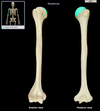
Clavicle
Junction of neck and anterior thorax (S-shaped bone).
Acromial end (clavicle)
Flattened lateral end that articulates with medial aspect of acromion via acromioclavicular joint

Sternal end (clavicle)
Location:
• Clavicle (medial end)
Description:
• Enlarged, medial end
• Projects above manubrium of sternum to deepen jugular notch
• Has smooth, articular surface
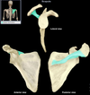
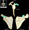
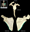
Scapula
Location:
• Posterior thorax
• Overlies ribs 2-7
Description:
• Large, triangular, flat bone
• Characteristic features include spine, acromion, coracoid process, and glenoid cavity
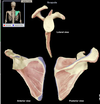
Spine (scapula)
Location:
• Scapula
Description:
• Prominent ridge on posterior surface of scapula
Comment:
• Provides attachment for trapezius and deltoid muscles
Acromion (scapula)
Location:
• Scapula
Description:
• Flattened, lateral part of scapular spine
Comment:
• Articulates with clavicle
• Subcutaneous superior point of shoulder (easily palpated)
• Provides attachment for trapezius and deltoid muscles
• Landmark for intramuscular injections

Glenoid cavity/fossa (scapula)
Location:
• Scapula
Description:
• Shallow depression at superior end of lateral border
Comment:
• Articulates with head of humerus to form glenohumeral (shoulder) joint
• Made “deeper” by rim of fibrocartilage (labrum)

Coracoid process (clavicle)
Location:
• Scapula (anterior)
Description:
• Prominent protuberance inferior to acromion of scapula
Comment:
• Provides attachment for pectoralis minor, coracobrachialis, and short head of biceps brachii muscles
Lateral border (clavicle)
Location:
• Scapula
• Between inferior angle and glenoid cavity
Description:
• Border of scapula inferior to glenoid cavity
• Superior end has infraglenoid tubercle
Medial border (clavicle)
Location:
• Scapula
Description:
• Border of scapula parallel to vertebral column
Also known as:
• Vertebral border of scapula
Comment:
• Provides attachment for levator scapulae, rhomboid major and rhomboid minor, and serratus anterior muscle

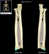
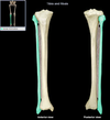
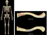
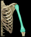
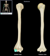
Humerus
Location:
• Arm
Description:
• Long bone
• Characteristic features include head, neck, greater and lesser tubercles, shaft, medial and lateral epicondyles, capitulum, and trochlea
Comment:
• Largest bone of upper limb
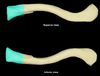
Head (humerus)
Location:
• Humerus (proximal)
Description:
• Rounded articular surface
• Continuous with anatomical neck
Comment:
• Articulates with glenoid cavity of scapula to form glenohumeral (shoulder) joint
Deltoid tuberosity (humerus)
Location:
• Humerus (shaft)
Description:
• Roughened area near mid-shaft on anterolateral surface
Comment:
• For attachment of deltoid muscle

Medial epicondyle (humerus)
Location:
• Humerus (distal)
Description:
• Medial subcutaneous projection near elbow
Comment:
• Provides attachment for hand flexor muscles
• Ulnar nerve subcutaneous on posterior aspect (“funny bone”

Lateral epicondyle (humerus)
Location:
• Humerus (distal)
Description:
• Small lateral projection near elbow
Comment:
• Provides attachment for hand extensor muscles

Trochlea (humerus)
Location:
• Humerus (distal)
Description:
• Grooved surface medial to capitulum
Comment:
• Articulates with trochlear notch of ulna
• Contributes to elbow joint

Capitulum (humerus)
Location:
• Humerus (distal)
Description:
• Dome-shaped surface lateral to trochlea
Comment:
• Articulates with head of radius
• Contributes to elbow joint
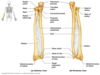
Ulna
Location:
• Forearm (medial) Ariculates with pinky!
LOOKS LIKE ICECREAM SCOOP
Description:
• Long, thin bone
• Articulates proximally with trochlea of humerus and radius
• Articulates distally with radius and carpal bones (lunate and triquetrum)
• Characteristic features include olecranon, trochlear notch, tuberosity, shaft, and styloid process
Comment:
• Fibrocartilage separates distal end from carpal bones

Trochlear notch (ulna)
Location:
• Ulna (proximal, anterior aspect)
• Between olecranon and coronoid process
Description:
• Prominent notch
• Has smooth articular surface
Function:
• Provides articulation with trochlea of humerus
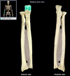
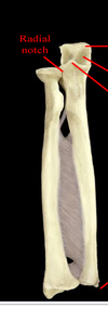
Radial notch (ulna)
Holds ulna
Smaller notch right below trochlear notch
Where radius attaches

Head (ulna)
Adjacent to styloid process of ulna. Distal to trochlear notch.

Styloid process (ulna)
Location:
• Ulna (distal)
Description:
• Pointed distal projection
Radius
Articulates with capitulum of humerus. Also articules with the thumb (lateral)
Head (radius)
Location:
• Radius (proximal)
Description:
• Disk-shaped, with concave superior surface
• Articulates with capitulum of humerus and radial notch of ulna