Basic Exam Flashcards
(888 cards)
What is the half life of Flumazenil?
Why is this important?
Half Life:
Midazolam: 1.7 - 2.6 hours (102 minutes - 156 minutes)
Flumazenil: 0.7 - 1.3 hours (42 min - 78 minutes)
Significantly shorter half life than benzodiazepine agonists.
This can lead to the recrudescence of sedation from benzodiazepines following the elimination of flumazenil.
Therefore, when administering flumazenil, careful attention needs to be paid to its duration of action relative to that of the benzodiazepines taken by the patient, and additional doses of flumazenil should be available.
What is the mechanism of action of Flumazenil?
*Caveat to Mechanism of Action*
Note that although flumazenil is generally considered a benzodiazepine antagonist, it exhibits a partial agonist effect.
In one study, propofol was potentiated by giving high doses of flumazenil, suggesting flumazenil has a mixed or partial agonist effect.
What is the half life of the common benzodiazepines?
Alprazolam, Diazepam, Lorazepam, Midazolam, and Temazepam
Half Life of Other Benzodiazepines (MALTD)
Midazolam is 1.7-2.6 hours (2 hours)
Alprazolam is 6-27 hours (A = Afternoon)
Temazepam is 10 hours (Tem = Ten)
Lorazepam is 11-22 hours (L = eLeven)
Diazepam is 20-50 hours (D = ~ 1 day)
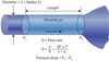
For Nitrous Oxide, a full tank will read:
What volume?
What pressure?
When will the pressure start to drop?
A full tank of N2O contains 1590 L at a pressure of ~745 psig.
Pressure within a tank of N2O will remain at ~745 psig until all liquefied gas is used up which is when the tank is ~16% full (253 L).
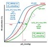
What acid base disturbances are common with different diuretics?
Acetazaloamide?
Thiazide?
Loops?
Potassium Sparing?
Thiazide and loop diuretics can cause an Hypokalemic Hypochloremic metabolic alkalosis.
Acetazolamide and potassium sparing can cause a hyperchloremic metabolic acidosis.
How does hyperventilation affect electrolytes?
Calcium?
Potassium?
Phosphate?
Sodium?
pH?
HCO3?
Chloride?
Lactate?
Respiratory alkalosis (ETCO2 of 30-35), such as from hyperventilation, can cause electrolyte abnormalities such as hypocalcemia, hypokalemia, and hypophosphatemia.
Loss of CO2 by 10 mmHg
Rise in pH (0.1)
Decreased HCO3 (2.0 mEq/L)
Decreased Potassium (0.4 mEq/L) - After will slowly return to normal
Decrease in Sodium
Increased Chloride (Balanced decrease in Bicarb)
Increased lactate (Balanced decrease in Bicarb)
What is the relationship between hyper/hypoventilation and calcium levels?
(Explain the physiology)
Hypocalcemia can occur with respiratory alkalosis.
In response to alkalosis, hydrogen ions bound to negatively charged plasma proteins, such as albumin, are released.
Calcium, being positively charged, can then bind to albumin and other proteins decreasing the serum calcium concentration (particularly the free/ionized, active fraction). This is the mechanism behind paresthesias that occur with hyperventilation.

What are the main determinants of myocardial oxygen demand?
1. Wall tension
2. Heart rate
3. Contractility
What is the formula for wall tension?
Wall tension (T) = (P * r) / (2 h)
P is the pressure within the ventricle
r is the radius of the ventricle
h is the thickness of the ventricular wall (Increase seen in hypertrophy)
What is the downside to developing left ventricular hypertrophy?
The ventricle hypertrophies to compensate for the increased wall tension due to pressure or volume overload, such as with aortic stenosis. However, this change comes at a cost. The hypertrophied ventricle is not as compliant due to increased diastolic pressures in the ventricle. This leads to a decrease in preload*, and the hypertrophied ventricle is now more *dependent on atrial contraction to maintain LVEDV
ex: (Atrial fibrillation with diastolic heart failure is a horrible combination)
What are the two determinants of acoustic impedence of ultrasound?
Acoustic Impedance (two factors determine)
1. Density of the medium(This is the main one)
2. Propagation speed of sound through the medium
Acoustic impedance is the product of the density of a medium and the propagation speed of sound through that medium. Ultrasound reflections that occur at the interface of different mediums are due to the changes in acoustic impedance. Since propagation speed changes slightly between biological mediums, acoustic impedance is primarily dependent upon density.
How does gentamicin affect neuromuscular blockade?
What is the mechanism behind this?
Prolongs neuromuscular blockade
Gentamicin inhibits prejunctional acetylcholine release and depresses postjunctional receptor sensitivity to acetylcholine, thus prolonging paralysis in a patient who received it along with a muscle relaxant.
It is believed that aminoglycoside antibiotics antagonize calcium ions by means of its involvement in the process of acetylcholine release by nerve impulses.
What are all the antibiotics that can prolong neuromuscular blockade?
Aminoglycosides
- Lincomycin, Clindamycin, Streptomycin, kanamycin, tobramycin, neomycin, and gentamicin
Polymyxins
Tetracyclines
Muscarinic receptor agonists can act through two mechanisms, directly on the muscarinic receptor or indirectly by inhibiting the breakdown of ACh causing more ACh to be available to bind to the muscarinic receptor.
1. What are the direct acting agents are choline esters?
2. And what are the alkaloids?
3. What are the anticholinergics?
The direct acting agents are choline esters (ACh, methacholine, carbachol, bethanechol)
direct acting agents have few clinical applications due to their very short half and longer activity can be achieved by methylating the choline moiety.
Alkaloids (pilocarpine, muscarine, arecoline or acetylcholinsterase inhibitors).
The indirect acting agents are acetylcholinesterase inhibitors (physostigmine, neostigmine, pyridostigmine, edrophonium, and echothiophate). These medications are often used to improve neuromuscular function in disease states where weakness occurs such as myasthenia gravis or to help reverse the action of nondepolarizing neuromuscular blocking agents.
Anticholinergic drugs are used commonly in anesthesia practice. These medications inhibit the action of ACh by reversibly binding at the muscarinic receptor. Antimuscarinic drugs used in anesthesia practice are atropine, scopolamine, and glycopyrrolate. Both atropine and scopolamine cross the blood-brain barrier, which can result in inhibition of vagal outflow from the central nervous system. At low doses, vagal outflow can potentially be augmented.
You have your line isolation monitor alarming. What is the next step?
When the line isolation monitor alarms, the first step should be to unplug the most recent electronic device that was plugged in.
What portion of the spinal cord is perfused by the anterior spinal artery?
How many are their?
What vessel is this from?
A single (not paired) anterior spinal artery supplies the anterior two-thirds of the spinal cord.
The spinal cord blood supply is anatomically divided into an anterior and a posterior blood supply. The anterior two-thirds of the spinal cord, primarily responsible for motor function, is perfused by the anterior spinal artery (ASA), which originates from the vertebral arteries and receives contributions from various radicular vessels that arise from intercostal arteries.
Is the posterior spinal artery paired?
What is the perfusion to the posterior spinal artery?
What does it arise from?
Two posterior spinal arteries (PSA) stem from the vertebral or posterior inferior cerebellar arteries (PICA). They comprise the blood supply to the posterior one-third of the spinal cord, primarily responsible for sensation, while concurrently receiving intercostal radicular vessel contributions.
What is the origination spinal levels of Artery of Adamkiewicz?
(List the regions from most to least common)
The artery of Adamkiewicz a radicular vessel contributing to anterior spinal cord blood supply
Most commonly originates within the T9 - T12 region
Followed by the L1 - L5 region
least commonly, within the T5 - T8 region
Compare adults vs. infants in terms of:
Spinal cord* vs. *Dural Sac Ending
In adults
Spinal cord ends at about L1-L2 (Conus Medullaris)
Dural sac ends at about S1-S2.
In an infant, these landmarks are shifted slightly caudad (Inferior)
Spinal cord ending at L3-L4
Dural sac terminating at S3-S4
When performing a spinal blockade in an adult, the iliac crest is commonly used as a landmark as it generally corresponds to the level of the L4 interspace is known as What Anatomical Landmark?
Tuffier’s line
What are the 3 drugs that can decrease post-operative delirium with Ketamine administration?
Benzodiazepines, propofol, and barbiturates can decrease ketamine-induced emergence delirium.
An important consideration when using ketamine relates to the high incidence of psychomimetic reactions early in the recovery period. The most common reactions include hallucinations, nightmares, altered cognition, and altered short-term memory. Repeated doses of ketamine may decrease the severity and incidence of emergence delirium, but one of the most effective strategies for prevention of emergence delirium is the use of midazolam approximately five minutes prior to an induction with ketamine.
How does magnesium and Calcium levels affect neuromuscular blockade?
Hypocalcemia potentiates neuromuscular blockade because calcium is important for the release of vesicles containing acetylcholine from nerves at the neuromuscular junction.
When calcium levels are low, less acetylcholine is released.
With less acetylcholine being released there is less competition at the acetylcholine receptor and neuromuscular blockers have a greater effect.
Hypermagnesemia acts similarly by blocking calcium from entering alpha motor neurons and preventing the release of some of the acetylcholine containing vesicles.
How do anti-convulsants affect neuromuscular blockade?
- *Shorten Non-Depolarizing Blockade:**
- Anticonvulsants (e.g. phenytoin, carbamazepine)
What effect does Ketamine have on neuromuscular blockade?
Prolongs