Basic Ultrasound Flashcards
(36 cards)
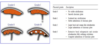
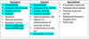
Levels of ultrasound procedures
- Standard/Basic
- Limited
- Specialized (when fetal anomaly suspected)
AWHONN Scope of Practice
1st trimester ultrasound
- +/- gestational sac
- Fetal #
- +/- cardiac activity
- Estimated GA via CRL
- +/- IUP
- Identify 1st tri complications:
- Anembryonic pregnancy
- Ectopic pregnancy
- Threatened, incomplete, complete, or misses AB
- Molar pregnancy
- Multiple gestation
AWHONN Scope of Practice
2nd+3rd trimester ultrasounds
- fetal #
- +/- cardiac activity
- Fetal presentation
- Placental location
- Amniotic fluid volume
- BPP and modified BPP
- Biometric measurements: EFA and EFW
- Cervical length measurements
- Adjunct to US guided procedures
Common ultrasound indications
- Evaluation of fetal growth
- Suspected fetal abnormality
- Adjunct to special procedures (le amnio)
- Uterine evaluation
Which 2 probes do we use the most in OB/GYN
- 3.5 MHz convex probe (abdomen, OB/GYN)
- 6.5 MHz transvaginal probe
Where do you orient the transducer notch?
- Notch oriented towards person’s head
- Left side of ultrasound screen = head
- Right side = feet
What are the different ultrasound modes?
- B-mode: “Brightness mode”
- 2D ultrasound in grey scale using real time.
- Used 99% of time
- M-mode: “Motion mode”
- Cursor line on fetal heart and shows motion of fetal heart, estimates FHR
- Doppler
- Color - shows direction of flow
- Towards transducer = red
- Away from transducer = blue
- Power - amplitude
- Spectral - velocity
- Color - shows direction of flow
Define: echogenic
The ability of a structure to produce echos
Define: anechoic
- No echos
- Appears black on ultrasound
Define: hypoechoic
- Less reflective and low amount of echoes when compared with neighboring structures
- Appears as varying shades of darker gray
Define: hyperechoic
- Highly reflective & echo rich when compared with neighboring structures
- Appears as varying shades of gray
- Echogenic is often used interchangeably
Define: isoechoic
- Having similar echogenicity to a neighboring structure
Define: homogenous (texture)
- Organ parenchyma is uniform in echogenicity
Define: inhomogenous/heterogenous (texture)
- Organ parenchyma is not uniform in echogenicity
Define: reverberation
- Artifacts that appear as multiple equally spaced lines along a ray line.
- Caused by the sound bouncing back and forth between tissue boundaries and then returning to the receiver
Define: acoustic enhancement
- Artifact with hyperechoic pattern to a poorly or non-attenuating structure or mass
- Often seen with fluid index
Define: shadowing
- Artifact appears as a hypoechoic pattern to highly attenuating structures (e.g., calcifications such as bone)
What is the ALARA principle?
- As Low As Reasonably Achievable
- Only perform when there is a valid medical indication, and use the lowest possible ultrasonic exposure setting to gain the necessary diagnostic info
Define: thermal index (TI)
- Measure of how much the ultrasound beam is theoretically heating the tissue. Its different for the types of tissue
- TIs (soft tissue): GYN and OB < 10 weeks
- TIb (bone): OB ultrasound > 10 weeks
- TI = <0.7 is the threshold for extended scanning
1st trimester gestational sac facts
- Visibility by tranvaginal and abdominal approaches
- Gestational sac characteristics (appearance, sizing, cardiac activity)
- What confirms IUP
- Visible as early as 4 - 4.5 transvaginal and 5 - 5.5 weeks abbdominal
- Thick, bright, double ring
- Sac > 2mm, fetal embryonic pole visible by transvaginal
- Embryon > 5 mm, cardiac + likely
- CRL > 7 mm, cardiac + definitely
- Yolk sac confirms IUP
*
Pregnancy Failure
- Gestational sac measuring >25 mm without an embryo within is diagnostic of a failed pregnancy
What biometry measures are used after 14 weeks?
- Biparietal diameter (BPD)
- Abdominal circumference (AC)
- Head circumference (HC)
Biparietal Diameter and Head Circumference Landmarks/Measurement
Landmarks
- Falx Cerebri Anteriorly and Posteriorly
- Thalami
- Cavum septum pellucidum
Measurement
- BPD
- Width of skull transversly through thalami
- Outer edge of parietal skull to inner edge
- HC
- Outer circumference edge of fetal skull (use ellipsis instead of calipers)
Abdominal Circumference
Landmarks/Measurement
Landmarks
- Junction of umbilical vein and left portal deep vein within liver
- Stomach
- Three ossification centers of spine
Measure
- OUTER transverse circumference