Biology New Information Flashcards
(271 cards)
Where is blood pumped into in the heart?
aorta, which branches into a series of arteries
What do the arterie the branch into?
arterioles and then into microscopic capilaries
What is exchanged across the capillary walls?
gases, nutrients and cellular waste products occurs via diffusion
How is blood brought back to the heart?
The capillaries then converge into venules and eventually into veins, which carry deoxygenated blood back toward the heart
Explain the heart sides (right and left)
Right side = deoxygenated blood into pulmonary circulation (toward the lungs)
Left side = oxygenated blood into systematic circulation (throughout the body)
What are the two upper chamber and two lower chambers of the heart called
Upper chambers = atria (thin walled)
lower chambers = ventricles (muscular)
The left ventricle is more muscular than the right because it is responsible for generating the forse that propels the systemic circulation and because it pumps agains the higher resistance

In patients with increased systemic resistance (artaries clogged), what happens to the heart?
the left ventricle can become hypertrophied (enlarged) which over time can lead to congestive heart failure and other cardiovascular diseases

Explain the process of blood flow through the heart
- Blood returning from the body first flows through the right atrium, then through the tricuspid valve into the right ventricle
- finally through the pulmonary semilunar valve into the pulmonary arteries to continue to the lungs
- Blood returning from the lungs flows through the pulmonary veins into the left atrium
- then through the mitral valve into the left ventricle
- Finally through the aortic semilunar valve into the systemic circulation

What are the valves in the heart, location and purpose?
-
Atrioventricular valves, located between the atria and ventricles on both sides of the heart, prevent backflow of blood into the atria
-
right side = tricuspid valve
- because it has three cusps
-
left side = mitral valve
- becaue it has two cusps
-
right side = tricuspid valve
-
Semilunar valves
- have three cusps
- located between the left ventricle and aorta and between right ventricle and pulmonary artery
What is the “lub-dub” sound from?
made by the successive closing of the atrioventricular and semilunar valves
What is the heart’s pumping cycle described as?
- two alternating phases: = heatbeat
- Systole
- Period during which the ventricles contract, forcing blood out of the heart into the pulmonary and systemic circulation
- Blood vessel contraction pressure
- Diastole
- period of cardiac muscle relaxation during which the blood drains into all four chambers
- pressure during cardiac relaxation
- Systole
- Together they make blood pressure measurement
How is cardiac output defined by?
The total volume of blood the left ventricle pumps out per minute
Cardiac output = heart rate (number of beats per minute) x stroke volume (volume of blood pumped out of the left ventricle per contraction
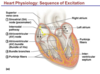
Explain how the heart rate is controlled by the body
- Cardiac muscle contracts rhythmically without stimulation from the nervous system, producing impulses that spread through its internal conduction system
- Ordinary cardiac contraction originates in, and is regulated by the sinoatrial (SA) node (the pacemaker) a small mass of specialized tissue located in the walls of the right atrium
- SA note spreads impulses through both atria, contracting simultaneously
- Impulses arrive at the atrioventricular (AV) node, which slowly conducts impulses to the rest of the heart, allowing enough time for atrial contraction and for the ventricles to fill with blood
- The impulses is then carried by the bundles of His (AV bundles), which branches into the right and left bundle branches
- Then through the Purkinje Fibers in the walls of both the ventricles, stimulating a strong contraction to rest of body
What innervates the heart
- The autonomic nervous system modifies the rate of the heart contraction
- The parasympathetic nervous system innervates the heart via the vagus nerves and causes a decrease in heart rate
- The sympathetic nervous system innervates the heart via the cervical and upper thoracic ganglia and causes an increase in heart rate
- Adrenal medulla exerts hormonal control via epinephrine (adrenaline) secretion, increasing heart rate
Name and describe the three types of blood vessels
-
Arteries = thick walled, muscular, elastic vessels that transport oxygenated blood from heart,
- except for the pulmonary arteries which transport deoxygenated blood
-
Veins = relatively thin walled, inelastic vessels that conduct deoxygenated blood to heart
- except for the pulmonary vein which transports oxygenated blood
- Most have valves, especially in legs, that prevent backflow
-
Capillaries = smallest diameter, red blood cells must often travel through them single file
- Location of where nutrients readily diffuse
Explain the lymph vessels
- secondary circulatory system distinct from the cardiovascular circulation
- vessels transport excess interstitial fluid, called lymph, to the cardiovascular system, thereby keeping fluid levels in the body consistant
- Smallest lymphatic vessels (lacteals) collect fats, in the form of chylomicrons, from the villi in the small intestine and deliver them into the bloodstream, bypassing the liver
- Lymph nodes are swellings along lymph vessels containing phagocytic cells (lymphocytes) that filter the lymph, removing and destroyign foreign particles and pathogens)
Explain the makeup of blood
- Contains four to six liters of blood
-
55% = liquid component
-
plasma
- aqueous mixture of nutrients, salts, repiratory gases, wastes, hormones, and blood proteins (eg. immunoglobulins, albumin, and fibronogen)
-
plasma
-
45% = cellular component
- erythrocytes, leukocytes and platelets
Explain Leukocytes
- Leukocytes = white blood cells (or WBCs)
- larger than erythrocytes and serves protective function
Explain Platelets
- Platelets
- cell fragments that lack nuclei and are involved in clot formation
- Many drugs inhibit platelet formation or adhesion to decrease clot development
- cell fragments that lack nuclei and are involved in clot formation
What are erythrocytes
-
Erythrocytes
- Red blood cells (RBC’s)
- oxygen-carrying components of blood
- contain ~250 million molecules of hemoglobin, each of which can bind up to four molecules of oxygen
- When hemoglobin binds to oxygen = oxyhemoglobin
- Primary form of oxygen transport in body
- Have distinct disk like shape that give them increased surface area for gas exchange and greater flexibility for movement
- Erythrocytes are formed from stem cells in the bone marrow; stimulated by erythropoietin, a hormone make in the kidnesy
What does it mean when you find immature erythrocytes circulating in the bloodstream?
- that is, before they have lost their organelles from the bone marrow -
- Can be an indicator of a number of disease states, such as hemolytic anemia, which is caused by a rapid destruction of red blood cells due to an infection or disorder
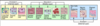
Explain ABO Blood types
-
Erythrocytes have characteristic cell-surface proteins (antigens)
-
Antigens = macromolecules that are foreign to the host organism and triggers an immune response
- two major antigens = ABO group and the Rh Factor
-
Antigens = macromolecules that are foreign to the host organism and triggers an immune response
- See attached for table with which Blood type has which antigen and antibody
- It is extremely important durign blood transfusions that donor and recipient blood types be appropriately matched

What is the universal recipient of blood?
Type AB
has neither anti-A nor anti-B

What is the universal Donor?
Type O
it will not elicit a response from the recipient’s immune system becaue it does not posses any surface antigens