Block 6 Flashcards

(506 cards)

1
Q

Innervation of pec major

A

Lateral pectoral nerve

2
Q

Action of pec major

A

Adductor and medial rotator of humerus

3
Q

Which of the following methods is most effective at destroying spores of the tubercle bacilli?

Immersion in 0.5% chlorhexidine in alcohol

Immersion in aqueous iodine

Heating in a hot air oven

Immersion in 0.1% sodium hypochlorite

Autoclaving

A

The tubercle bacilli has a waxy outer membrane that renders it more resistant to sterilisation and cleaning methods. Whilst 0.1% sodium hypochlorite will destroy many microbes it is less reliable in destroying tubercle bacilli. Hot air ovens provide less reliable pathogen destruction than autoclaving, but may be indicated in situations where the equipment is sensitive to the autoclaving process. From the list of options above, autoclaving will most reliably destroy tubercle bacilli.

4
Q

Def: cleaning

A

Removal of physical debris

5
Q

Def: disinfection

A

Reduction in number of viable organisms

6
Q

Sterilisation

A

Removal of all organisms and spores

7
Q

Sterilisation technique options

A

Autoclaving

Glutaraldehyde solution

Ethylene oxide

Gamma irradiation

8
Q

A 59 year old man is undergoing an extended right hemicolectomy for a carcinoma of the splenic flexure of the colon. The surgeons divide the middle colic vein close to its origin. Into which of the following structures does this vessel primarily drain?

Superior mesenteric vein

Portal vein

Inferior mesenteric vein

Inferior vena cava

Ileocolic vein

A

The middle colonic vein drains into the SMV, if avulsed during mobilisation then dramatic haemorrhage can occur and be difficult to control.

9
Q

A 65 year old male with known nasopharyngeal carcinoma presents with double vision over a few weeks. On examination he is found to have left eye proptosis and it is down and out. He reports pain on attempting to move the eye. There is an absent corneal reflex. What is the most likely diagnosis?

Posterior communicating artery aneurysm

Cavernous sinus syndrome

Optic nerve tumour

Migraine

Cerebral metastases

A

Cavernous sinus syndrome is most commonly caused by cavernous sinus tumours. In this case, the nasopharyngeal malignancy has locally invaded the left cavernous sinus. Diagnosis is based on signs of pain, opthalmoplegia, proptosis, trigeminal nerve lesion (opthalmic branch) and Horner’s syndrome.

10
Q

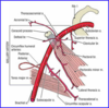
Medial relations of the cavernous sinus

A

Pituitary fossa

Sphenoid sinus

11
Q

Lateral relations of the cavernous sinus

A

Temporal bone

12
Q

Lateral wall components of the cavernous sinus

A

(from top to bottom:)
Oculomotor nerve
Trochlear nerve
Ophthalmic nerve
Maxillary nerve

13
Q

Contents of the cavernous sinus

A

(from medial to lateral:)
Internal carotid artery (and sympathetic plexus)
Abducens nerve

14
Q

Blood supply of the cavernous sinus

A

Ophthalmic vein, superficial cortical veins, basilar plexus of veins posteriorly.

Drains into the internal jugular vein via: the superior and inferior petrosal sinuses

15
Q

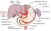
In patients with an annular pancreas where is the most likely site of obstruction?

The first part of the duodenum

The second part of the duodenum

The fourth part of the duodenum

The third part of the duodenum

The duodeno-jejunal flexure

A

The pancreas develops from two foregut outgrowths (ventral and dorsal). During rotation the ventral bud and adjacent gallbladder and bile duct lie together and fuse. When the pancreas fails to rotate normally it can compress the duodenum with development of obstruction. Usually occurring as a result of associated duodenal malformation. The second part of the duodenum is the commonest site.

16
Q

Theme: Chest pain

A.Achalasia

B.Pulmonary embolus

C.Dissection of thoracic aorta

D.Boerhaaves syndrome

E.Gastro-oesophageal reflux

F.Carcinoma of the oesophagus

G.Oesophageal candidiasis

Please select the most likely cause for chest pain for the scenario given. Each option may be used once, more than once or not at all.

41.A 43 year old man who has a long term history of alcohol misuse is admitted with a history of an attack of vomiting after an episode of binge drinking. After vomiting he developed sudden onset left sided chest pain, which is pleuritic in nature. On examination he is profoundly septic and drowsy with severe epigastric tenderness and left sided chest pain.

A 22 year old man is admitted with severe retrosternal chest pain and recurrent episodes of dysphagia. These occur sporadically and often resolve spontaneously. On examination there are no physical abnormalities and the patient seems well.

An obese 53 year old man presents with symptoms of recurrent retrosternal discomfort and dyspepsia. This is typically worse at night after eating a large meal. On examination there is no physical abnormality to find.

A

Boerhaaves syndrome

In patients with Boerhaaves the rupture is often on the left side. The story here is typical. All patients should have a contrast study to confirm the diagnosis and the affected site prior to thoracotomy.

Achalasia

Achalasia may produce severe chest pain and many older patients may undergo cardiac investigations prior to endoscopy.
Endoscopic injection with botulinum toxin is a popular treatment (although the benefit is not long lasting). Cardiomyotomy together with an antireflux procedure is a more durable alternative.

Gastro-oesophageal reflux

Patients with GORD often have symptoms that are worse at night. In this age group an Upper GI endoscopy should probably be performed.

17
Q

Tearing interscapular pain

Discrepancy in arterial blood pressures taken in both arms

May show mediastinal widening on chest x-ray

A

Dissection of thoracic aorta

18
Q

Spectrum of oesophageal motility disorders

Caused by uncoordinated contractions of oesphageal muscles

May show “nutcracker oesophagus” on barium swallow

Symptoms include dysphagia, retrosternal discomfort and dyspepsia

A

Diffuse oesophageal spasm

19
Q

Common cause of retrosternal discomfort

Usually associated with symptoms of regurgitation, odynophagia and dyspepsia

Symptoms usually well controlled with PPI therapy

Risk factors include obesity, smoking and excess alcohol consumption

A

Gastro-oesphageal reflux

20
Q

Spontaneous rupture of the oesophagus

Caused by episodes of repeated vomiting often in association with alcohol excess

Typically there is an episode of repetitive vomiting followed by severe chest and epigastric pain

Diagnosis is by CT and contrast studies

Treatment is surgical; during first 12 hours primary repair, beyond this usually creation of controlled fistula with a T Tube, delay beyond 24 hours is associated with fulminent mediastinitis and is usually fatal.

A

Boerhaaves syndrome

21
Q

Difficulty swallowing, dysphagia to both liquids and solids and sometimes chest pain

Usually caused by failure of distal oesphageal inhibitory neurones

Diagnosis is by pH and manometry studies together with contrast swallow and endoscopy

Treatment is with either botulinum toxin, pneumatic dilatation or cardiomyotomy

A

Achalasia

22
Q

Theme: Nerve Injury

A.Median nerve

B.Ulnar nerve

C.Radial nerve

D.Musculocutaneous nerve

E.Axillary nerve

F.Anterior interosseous nerve

G.Posterior interosseous nerve

For each scenario please select the most likely underlying nerve injury. Each option may be used once, more than once or not at all.

44.A 10 year old boy is admitted to casualty following a fall. On examination there is deformity and swelling of the forearm. The ability to flex the fingers of the affected limb is impaired. However, there is no sensory impairment. Imaging confirms a displaced upper forearm fracture

A well toned weight lifter attends clinic reporting weakness of his left arm. There is weakness of flexion and supination of the forearm.

An 18 year old girl sustains an Holstein-Lewis fracture. Which nerve is at risk?

A

Anterior interosseous nerve

Forearm fractures may be complicated by neurovascular compromise. The anterior interosseous nerve may be affected. It has no sensory supply so the defect is motor alone.

Musculocutaneous nerve

Musculocutaneous nerve compression due to entrapment of the nerve between biceps and brachialis. Elbow flexion and supination of the arm are affected. This is a rare isolated injury.

Radial nerve

Proximal lesions affect the triceps. Also paralysis of wrist extensors and forearm supinators occur. Reduced sensation of dorsoradial aspect of hand and dorsal 31/2 fingers. Holstein-Lewis fractures are fractures of the distal humerus with radial nerve entrapment.

23
Q

Location of brachial plexus roots?

A

Posterior triangle

24
Q

Passage of the brachial plexus roots

A

Between scalenus anterior and medius

25
Location of brachial plexus trunks
Posterior to middle third of clavicle.
26
Relationship of the upper and middle third trunks to the subclavian artery?
Superior
27
Relation of the lower brachial plexus trunk to the subclavian artery
Posterior
28
Where are the divisions of the brachial plexus?
Apex of axilla
29
Draw the brachial plexus
https://www.youtube.com/watch?v=Z\_Y\_kVdH9zE
30
A 44 year old man recieves a large volume transfusion of whole blood. The whole blood is two weeks old. Which of the following best describes its handling of oxygen? It will have a low affinity for oxygen Its affinity for oxygen is unchanged It will more readily release oxygen in metabolically active tissues than fresh blood The release of oxygen in metabolically active tissues will be the same as fresh blood It will have an increased affinity for oxygen
Stored blood has less 2,3 DPG and therefore has a higher affinity for oxygen, this reduces its ability to release it at metabolising tissues.
31
Haldane effect?
Left shit- increased saturation of Hb with oxygen for given O2 tension i.e. reduced delivery to tissue
32
Bohr shift
Shifts to right = for given oxygen tension there is reduced saturation of Hb with oxygen i.e. Enhanced oxygen delivery to tissues
33
Factors causing haldane effect
HbF, methaemoglobin, carboxyhaemoglobin low [H+] (alkali) low pCO2 low 2,3-DPG low temperature
34
Factors causing Bohr effect
raised [H+] (acidic) raised pCO2 raised 2,3-DPG\* raised temperature
35
A 32 year old male is receiving a blood transfusion after being involved in a road traffic accident. A few minutes after the transfusion he complains of loin pain. His observations show temperature 39 oC, HR 130bpm and blood pressure is 95/40mmHg. What is the best test to confirm his diagnosis? USS abdomen Direct Coomb's test Blood cultures Blood film Sickle cell test
The diagnosis is of an acute haemolytic transfusion reaction, normally due to ABO incompatibility. Haemolysis of the transfused cells occurs causing the combination of shock, haemoglobinaemia and loin pain. This may subsequently lead to disseminated intravascular coagulation. A Coomb's test should confirm haemolysis. Other tests for haemolysis include: unconjugated bilirubin, haptoglobin, serum and urine free haemoglobin. Note that delayed haemolytic reactions are normally associated with antibodies to the Rh system and occur 5-10 days after transfusion.
36
A 42 year old female presents with symptoms of biliary colic and on investigation is identified as having gallstones. Of the procedures listed below, which is most likely to increase the risk of gallstone formation? Partial gastrectomy Jejunal resection Liver lobectomy Ileal resection Left hemicolectomy
Bile salt reabsorption occurs at the ileum. Therefore cholesterol gallstones form as a result of ileal resection.
37
Theme: Management of skin lesions A.Excision biopsy B.Excision with 0.5 cm margin C.Excision with 2 cm margin D.Shave biopsy and cautery E.Punch biopsy F.Excision and full thickness skin graft G.Discharge For each skin lesion please select the most appropriate management option. Each option may be used once, more than once, or not at all. 2.A 22 year old women presents with a newly pigmented lesion on her right shin, it has regular borders and normal appearing dermal appendages. However, she reports a recent increase in size. A 58 year old lady presents with changes that are suspicious of lichen sclerosis of the perineum. A 73 year old man presents with a 1.5cm ulcerated basal cell carcinoma on his back.
Excision biopsy Lesion bearing normal dermal appendages and regular borders are likely to be a benign pigmented naevi. Therefore diagnostic and not radical excision is indicated. Punch biopsy Punch biopsies are a useful option for obtaining a full thickness tissues sample with minimal tissue disruption. In this situation the other differential would be AIN or VIN and punch biopsies would be useful in distinguishing thes Excision with 0.5 cm margin A small lesion such as this is adequately treated by local excision. The British Association of Dermatology guidelines suggest that excision of conventional BCC (\<2cm) with margins of 3-5mm have locoregional control rates of 85%. Morpoeic lesions have higher local recurrence rates.
38
When should UFH be considered in PE?
(a) as a first dose bolus, (b) in massive PE, or (c) where rapid reversal of effect may be needed.
39
Management of massive PE with cardiovascular compromise
Thrombolysis is 1st line for massive PE (ie circulatory failure) and may be instituted on clinical grounds alone if cardiac arrest is imminent; a 50 mg bolus of alteplase is recommended. Invasive approaches (thrombus fragmentation and IVC filter insertion) should be considered where facilities and expertise are readily available.
40
A 32 year old man is diagnosed as having a carcinoma of the caecum. On questioning, his mother developed uterine cancer at the age of 39 and his maternal uncle died from colonic cancer aged 38. His older brother developed a colonic cancer with micro satellite instability aged 37. What is the most appropriate operative treatment? Limited ileocaecal resection Right hemicolectomy Extended right hemicolectomy Panproctocolectomy Sub total colectomy
Panproctocolectomy The likely diagnosis is one of a familial cancer syndrome and now that he has developed a colonic cancer the safest operative strategy is a total colectomy and end ileostomy.
41
A laceration of the wrist produces a median nerve transection. The wound is clean and seen immediately after injury. Collateral soft tissue damage is absent. The patient asks what the prognosis is. You indicate that the nerve should regrow at approximately: 0.1 mm per day 1 mm per day 5 mm per day 1 cm per day None of the above
Transection of a peripheral nerve results in hemorrhage and retraction of the severed nerve ends. Almost immediately, degeneration of the axon distal to the injury begins. Degeneration also occurs in the proximal fragment back to the first node of Ranvier. Phagocytosis of the degenerated axonal fragments leaves neurilemmal sheath with empty cylindrical spaces where the axons were. Several days following the injury, axons from the proximal fragment begin to regrow. If they make contact with the distal neurilemmal sheath, regrowth occurs at about the rate of 1 mm/day. However, if associated trauma, fracture, infection, or separation of neurilemmal sheath ends precludes contact between axons, growth is haphazard and a traumatic neuroma is formed. When neural transaction is associated with widespread soft tissue damage and hemorrhage (with increased probability of infection), many surgeons choose to delay reapproximation of the severed nerve end for 3 to 4 weeks.
42
Seddon classification of nerve injury
Neuropraxia Axonotmesis Neurotmesis
43
Neuropraxia
Nerve intact but electrical conduction is affected Full recovery Autonomic function preserved Wallerian degeneration does not occur
44
Axonotmesis
Axon is damaged and the myelin sheath is preserved. The connective tissue framework is not affected. Wallerian degeneration occurs.
45
Neurotmesis
Disruption of the axon, myelin sheath and surrounding connective tissue. Wallerian degeneration occurs.
46
Axonal degeneration distal to the site of injury. Typically begins 24-36 hours following injury. Axons are excitable prior to degeneration occurring. Myelin sheath degenerates and is phagocytosed by tissue macrophages.
Wallerian Degeneration
47
Theme: Head injury A.Subdural haematoma B.Extradural haematoma C.Subarachnoid haemorrhage D.Basal skull fracture E.Intracerebral haematoma F.Le fort 1 fracture of maxilla G.Le fort fracture 3 affecting maxilla H.Mandibular fracture What is the most likely diagnosis for the scenario given? Each option may be used once, more than once or not at all. 9.A 32 year old female hits her head on the steering wheel during a collision with another car. She has periorbital swelling and a flattened appearance of the face. A 29 year bouncer is hit on the side of the head with a bat. He now presents to A&E with odd behaviour and complaining of a headache. Whilst waiting for a CT scan he becomes drowsy and unresponsive. A 40 year old alcoholic presents with worsening confusion over 2 weeks. He has weakness of the left side of the body.
Le fort fracture 3 affecting maxilla The flattened appearance of the face is a classical description of the dish/pan face associated with Le fort fracture 2 or 3 of the maxilla. Extradural haematoma The middle meningeal artery is prone to damage when the temporal side of the head is hit. Note that there may NOT be any initial LOC or lucid interval Subdural haematoma Subdural haematomas can have a history over weeks/months. It is common in alcoholics due to cerebral atrophy causing increased stretching of veins.
48
ICP monitoring in head injury
GCS 3-8 with normal CT scan is appropriate Mandatory in those with abnormal CT scan
49
What is minimal CPP in adults?
70mmHg in adults 40-70 in children
50
Management of depressed skull fractures
Depressed skull fractures that are open require formal surgical reduction and debridement, closed injuries may be managed non operatively if there is minimal displacement.
51
Main component of colloid.
Thyroglobulin It is high molecular weight protein that acts as a storage form of thyroid hormones.
52
Which of the following is not true of hyper acute solid organ transplant rejection? It may occur during the surgical procedure itself. May occur as a result of blood group A, B or O incompatibility. May be due to pre existing anti HLA antibodies. On biopsy will typically show neo intimal hyperplasia of donor arterioles. Complement system activation is one of the key mediators.
On biopsy will typically show neo intimal hyperplasia of donor arterioles. These changes are more often seen in the chronic setting. Thrombosis is more commonly seen in the hyperacute phase.
53
Theme: Management of abdominal aortic aneurysms A.Immediate laparotomy B.Immediate CT C.AAA repair during next 48 hours D.USS in 6 months E.CT scan during next 4 weeks F.Endovascular aortic aneurysm repair G.Discharge H.Palliate I.None of the above Please select the most appropriate management for the scenario given. Each option may be used once, more than once or not at all. 16.A 66 year old man is referred via the aneurysm screening programme with an abdominal aortic aneurysm measuring 4.4 cm. Apart from well controlled type 2 DM he is otherwise well A 72 year old man has a CT scan for abdominal discomfort and the surgeon suspects AAA. This shows a 6.6cm aneurysm with a 3.5cm neck and it continues to involve the right common iliac. The left iliac is occluded. He is hypertensive and has Type 2 DM which is well controlled. An 89 year old man presents with hypotension and collapse and is found by the staff in the toilet of his care home. He is moribund and unable to give a clear history. He had suffered a cardiac arrest in the ambulance but has since been resuscitated and now has a Bp of 95 systolic. He has an obviously palpable AAA.
USS in 6 months At this point continue with ultrasound surveillance AAA repair during next 48 hours Assuming he is fit enough. This would be a typical 'open ' case as the marked iliac disease would make EVAR difficult Palliate He will not survive aortic surgery and whilst some may disagree, I would argue that taking this case to theatre would be futile
54
Difference between true and false aneurysm
They may occur as either true or false aneurysm. With the former all 3 layers of the arterial wall are involved, in the latter only a single layer of fibrous tissue forms the aneurysm wall.
55
Epidemiology of AAA
True abdominal aortic aneurysms have an approximate incidence of 0.06 per 1000 people. They are commonest in elderly men and for this reason the UK is now introducing the aneurysm screening program with the aim of performing an abdominal aortic ultrasound measurement in all men aged 65 years.
56
Causes of AAA
The commonest group is those who suffer from standard arterial disease, i.e. Those who are hypertensive and have been or are smokers. Other patients such as those suffering from connective tissue diseases such as Marfan's may also develop aneurysms. In patients with abdominal aortic aneurysms the extracellular matrix becomes disrupted with a change in the balance of collagen and elastic fibres.
57
Rupture of AAA
20% rupture anteriorly into the peritoneal cavity with very poor prognosis 80% rupture posteriorly into the retroperitoneal space
58
Risk of AAA rupture
The risk of rupture is related to aneurysm size, only 2% of aneurysms measuring less than 4cm in diameter will rupture over a 5 year period. This contrasts with 75% of aneurysms measuring over 7cm in diameter.
59
Imaging in AAA
most vascular surgeons will subject patients with an aneurysm size of 5cm or greater to CT scanning of the chest, abdomen and pelvis with the aim of delineating anatomy and planning treatment. Depending upon co-morbidities, surgery is generally offered once the aneurysm is between 5.5cm and 6cm.
60
Indications for surgery in AAA
Symptomatic aneurysms (80% annual mortality if untreated) Increasing size above 5.5cm if asymptomatic Rupture (100% mortality without surgery)
61
Procedure in AAA repair
GA Invasive monitoring (A-line, CVP, catheter) Incision: Midline or transverse Bowel and distal duodenum mobilised to access aorta. Aneurysm neck and base dissected out and prepared for cross clamp Systemic heparinisation Cross clamp (proximal first) Longitudinal aortotomy Atherectomy Deal with back bleeding from lumbar vessels and inferior mesenteric artery Insert graft either tube or bifurcated depending upon anatomy Suture using Prolene (3/0 for proximal , distal anastomosis suture varies according to site) Clamps off: End tidal CO2 will rise owing to effects of reperfusion, at this point major risk of myocardial events. Haemostasis Closure of aneurysm sac to minimise risk of aorto-enteric fistula Closure: Loop 1 PDS or Prolene to abdominal wall Skin- surgeons preference ITU
62
Complications of AAA repair
Greatest risk of complications following emergency repair Complications: Embolic- gut and foot infarcts Cardiac - owing to premorbid states, re-perfusion injury and effects of cross clamp Wound problems Later risks related to graft- infection and aorto-enteric fistula
63
Management of suprarenal AAA
These patients will require a supra renal clamp and this carries a far higher risk of complications and risk of renal failure.
64
Management of ruptured AAA
Stratified based on haemodynamic status Compromise- laparotomy Stable- CT ?rupture. Operative details are similar to elective repair although surgery should be swift, blind rushing often makes the situation worse. Plunging vascular clamps blindly into a pool of blood at the aneurysm neck carries the risk of injury the vena cava that these patients do not withstand. Occasionally a supracoeliac clamp is needed to effect temporary control, although leaving this applied for more than 20 minutes tends to carry a dismal outcome.
65
Outcome of retroperitoneal AAA rupture
These patients will tend to develop retroperitoneal haematoma. This can be disrupted if BP is allowed to rise too high so aim for BP 100mmHg.
66
AAA features suitable for EVAR
Long neck Straight iliac vessels Healthy groin vessels (fenestrated grafts can allow suprarenal AAA to be treated)
67
Procedure in EVAR AAA
GA Radiology or theatre Bilateral groin incisions Common femoral artery dissected out Heparinisation Arteriotomy and insertion of guide wire Dilation of arteriotomy Insertion of EVAR Device Once in satisfactory position it is released Arteriotomy closed once check angiogram shows good position and no endoleak
68
Complications of EVAR AAA
Endoleaks depending upon site are either Type I or 2. These may necessitate re-intervention and all EVAR patients require follow up . Details are not needed for MRCS.
69
A 50 year old lady presents with pain in her proximal femur. Imaging demonstrates a bone metastasis from an unknown primary site. CT scanning with arterial phase contrast shows that the lesion is hypervascular. From which of the following primary sites is the lesion most likely to have originated? Breast Renal Bronchus Thyroid Colon
Renal metastases have a tendency to be hypervascular. This is of considerable importance if surgical fixation is planned.
70
The typical tumours that spread to bone include:
Breast Bronchus Renal Thyroid Prostate
71
Commonest bony met sites
Vertebrae (usually thoracic) Proximal femur Ribs Sternum Pelvis Skull
72
Which of the following is true regarding the Salmonella species? Rose spots appear in all patients with typhoid They are normally present in the gut as commensals Subsequent chronic biliary infection occurs in 75% of cases A relative bradycardia is often seen in typhoid fever Salmonella typhi can be categorised into type A, B and C
A relative bradycardia is often seen in typhoid fever
73
initially systemic upset as above relative bradycardia abdominal pain, distension constipation: although Salmonella is a recognised cause of diarrhoea, constipation is more common in typhoid rose spots: present on the trunk in 40% of patients, and are more common in paratyphoid
Salmonella
74
Possible Cx of salmonella infection
osteomyelitis (especially in sickle cell disease where Salmonella is one of the most common pathogens) GI bleed/perforation meningitis cholecystitis chronic carriage (1%, more likely if adult females)
75
A 43 year old lady undergoes a day case laparoscopic cholecystectomy. The operation is more difficult than anticipated and a drain is placed to the operative site. Whilst in recovery, the patient loses 1800ml of frank blood into the drain. Which of the following will not occur? Release of aldosterone via the Bainbridge reflex Reduced urinary sodium excretion Increase in sympathetic discharge to ventricular muscle Fall in parasympathetic discharge to the sino atrial node Decreased stimulation from atrial pressure receptors
The Bainbridge reflex is the increase in heart rate mediated via atrial stretch receptors that occurs following a rapid infusion of blood.
76
A 48 year old lady has a metallic heart valve and requires a paraumbilical hernia repair. Perioperatively she is receiving intra venous unfractionated heparin. To perform the surgery safely a normal coagulation state is required. Which of the following strategies is routine standard practice? Administration of 10 mg of vitamin K the night prior to surgery and stopping the heparin infusion 6 hours pre operatively Stopping the heparin infusion 6 hours pre operatively Stop the heparin infusion on induction of anaesthesia Stopping the heparin infusion 6 hours pre operatively and administration of intravenous protamine sulphate on commencing the operation None of the above
Patients with metallic heart valves will generally stop unfractionated heparin 6 hours pre operatively. Unfractionated heparin is generally cleared from the circulation within 2 hours so this will allow plenty of time and is the method of choice in the elective setting. Protamine sulphate will reverse heparin but is associated with risks of anaphylaxis and is thus not generally used unless immediate reversal of anticoagulation is needed, e.g. coming off bypass.
77
MOA heparin
Causes the formation of complexes between antithrombin and activated thrombin/factors 7,9,10,11 & 12
78
Which of the following statements relating to biliary atresia is untrue? It most commonly presents as prolonged conjugated jaundice in the neonatal period. Evidence of portal hypertension at diagnosis is seldom present in the UK. It may be confused with Alagille syndrome. The Kasai procedure is best performed in the first 8 weeks of life. Survival following a successful Kasai procedure is approximately 45% at 5 years.
Biliary atresia usually presents with obstructed jaundice. A Kasai procedure is best performed in the first 8 weeks of life. If a Kasai procedure is successful most patients will not require liver transplantation. 45% of patients post Kasai procedure will require transplantation. However, overall survival following a successful Kasai procedure is 80%.
79
Alagille syndrome
Alagille syndrome autosomal dominant disorder characterised by presence of paucity of bile ducts and cardiac defects. Only the embryonic form of biliary atresia is associated with cardiac and other embryological defects.
80
Biliary atresia
Biliary tree lumen is obliterated by an inflammatory cholangiopathy causing progressive liver damage
81
Infant well in 1st few weeks of life No family history of liver disease Jaundice in infants \> 14 days in term infants (\>21 days in pre term infants) Pale stool, yellow urine (colourless in babies) Associated with cardiac malformations, polysplenia, situs inversus
Biliary atresia
82
Ix in biliary atresia
Conjugated bilirubin (prolonged physiological jaundice or breast milk jaundice will cause a rise in unconjugated bilirubin, whereas those with obstructive liver disease will have a rise in conjugated bilirubin) Ultrasound of the liver (excludes extrahepatic causes, in biliary atresia infant may have tiny or invisible gallbladder) Hepato-iminodiacetic acid radionuclide scan (good uptake but no excretion usually seen)
83
Mx of biliary atresia
Early recognition is important to prevent liver transplantation. Nutritional support. Roux-en-Y portojejunostomy (Kasai procedure). If Kasai procedure fails or late recognition, a liver transplant becomes the only option.
84
A 23 year old man is stabbed in the chest approximately 10cm below the right nipple. In the emergency department a abdominal ultrasound scan shows a large amount of intraperitoneal blood. Which of the following statements relating to the likely site of injury is untrue? Part of its posterior surface is devoid of peritoneum. The quadrate lobe is contained within the functional right lobe. Its nerve supply is from the coeliac plexus. The hepatic flexure of the colon lies posterio-inferiorly. The right kidney is closely related posteriorly.
The right lobe of the liver is the most likely site of injury. Therefore the answer is B as the quadrate lobe is functionally part of the left lobe of the liver. The liver is largely covered in peritoneum. Posteriorly there is an area devoid of peritoneum (the bare area of the liver). The right lobe of the liver has the largest bare area (and is larger than the left lobe).
85
A 22 year old man is involved in a fight and sustains a skull fracture with an injury to the middle meningeal artery. A craniotomy is performed, and with considerable difficulty the haemorrhage from the middle meningeal artery is controlled by ligating it close to its origin. What is the most likely sensory impairment that the patient may notice post operatively? Parasthesia of the ipsilateral external ear Loss of taste sensation from the anterior two thirds of the tongue Parasthesia overlying the angle of the jaw Loss of sensation from the ipsilateral side of the tongue Loss of taste from the posterior two thirds of the tongue
The auriculotemporal nerve is closely related to the middle meningeal artery and may be damaged in this scenario. The nerve supplied sensation to the external ear and outermost part of the tympanic membrane. The angle of the jaw is innervated by C2,3 roots and would not be affected. The tongue is supplied by the glossopharyngeal nerve.
86
Course of the middle meningeal artery?
Middle meningeal artery is typically the third branch of the first part of the maxillary artery, one of the two terminal branches of the external carotid artery. After branching off the maxillary artery in the infratemporal fossa, it runs through the foramen spinosum to supply the dura mater (the outermost meninges) .
87
What are the other arteries supplying the meninges?
Anterior and posterior meningeal arteries
88
Where is the MMA vulnerable to injury?
Where it runs beneath the pterion
89
What nerve is closely associated to the MMA?
The middle meningeal artery is intimately associated with the auriculotemporal nerve which wraps around the artery making the two easily identifiable in the dissection of human cadavers and also easily damaged in surgery.
90
A 72 year old man presents with haemoptysis and undergoes a bronchoscopy. The carina is noted to be widened. At which level does the trachea bifurcate? T3 T5 T7 T2 T8
The trachea bifurcates at the level of the fifth thoracic vertebra. Or the sixth in tall subjects.
91
Arterial and venous supply of the trachea?
Inferior thyroid arteries and the thyroid venous plexus.
92
Theme: Hernias A.Umbilical hernia B.Para umbilical hernia C.Morgagni hernia D.Littres hernia E.Bochdalek hernia F.Richters hernia G.Obturator hernia Please select the hernia that most closely matches the description given. Each option may be used once, more than once or not at all. 27.A 1 day old infant is born with severe respiratory compromise. On examination he has a scaphoid abdomen and an absent apex beat. A 2 month old infant is troubled by recurrent colicky abdominal pain and intermittent intestinal obstruction. On imaging the transverse colon is herniated into the thoracic cavity, through a mid line defect. A 78 year old lady is admitted with small bowel obstruction, on examination she has a distended abdomen and the leg is held semi flexed. She has some groin pain radiating to the ipsilateral knee.
Bochdalek hernia The large hernia may displace the heart although true dextrocardia is not present. The associated pulmonary hypoplasia will compromise lung development. Morgagni hernia Morgagni hernia may contain the transverse colon. Unless there is substantial herniation, pulmonary hypoplasia is uncommon. As a result, major respiratory compromise is often absent. Obturator hernia The groin swelling in obturator hernia is subtle and hard to elicit clinically. There may be pain in the region of sensory distribution of the obturator nerve. The defect is usually repaired from within the abdomen.
93
Interparietal hernia occurring at the level of the arcuate line Rare May lie beneath internal oblique muscle. Usually between internal and external oblique Equal sex distribution Position is lateral to rectus abdominis Both open and laparoscopic repair are possible, the former in cases of strangulation
Spigelian hernia
94
Boundaries of the lumbar traingle?
``` Crest of ilium (inferiorly) External oblique (laterally) Latissimus dorsi (medially) ```
95
Treatment of lumbar hernia
- Direct anatomical repair with or without mesh re-enforcement is the procedure of choice
96
Herniation through the obturator canal Commoner in females Usually lies behind pectineus muscle Elective diagnosis is unusual most will present acutely with obstruction When presenting acutely most cases with require laparotomy or laparoscopy (and small bowel resection if indicated)
Obturator hernia
97
Condition in which part of the wall of the small bowel (usually the anti mesenteric border) is strangulated within a hernia (of any type) They do not present with typical features of intestinal obstruction as lumenal patency is preserved Where vomiting is prominent it usually occurs as a result of paralytic ileus from peritonitis (as these hernias may perforate)
Richters hernia
98
Occur through sites of surgical access into the abdominal cavity Most common following surgical wound infection To minimise following midline laparotomy Jenkins Rule should be followed and this necessitates a suture length 4x length of incision with bites taken at 1cm intervals, 1 cm from the wound edge Repair may be performed either at open surgery or laparoscopically and a wide variety of techniques are described
Incisional hernia
99
Typically congenital diaphragmatic hernia 85% cases are located in the left hemi diaphragm Associated with lung hypoplasia on the affected side More common in males Associated with other birth defects May contain stomach May be treated by direct anatomical apposition or placement of mesh. In infants that have severe respiratory compromise mechanical ventilation may be needed and mortality rate is high
Bochdalek hernia
100
Rare type of diaphragmatic hernia (approx 2% cases) Herniation through foramen of Morgagni Usually located on the right and tend to be less symptomatic More advanced cases may contain transverse colon As defects are small pulmonary hypoplasia is less common Direct anatomical repair is performed
Morgagni Hernia
101
Hernia through weak umbilicus Usually presents in childhood Often symptomatic Equal sex incidence 95% will resolve by the age of 2 years Surgery performed after the third birthday
Umbilical hernia
102
Usually a condition of adulthood Defect is in the linea alba More common in females Multiparity and obesity are risk factors Traditionally repaired using Mayos technique - overlapping repair, mesh may be used though not if small bowel resection is required owing to acute strangulation
Paraumbilical hernia
103
Hernia containing Meckels diverticulum Resection of the diverticulum is usually required and this will preclude a mesh repair
Littres hernia
104
A 23 year old man is injured during a game of rugby. He suffers a fracture of the distal third of his clavicle, it is a compound fracture and there is evidence of arterial haemorrhage. Which of the following vessels is most likely to be encountered first during subsequent surgical exploration? Posterior circumflex humeral artery Axillary artery Thoracoacromial artery Sub scapular artery Lateral thoracic artery
The thoracoacromial artery arises from the second part of the axillary artery. It is a short, wide trunk, which pierces the clavipectoral fascia, and ends, deep to pectoralis major by dividing into four branches.
105
Passage of the thoraco-acromial artery
The thoracoacromial artery (acromiothoracic artery; thoracic axis) is a short trunk, which arises from the forepart of the axillary artery, its origin being generally overlapped by the upper edge of the Pectoralis minor. Projecting forward to the upper border of the Pectoralis minor, it pierces the coracoclavicular fascia and divides into four branches: pectoral, acromial, clavicular, and deltoid.
106
Branches of the thoraco-acromial artery? CAPD
Pectoral Acromial Clavicular Deltoid
107
Descends between the two Pectoral muscles, and is distributed to them and to the breast, anastomosing with the intercostal branches of the internal thoracic artery and with the lateral thoracic.
Pectoral branch of the thoracoacromial artery
108
Runs laterally over the coracoid process and under the Deltoid, to which it gives branches; it then pierces that muscle and ends on the acromion in an arterial network formed by branches from the suprascapular, thoracoacromial, and posterior humeral circumflex arteries.
Acromial branch
109
Runs upwards and medially to the sternoclavicular joint, supplying this articulation, and the Subclavius.
Clavicular branch of the thoracoacromial
110
Arising with the acromial, it crosses over the Pectoralis minor and passes in the same groove as the cephalic vein, between the Pectoralis major and Deltoid, and gives branches to both muscles.
Deltoid branch of the thoracoacromial artery
111
The following are true of the femoral nerve, except: It is derived from L2, L3 and L4 nerve roots It supplies sartorius It supplies quadriceps femoris It gives cutaneous innervations via the saphenous nerve It supplies adductor longus
Adductor longus is supplied by the obturator nerve.
112
Femoral nerve roots
L2, 3, 4
113
Innervated by femoral nerve?
Pectineus Sartorius Quadriceps femoris Vastus lateralis/medialis/intermedius
114
Branches of the femoral nerve
Medial cutaneous nerve of thigh Saphenous nerve Intermediate cutaneous nerve of thigh
115
Femoral nerve supply MISVQ Scan for PE
M edial cutaneous nerve of the thigh I ntermediate cutaneous nerve of the thigh S aphenous nerve V astus Q uadriceps femoris S artorius PE ectineus
116
ABC's of Non- GI causes of vomiting
Acute renal failure Brain (Increased ICP) Cardiac (Inferior MI) DKA Ears (labyrinthitis) Foreign substances (Tylenol, theo, etc) Glaucoma Hyperemesis Gravidarum Infections (pyelonephritis, meningitis)
117
Approximately what proportion of salivary secretions is provided by the submandibular glands? 10% 70% 40% 90% 20%
Although they are small, the submandibular glands provide the bulk of salivary secretions and contribute 70%, the sublingual glands provide 5% and the remainder from the parotid.
118
Which of the following statements relating to gastric cancer is untrue? It is associated with chronic helicobacter pylori infection 5% of gastric malignancies are due to lymphoma In the Lauren classification the diffuse type of adenocarcinoma typically presents as a large exophytic growth in the antrum Smoking is a risk factor It is associated with acanthosis nigricans
The Lauren classification describes a diffuse type of adenocarcinoma (Linitis plastica type lesion) and an intestinal type. The diffuse type is often deeply infiltrative and may be difficult to detect on endoscopy. Barium meal appearances can be characteristic
119
Barium meal appearances of linitis plastica:
Due to the increased rigidity of the wall, the stomach cannot be adequately distended, with only a narrow lumen identified. The normal mucosal fold pattern is absent, either distorted, thickened or nodular.
120
Treatment of gastric cancer \>5-10cm from the GOJ
Sub total gastrectomy
121
Treatment of gastric cancer if tumour \<5cm from GOJ
Total gastrectomy
122
Prognosis in RO resection of gastric cancer
54%
123
5ys in early gastric cancer
91%
124
5ys in Stage 3 gastric cancer
18%
125
Procedure in Total gastrectomy, lymphadenectomy and Roux en Y anastomosis
General anaesthesia Prophylactic intravenous antibiotics Incision: Rooftop. Perform a thorough laparotomy to identify any occult disease. Mobilise the left lobe of the liver off the diaphragm and place a large pack over it. Insert a large self retaining retractor e.g. omnitract or Balfour (take time with this, the set up should be perfect). Pack the small bowel away. Begin by mobilising the omentum off the transverse colon. Proceed to detach the short gastric vessels. Mobilise the pylorus and divide it at least 2cm distally using a linear cutter stapling device. Continue the dissection into the lesser sac taking the lesser omentum and left gastric artery flush at its origin. The lymph nodes should be removed en bloc with the specimen where possible. Place 2 stay sutures either side of the distal oesophagus. Ask the anaesthetist to pull back on the nasogastric tube. Divide the distal oesophagus and remove the stomach. The oesphago jejunal anastomosis should be constructed. Identify the DJ flexure and bring a loop of jejunum up to the oesophagus (to check it will reach). Divide the jejunum at this point. Bring the divided jejunum either retrocolic or antecolic to the oesophagus. Anastamose the oesophagus to the jejunum, using either interrupted 3/0 vicryl or a stapling device. Then create the remainder of the Roux en Y reconstruction distally. Place a jejunostomy feeding tube. Wash out the abdomen and insert drains (usually the anastomosis and duodenal stump). Help the anaesthetist insert the nasogastric tube (carefully!) Close the abdomen and skin. Enteral feeding may commence on the first post-operative day. However, most surgeons will leave patients on free NG drainage for several days and keep them nil by mouth.
126
Which is the least likely to cause hyperuricaemia? Severe psoriasis Lesch-Nyhan syndrome Amiodarone Diabetic ketoacidosis Alcohol
Amiodarone Decreased tubular secretion of urate occurs in patients with acidosis (eg, diabetic ketoacidosis, ethanol or salicylate intoxication, starvation ketosis). The organic acids that accumulate in these conditions compete with urate for tubular secretion.
127
Mnemonic of the drugs causing hyperuricaemia as a result of reduced excretion of urate Can't leap
C iclosporin A lcohol N icotinic acid T hiazides L oop diuretics E thambutol A spirin P yrazinamide
128
Causes of increased uric acid synthesis
Lesch-Nyhan disease Myeloproliferative disorders Diet rich in purines Exercise Psoriasis Cytotoxics
129
Causes of decreased uric acid excretion
Drugs: low-dose aspirin, diuretics, pyrazinamide Pre-eclampsia Alcohol Renal failure Lead
130
Theme: Right iliac fossa pain A.Open Appendicectomy B.Laparoscopic appendicectomy C.Laparotomy D.CT Scan E.Colonoscopy F.Ultrasound scan abdomen/pelvis G.Active observation For each scenario please select the most appropriate management option from the list. Each option may be used once, more than once or not at all. 37.A 21 year old women is admitted with a 48 hour history of worsening right iliac fossa pain. She has been nauseated and vomited twice. On examination, she is markedly tender in the right iliac fossa with localised guarding. Vaginal examination is unremarkable. Urine dipstick (including beta HCG) is negative. Blood tests show a WCC of 13.5 and CRP 70. An 8 year old boy presents with a 4 hour history of right iliac fossa pain with nausea and vomiting. He has been back at school for two days after being kept home with a flu like illness. On examination he is tender in the right iliac fossa, although his abdomen is soft. Temperature is 38.3oc. Blood tests show a CRP of 40 and a WCC of 8.1. A 21 year old women presents with right iliac fossa pain. She reports some bloodstained vaginal discharge. She has a HR of 65 bpm.
Laparoscopic appendicectomy She is likely to have appendicitis. In women of this age there is always diagnostic uncertainty. With a normal vaginal exam laparoscopy would be preferred over USS Active observation This is mesenteric adenitis. Note history of flu like illness and temp \> 38o c. The decision as to how to manage this situation is based on the abdominal findings. Patients with localising signs such as guarding or peritonism should undergo surgery. Ultrasound scan abdomen/pelvis This patient is suspected of having an ectopic pregnancy. She needs an urgent β HCG and USS of the pelvis. If she were haemodynamically unstable then laparotomy would be indicated.
131
Which of the following nerves conveys sensory information from the laryngeal mucosa? Glossopharyngeal Laryngeal branches of the vagus Ansa cervicalis Laryngeal branches of the trigeminal None of the above
The laryngeal branches of the vagus supply sensory information from the larynx.
132
Location of the larynx?
C3-C6
133
What are the paired cartilaginous segments of the larynx?
arytenoid, corniculate and cuneiform.
134
What are the single cartilaginous segments of the larynx?
single; thyroid, cricoid and epiglottic (cricoid forms a complete ring)
135
Extent of the laryngeal cavity?
From the laryngeal inlet to the inferior border of the cricoid cartilage
136
What are the divisions of the laryngeal cavity?
Laryngeal vestibule Laryngeal ventricle Infraglottic cavity
137
Laryngeal vestibule
Superior to the vestibular folds
138
Laryngeal ventricle
Lies between vestibular folds and superior to the vocal cords
139
Infraglottic cavity
From the vocal cords to the inferior border of the cricoid cartilage
140
Components of the vocal cord?
Vocal ligament Vocalis muscle (most medial part of thyroarytenoid muscle)
141
Components of the glottis
The glottis is composed of the vocal folds, processes and rima glottidis. The rima glottidis is the narrowest potential site within the larynx, as the vocal cords may be completely opposed, forming a complete barrier.
142
What is the only muscle of the larynx not innervated by the recurrent laryngeal nerve?
Circothyroid
143
Muscles of the larynx
Posterior cricoarytenoid Lateral cricoarytenoid Thyroarytenoid Transverse and oblique arytenoids Vocalis Cricothyroid
144
Action of the posterior cricoarytenoid
Abducts the vocal fold
145
Action of the lateral cricoarytenoid
Adducts vocal fold
146
Action of thyroarytenoid
Relaxes vocal fold
147
Action of transverse and oblique arytenoids
Closure of intercartilaginous part of the rima glottidis
148
Action of vocalis
Relaxes posterior vocal ligament, tenses anterior part
149
Action of cricothyroid
Tenses vocal fold
150
Origin and insertion of Posterior cricoarytenoid
Posterior aspect of lamina of cricoid Muscular process of arytenoid
151
Origin and insertion of Lateral cricoarytenoid
Arch of cricoid Muscular process of arytenoid
152
Origin and insertion of thyroarytenoid
Posterior aspect of thyroid cartilage Muscular process of arytenoid
153
Origin and insertion of transverse and oblique arytenoids
Arytenoid cartilage Contralateral arytenoid
154
Origin and insertion of vocalis
Depression between lamina of thyroid cartilage Vocal ligament and vocal process of arytenoid cartilage
155
Origin and insertion of cricothyroid
Anterolateral part of cricoid Inferior margin and horn of thyroid cartilage
156
Arterial supply of the larynx
Arterial supply is via the laryngeal arteries, branches of the superior and inferior thyroid arteries
157
To which nerve is the superior laryngeal artery closely related?
Internal laryngeal nerve
158
To which nerve is the inferior laryngeal artery related?
Inferior laryngeal nerve
159
Venous drainage of the larynx
Superior laryngeal vein-\> superior thyroid vein Inferior laryngeal vein-\> middle or thyroid venous plexus
160
Lymphatic drainage of the vocal cords
No lymphatic drainage and act as a lymphatic watershed
161
Lymphatic drainage of the supraglottic part of the larynx
Upper deep cervical nodes
162
Lymphatic drainage of the subglottic part
Prelaryngeal and pretracheal nodes and inferior deep cervical nodes
163
Which of the following nerves passes through the greater sciatic foramen and innervates the perineum? Pudendal Sciatic Superior gluteal Inferior gluteal Posterior cutaneous nerve of the thigh
The pudendal nerve innervates the perineum. It passes between piriformis and coccygeus medial to the sciatic nerve.
164
What are the three divisions of the pudendal nerve?
3 divisions of the pudendal nerve: Rectal nerve Perineal nerve Dorsal nerve of penis/ clitoris All these pass through the greater sciatic foramen.
165
What are the gluteal muscles and their action?
Gluteus maximus, medius, minimus. All extend and abduct the hip
166
Insertion of gluteus maximus
Inserts into gluteal tuberosity of the femur and iliotibial tract
167
Attachment of gluteus medius
Attach to lateral greater trochanter
168
Attachment of gluteus minimis
Attach to anterior greater trochanter
169
What are the deep lateral hip rotators?
Piriformis Gemeilli Obturator internus Quadratus femoris
170
What are the nerve roots of the superior gluteal nerve?
L5 S1
171
What muscles are innervated by the superior gluteal nerve?
Gluteus medius Gluteus minimis Tensor fascia lata
172
What innervates gluteus maximus?
Inferior gluteal nerve
173
Damage to which nerve causes a Trendelenberg gait?
Damage to the superior gluteal nerve will result in the patient developing a Trendelenberg gait. Affected patients are unable to abduct the thigh at the hip joint. During the stance phase, the weakened abductor muscles allow the pelvis to tilt down on the opposite side. To compensate, the trunk lurches to the weakened side to attempt to maintain a level pelvis throughout the gait cycle. The pelvis sags on the opposite side of the lesioned superior gluteal nerve.
174
A 60-year-old man presents with lower urinary tract symptoms and is offered a PSA test. Which one of the following could interfere with the PSA level? Vigorous exercise in the past 48 hours Poorly controlled diabetes mellitus Drinking more than 4 units of alcohol in the past 48 hours Smoking Recent cholecystectomy
Vigorous exercise in the past 48 hours
175
What is prostate specific antigen?
Serine protease enzyme produced by normal and malignant prostate epithelial cells
176
What are the age-adjusted upper limits for PSA?
50-59- 3 60-69- 4 \>70- 5 aide memoire for upper PSA limit: (age - 20) / 10
177
Other causes of raised PSA
benign prostatic hyperplasia (BPH) prostatitis and urinary tract infection (NICE recommend to postpone the PSA test for at least 1 month after treatment) ejaculation (ideally not in the previous 48 hours) vigorous exercise (ideally not in the previous 48 hours) urinary retention instrumentation of the urinary tract
178
Specificity and sensitivity of PSA
around 33% of men with a PSA of 4-10 ng/ml will be found to have prostate cancer. With a PSA of 10-20 ng/ml this rises to 60% of men around 20% with prostate cancer have a normal PSA various methods are used to try and add greater meaning to a PSA level including age-adjusted upper limits and monitoring change in PSA level with time (PSA velocity or PSA doubling time)
179
Which of the following is true in relation to the sartorius muscle? Innervated by the deep branch of the femoral nerve Inserts at the fibula It is the shortest muscle in the body Forms the Pes anserinus with Gracilis and semitendinous muscle Causes extension of the knee
Forms the Pes anserinus with Gracilis and semitendinous muscle It is innervated by the superficial branch
180
Origin and insertion of sartorius
Anterior superior iliac spine Medial surface of the of the body of the tibia (upper part). It inserts anterior to gracilis and semitendinosus
181
Innervation of sartorius
Superficial branch of femoral nerve
182
Action of sartorius
Flexor of the hip and knee, slight abducts the thigh and rotates it laterally It assists with medial rotation of the tibia on the femur. For example it would play a pivotal role in placing the right heel onto the left knee ( and vice versa)
183
Important relations of sartorius
The middle third of this muscle, and its strong underlying fascia forms the roof of the adductor canal , in which lie the femoral vessels, the saphenous nerve and the nerve to vastus medialis.
184
Which of the following is a permanent suture material best suited for interrupted mattress dermal closure? 2/0 Polydiaxone 3/0 Polydiaxone 4/0 Polyglycolic acid 1/0 Dexon 3/0 Polypropylene
Of the sutures listed only prolene is a permanent suture material. It is a good agent for skin closure as it does not incite an inflammatory response and thus provides good cosmesis.
185
Features of suture size
The higher the index number the smaller the suture i.e. : 6/0 prolene is finer than 1/0 prolene. Finer sutures have less tensile strength. For example 6/0 prolene would not be a suture suitable for abdominal mass closure but would be ideal for small calibre distal arterial anastomoses.
186
Theme: Nerve lesions A.Sciatic nerve B.Peroneal nerve C.Tibial Nerve D.Obturator nerve E.Ilioinguinal nerve F.Femoral nerve G.None of the above Please select the most likely nerve injury for the scenario given. Each option may be used once, more than once or not at all 9.A 56 year old man undergoes a low anterior resection with legs in the Lloyd-Davies position. Post operatively he complains of foot drop. A 23 year old man complains of severe groin pain several weeks after a difficult inguinal hernia repair. A 72 year old man develops a foot drop after a revision total hip replacement.
Peroneal nerve Positioning legs in Lloyd- Davies stirrups can carry the risk of peroneal nerve neuropraxia if not done carefully. Ilioinguinal nerve The ilioinguinal nerve may have been entrapped in the mesh causing a neuroma. Sciatic nerve This may be done by a number of approaches, in this scenario a posterior approach is the most likely culprit.
187
Muscles in the anterior compartment of the leg
Tibialis anterior EDL Peroneus tertius EHL
188
Innervation of the anterior compartment tof the lower limb?
Deep peroneal nerve
189
Muscles in the peroneal compartment of the lower limb
Peroneus longus Peroneus brevis
190
Innervation of the peroneal compartment of the lower leg
Superficial peroneal nerve
191
Muscles in the superficial posterior compartment of the lower limb
Gastrocnemius Plantaris (10%) Soleus
192
Innervation of the superficial posterior compartment of the lower limb
Tibial nerve
193
Muscles in the deep posterior compartment of the lower limb
FDL FHL Tibialis posterior
194
Innervation of the deep posterior compartment of the lower limb
Tibial
195
Tibialis anterior
Dorsiflexes ankle joint, inverts foot
196
Extensor digitorum longus
Extends lateral four toes, dorsiflexes ankle joint
197
Peroneus tertius
Dorsiflexes ankle, everts foot
198
Extensor hallucis longus
Dorsiflexes ankle joint, extends big toe
199
Peroneus longus
Everts foot, assists in plantar flexion
200
Peroneus brevis
Plantar flexes the ankle joint
201
Gastrocnemius
Plantar flexes the foot, may also flex the knee
202
Soleus
Plantar flexor
203
Flexor digitorum longus
Flexes the lateral four toes
204
Flexor hallucis longus
Flexes the great toe
205
Tibialis posterior
Plantar flexor, inverts the foot
206
Which of the symptoms below is least typical of pancreatic cancer? Painless jaundice Hyperamylasaemia Hyperglycaemia Weight loss Classical Courvoisier syndrome
Raised serum amylase is relatively uncommon. The typical Courvoisier syndrome typically occurs in 20% and hyperglycaemia occurs in 15-20%.
207
A 53 year old man has a 1.5cm polyp identified and completely removed during a colonoscopy. Histology confirms a low grade adenoma. What is the correct follow up? Discharge. Repeat endoscopy in 5 years. Repeat endoscopy in 3 years. Segmental resection of the affected area. Barium enema at 5 years.
It would be unsafe to discharge. Follow up with barium enemas for polyps is counter intuitive. In the UK NICE guidance (2011) this patient would only be classified as high risk if other adenomas were present, or the removal incomplete, in which case a repeat endoscopy at 1 year would be required. Otherwise the patient is at intermediate risk and repeat endoscopy at 3 years is warranted.
208
Colonic polyp follow up 1 or 2 adenomas less than 1cm
Low risk No follow up or re-colonoscopy at 5 years
209
Follow up of colonic polyps 3 or 4 small adenomas or 1 adenoma greater than 1cm
Re-scope at 3 years
210
Follow up of colonic polyps More than 5 small adenomas or more than 3 with 1 of them greater than 1cm
Re scope at 1 year
211
A 19 year old soldier has just returned from a prolonged marching exercise and presents with a sudden onset, severe pain, in the forefoot. Clinical examination reveals tenderness along the second metatarsal. Plain x-rays are taken of the area, these demonstrate callus surrounding the shaft of the second metatarsal. What is the most likely diagnosis? Stress fracture Mortons neuroma Osteochondroma Acute osteomyelitis Freiberg's disease
A short history of pain together with clinical examination and radiological signs affecting the second metatarsal favour a stress fracture. The fact that callus is present suggests that immobilisation is unlikely to be beneficial. Freibergs disease is an anterior metatarsalgia affecting the head of the second metarsal, it typically occurs in the pubertal growth spurt. The initial injury was thought to be due to stress microfractures at the growth plate. The key feature in the history which distinguishes the injury as being stress fracture is the radiology. In Freibergs disease the x-ray changes include; joint space widening, formation of bony spurs, sclerosis and flattening of the metatarsal head.
212
A 23 year old lady has Graves disease that has relapsed on stopping anti thyroid drugs, radioiodine is offered as the next treatment by the endocrinologists. Which statement is false? Close contact with children is not permitted for up to 4 weeks following treatment 15% of patients with opthalmopathy will see worsening of eye signs Symptomatic improvement takes 6-8 weeks Up to 80% of patients will become hypothyroid It increases the risk of parathyroid carcinoma
Radio-iodine- may worsen opthalmopathy, contraindicated in pregnancy and those wishing to concieve within 6 months. No increased risk of parathyroid carcinoma
213
Features of surgery for hyperthyroidism
Symptomatic improvement in 10d No effect on ophthalmopathy Risk of damage to adjacent anatomical structures No restrictions on contact
214
Features of radioiodine for hyperthyroidism
Symptomatic improvement takes up to 2 months Eye signs may worsen No risk of anatomical damage No contact with children for 4 weeks
215
Complications of thyroid surgery
Anatomical such as recurrent laryngeal nerve damage. Bleeding. Owing to the confined space haematoma's may rapidly lead to respiratory compromise owing to laryngeal oedema. Damage to the parathyroid glands resulting in hypocalcaemia.
216
Theme: Haematuria A.Benign prostatic hyperplasia B.Ureteric calculus C.Pyelonephritis D.Prostatitis E.Cystitis F.Prostate cancer Please select the most likely source of haematuria for the scenarios given. Each option may be used once, more than once or not at all. 16.A 67 year old man presents with recurrent episodes of haematuria, typically at the end of the urinary stream, he has been suffering from occasional fevers and has noticed pus on the urethral meatus on occasion. On examination the prostate has no discernable masses but is tender. A 23 year old girl is admitted with loin pain and a fever, she has noticed haematuria for the past week accompanied by dysuria, this was treated empirically with trimethoprim. A 56 year old man is admitted with severe loin to groin pain associated with haematuria. He was well until 1 week ago when he was unwell with diarrhoea and vomiting.
Prostatitis This is most likely prostatitis and the bleeding at the end of micturition suggests a distal problem. Treatment is usually with prolonged courses of antibiotics. Pyelonephritis This is most likely pyelonephritis and partially treated cystitis is a common cause. Ureteric calculus Ureteric stones may develop in a background of dehydration.
217
A 38 year old man falls onto an outstretched hand. Following the accident he is examined in the emergency department. On palpating his anatomical snuffbox there is tenderness noted in the base. What is the most likely injury in this scenario? Rupture of the tendon of flexor pollicis Scaphoid fracture Distal radius fracture Rupture of flexor carpi ulnaris tendon None of the above
A fall onto an outstretched hand is a common mechanism of injury for a scaphoid fracture. This should be suspected clinically if there is tenderness in the base of the anatomical snuffbox. A tendon rupture would not result in bony tenderness.
218
A 25 year old man sustains a severe middle cranial fossa basal skull fracture. Once he has recovered it is noticed that he has impaired tear secretion. This is most likely to be the result of damage to which of the following? Stellate ganglion Ciliary ganglion Otic ganglion Trigeminal nerve Greater petrosal nerve
The greater petrosal nerve may be injured and carries fibres for lacrimation (see below).
219
def: metaplasia
Definition: reversible change of differentiated cells to another cell type. May represent an adaptive substitution of cells that are sensitive to stress by cell types better able to withstand the adverse environment. Can be a normal physiological response (ossification of cartilage to form bone)
220
What is the most common epithelial metaplasia
Columnar cells to squamous cells (smoking causes ciliated columnar cells to be replaced by squamous epithelial cells)
221
Metaplasia in Barrett's
Squamous to columnar cells
222
A 43 year old lady presents with urinary incontinence. At which of the following locations is Onufs nucleus likely to be found? Medulla oblongata Anterior horn of L5 nerve roots Micturition centre in the Pons Anterior horn of S2 nerve roots None of the above
Onufs nucleus is located in the anterior horn of S2 and is the origin of neurones to the external urethral sphincter.
223
Commonest causes of urinary incontinence
Stress urinary incontinence (50%) Urge incontinence (15%) Mixed (35%)
224
Innervation of the external urethral sphincter
More functionally important in maintenance of urinary continence in females. Innervated by the pudendal nerve
225
Which of the following structures passes through the quadrangular space near the humeral head? Axillary artery Radial nerve Axillary nerve Median nerve Transverse scapular artery
Axillary nerve The quadrangular space is bordered by the humerus laterally, subscapularis superiorly, teres major inferiorly and the long head of triceps medially. It lies lateral to the triangular space. It transmits the axillary nerve and posterior circumflex humeral artery.
226
Structures transmitted by the quadrangular space
Axillary nerve Posterior circumflex artery
227
Theme: Bowel cancer management A.Loop colostomy B.Loop ileostomy C.Ileo-colic bypass D.Hartman's procedure E.Sub total colectomy F.Right hemicolectomy G.Left hemicolectomy H.Abdomino-perineal excision of the colon and rectum I.Anterior resection Please select the most appropriate management option for the scenario given. Each option may be used once, more than once or not at all. 24.A 67 year old man is admitted with acute abdominal pain. He has features of large bowel obstruction. At laparotomy he has a carcinoma of the sigmoid colon and perforation of the caecum. A 89 year old lady is admitted with large bowel obstruction. She has tenderness of the right side of her abdomen and CT scanning shows a sigmoid lesion with liver metastasis. Her caecum measures 11cm. A patient has a tumour 10cm from the anal verge. Staging investigations show localised disease only.
Sub total colectomy Large bowel obstruction will typically result in caecal perforation once the caecal diameter exceeds 10cm. Once this has occurred the only realistic option is a sub total colectomy and end ileostomy. Loop colostomy A loop colostomy is the safest option. A stent would be ideal (but is not on the list). Anterior resection This should be manageable with a low anterior resection. A covering loop ileostomy should be constructed to mitigate the effects of any anastomotic leakage. The functional effects of low anterior resection can be variable and some patients with poor pre-operative anal function (e.g. faecal incontinence) may be better served with a non restorative procedure (such as a low Hartmans type resection/ low anterior resection and end colostomy). Loop colostomy remains the traditional method for relieving inoperable large bowel obstruction. Colonic stents are becoming increasing popular alternatives, especially as a bridge to surgery.
228
Theme: Bleeding disorders A.Vitamin K deficiency B.von Willebrand's disease C.Acquired haemophilia D.Haemophilia B E.Protein C deficiency F.Disseminated intravascular coagulation G.Factor V Leiden H.Excess heparin I.Warfarin overdose J.Antiphospholipid syndrome What is the most likely diagnosis for the scenario given? Each option may be used once, more than once or not at all. 28.A 33 year old female is admitted for varicose vein surgery. She is fit and well. After the procedure she is persistently bleeding. She is known to have menorrhagia. Investigations show a prolonged bleeding time and increased APTT. She has a normal PT and platelet count. A 70 year old heavy smoker presents with 3 weeks of haematuria and bruising. He is normally fit and well. He is on no medications. His results reveal: Hb 9.0 WCC 11 Pl 255 PT 16 (normal) APTT 58 (increased) Thrombin time 20 (normal). A 28 year old female is attends the gynaecology unit for a D+C following an incomplete miscarriage. She has previously had recurrent pulmonary embolic events. After the procedure she is persistently bleeding. Her APTT is 52 (increased).
von Willebrand's disease Bleeding post operatively, epistaxis and menorrhagia may indicate a diagnosis of vWD. Haemoarthroses are rare. The bleeding time is usually normal in haemophilia (X-linked) and vitamin K deficiency. Acquired haemophilia This patient has Factor 8 acquired disorder. He is likely to have developed a lung malignancy (smoker) and as a result aquired a haemophilia disorder. The elderly, pregnancy, malignancy and autoimmune conditions are associated with acquired haemophilia. Prolonged APTT is key to the diagnosis. Management involves steroids. Antiphospholipid syndrome A combination of thromboembolism and bleeding in a young woman should raise the possibility of antiphospholipid syndrome. Other features may include foetal loss, venous and arterial thrombosis and thrombocytopenia. A Lupus anticoagulant may be present and the APTT is prolonged.
229
Which of the following pairings of foramina and their contents is not correct? Superior orbital fissure and the oculomotor nerve Foramina rotundum and the maxillary nerve Jugular foramen and the hypoglossal nerve Foramina spinosum and the middle meningeal artery Carotid canal and the internal carotid artery
The hypoglossal nerve passes through the hypoglossal canal.
230
Which of the following is associated with reduced lung compliance? Older age Emphysematous type COPD Decline in pulmonary blood flow Adopting a vertical posture Adjusting a ventilator to maintain high lung volumes
Increased lung compliance = Older age, COPD Lung compliance is a measure of the ease of expansion of the lungs and thorax, determined by pulmonary volume and elasticity. A high degree of compliance indicates a loss of elastic recoil of the lungs, as in old age or emphysema. This increased lung compliance is due to loss of supportive tissue around the airways. While a normal lung has a high passive elastic recoil, the sick lung has a decreased elasticity (i.e. decreased transpulmonary pressure) which leads to increased lung compliance. Decreased compliance means that a greater change in pressure is needed for a given change in volume, as in atelectasis, pulmonary fibrosis, pneumonia, or lack of surfactant.
231
A 55 year old man with carcinoma of the larynx is undergoing a difficult laryngectomy. The surgeons divide the thyrocervical trunk, from which of the following vessels does this structure most commonly originate? Subclavian artery Common carotid artery Vertebral artery External carotid artery Internal carotid artery
The thyrocervical trunk is a branch of the subclavian artery. It arises from the first part between the subclavian artery and the inner border of scalenus anterior. It branches off the subclavian distal to the vertebral artery.
232
Def: thoracic outlet
Where the subclavian artery and vein and the brachial plexus exit the thorax and enter the arm.
233
What is the most anterior structure at the thoracic outlet?
Subclavian vein is the most anterior structure and is immediately anterior to scalenus anterior and its attachment tto the first rib
234
Where does the subclavian artery leave the thorax?
Scalenus anterior has 2 parts, subclavian artery leaves the thorax by passing over the first rib and between these 2 portions
235
What lies direcetly posterior to the subclavian artery at the thoracic outlet?
Lowest trunk of the brachial plexus, formed by union of C8 and T1 lies directly posterior and is in contact with the superior surface of the first rib
236
Describe the arrangement of structures at the thoracic outlet
Subclavian vein is the most atnerior, anterior to the anterior portion of scalene Artery and brachial plexus lie inbetween the two heads of scalane. Artery is more anterior than plexus Lowest root of brachial plexus lies on first rib.
237
Theme: Intravenous access A.14 G peripheral cannula B.Intraosseous infusion C.Triple lumen central line (internal jugular route) D.Triple lumen central line (femoral vein route) E.Swann Ganz Catheter F.Swann Ganz Introducer (7G) G.22 G peripheral cannula H.Hickman line Please select the most appropriate modality of intravenous access for the scenario given. Each option may be used once, more than once or not at all. 34.A 45 year old man with liver cirrhosis is admitted with a brisk upper GI bleed. Multiple infusions are required and he is peripherally shut down. A 3 year old is injured in a road traffic accident and is hypotensive and tachycardic due to a suspected splenic injury, she is peripherally shut down. A 73 year old man with Dukes C colonic cancer requires a long course of chemotherapy. He has poor peripheral veins.
Triple lumen central line (femoral vein route) A central line is the most sensible option. He is highly likely to be coagulopathic and a femoral insertion route is safest in these circumstances. Multiple infusions and absence of peripheral veins are the compelling indications for central access in this case. Intraosseous infusion Intraosseous infusions are the preferred route in this situation as peripheral cannulation will be difficult and unreliable. Hickman line A Hickman line is the most reliable long term option. Most Hickman lines are inserted under local anaesthesia with image guidance. They have a cuff that usually becomes integrated with the surrounding tissues. This requires a brief dissection during line removal.
238
The following structures are closely related to the brachiocephalic artery except: Trachea posteriorly Right brachiocephalic vein Inferior thyroid vein Right recurrent laryngeal nerve None of the above
There is no brachiocephalic artery on the left, however the left brachiocephalic vein lies anteriorly to the roots of all the 3 great arteries (including the brachiocephalic artery). The right recurrent laryngeal nerve has no relation to the brachiocephalic artery.
239
Features of the brachiocephalic artery
The brachiocephalic artery is the largest branch of the aortic arch. From its aortic origin it ascends superiorly, it initially lies anterior to the trachea and then on its right hand side. It branches into the common carotid and right subclavian arteries at the level of the sternoclavicular joint.
240
Path of the brachiocephalic artery
Origin- apex of the midline of the aortic arch Passes superiorly and posteriorly to the right Divides into the right subclavian and right common carotid artery
241
Branches of the brachiocephalic artery
Normally none but may have the thyroidea ima artery
242
Anterior relations of the brachiocephalic artery
Sternohyoid Sternothyroid Thymic remnants Left brachiocephalic vein Right inferior thyroid veins
243
Posterior relations of the brachiocephalic artery
Trachea Right pleura
244
Right lateral relations of the brachiocephalic artery
Right brachiocephalic vein Superior part of SVC
245
Left lateral relations of the brachiocephalic artery
Thymic remnants Origin of left common carotid Inferior thyroid veins Trachea (higher level)
246
Which of the following structures separates the ulnar artery from the median nerve? Brachioradialis Pronator teres Tendon of biceps brachii Flexor carpi ulnaris Brachialis
It lies deep to pronator teres and this separates it from the median nerve.
247
Path of the ulnar artery
Starts: middle of antecubital fossa Passes obliquely downward, reaching the ulnar side of the forearm at a point about midway between the elbow and the wrist. It follows the ulnar border to the wrist, crossing over the flexor retinaculum. It then divides into the superficial and deep volar arches.
248
Ulnar artery is deep to?
Prontaor teres Flexor carpi radialis Palmaris longus
249
Ulnar artery lies on
Brachialis Flexor digitorum profundus
250
Relation of the ulnar artery to the flexor retinaculum
Superficial to the flexor retinaculum at the wrist
251
Relation between the median nerve and the ulnar artery
The median nerve is in relation with the medial side of the artery for about 2.5 cm. And then crosses the vessel, being separated from it by the ulnar head of the Pronator teres
252
Relation of the ulnar nerve to the ulnar artery
The ulnar nerve lies medially to the lower two-thirds of the artery
253
Branch of the ulnar artery in the forearm
Anterior interosseous artery
254
Which muscle is supplied by the superficial peroneal nerve? Peroneus tertius Sartorius Adductor magnus Peroneus brevis Gracilis
Peroneus brevis
255
Superficial peroneal nerve supplies
Lateral compartment of leg: peroneus longus, peroneus brevis (action: eversion and plantar flexion) Sensation over dorsum of the foot (except the first web space, which is innervated by the deep peroneal nerve)
256
Path of the superficial peroneal nerve
Passes between peroneus longus and peroneus brevis along the length of the proximal one third of the fibula 10-12 cm above the tip of the lateral malleolus, the superficial peroneal nerve pierces the fascia 6-7 cm distal to the fibula, the superficial peroneal nerve bifurcates into intermediate and medial dorsal cutaneous nerves
257
A 65-year-old Asian female presents with an extracapsular neck of femur fracture. Investigations show: Calcium2.07 mmol/l (2.20-2.60 mmol/l) Phosphate0.66 mmol/l (0.8-1.40 mmol/l) ALP256 IU/l (44-147 IU/l) What is the most likely diagnosis? Bone tuberculosis Hypoparathyroidism Myeloma Osteomalacia Paget's disease
Osteomalacia low: calcium, phosphate raised: alkaline phosphatase The low calcium and phosphate combined with the raised alkaline phosphatase point towards osteomalacia.
258
Overview of osteomalacia
normal bony tissue but decreased mineral content rickets if when growing osteomalacia if after epiphysis fusion
259
Types of osteomalacia
vitamin D deficiency e.g. malabsorption, lack of sunlight, diet renal failure drug induced e.g. anticonvulsants vitamin D resistant; inherited liver disease, e.g. cirrhosis
260
Ix in osteomalacia
low calcium, phosphate, 25(OH) vitamin D raised alkaline phosphatase x-ray: children - cupped, ragged metaphyseal surfaces; adults - translucent bands (Looser's zones or pseudofractures)
261
low: calcium, phosphate raised: alkaline phosphatase
Osteomalacia
262
Treatment of osteomalacia
Calcium with vitamin D
263
Consent Form 1
For competent adults who are able to consent for themselves where consciousness may be impaired (e.g. GA)
264
Consent Form 2
For an adult consenting on behalf of a child where consciousness is impaired
265
Consent Form 3
For an adult or child where consciousness is not impaired
266
Consent Form 4
For adults who lack capacity to provide informed consent
267
Features of capacity
1. Understand and retain information 2. Patient believes the information to be true 3. Patient is able to weigh the information to make a decision All patients must be assumed to have capacity
268
Which of the following statements relating to alveolar ventilation is untrue? Anatomical dead space is measured by helium dilution Physiological dead space is increased in PE Alveolar ventilation is defined as the volume of fresh air entering the alveoli per minute Anatomical dead space is increased by adrenaline Type 2 pneumocytes in the alveoli secrete surfactant
Anatomical dead space is measured by Fowlers method. A patient inhales 100% oxygen to empty the conducting zone gases of nitrogen and then exhales through a mouthpiece which analyses the nitrogen concentration at the mouth. Initially the exhaled gases contain no nitrogen as this is dead space gas; the nitrogen concentration will increase as the alveolar gases are exhaled. Nitrogen which is measured following the breath of 100% oxygen must then have come only from gas exchanging areas of the lung and not dead space
269
def: minute ventilation
Minute ventilation is the total volume of gas ventilated per minute. MV (ml/min)= tidal volume x Respiratory rate (resps/min).
270
What are the 2 types of deadspace in ventilation
Anatomical dead space Physiological deadspace
271
Alveolar ventilation=
Alveolar ventilation is the volume of fresh air entering the alveoli per minute. Alveolar ventilation = minute ventilation - Dead space volume Next question
272
Features of anatomical dead space
150ml Volume of gas in the respiratory tree not involved in gaseous exchange: mouth, pharynx, trachea, bronchi up to terminal bronchioles Measured by Fowlers method Increased by: Standing, increased size of person, increased lung volume and drugs causing bronchodilatation e.g. Adrenaline
273
Phsyiological dead space
Physiological dead space: normal 150 mls, increases in ventilation/perfusion mismatch e.g. PE, COPD, hypotension Volume of gas in the alveoli and anatomical dead space not involved in gaseous exchange.
274
What is the most common cause of hypercalcaemia in the UK in hospitalised patients? Thiazide use Metastatic malignancy Primary hyperparathyroidism Osteogenic sarcoma Sarcoidosis
Metastatic cancer accounts for most cases of hypercalcaemia in hospitalised patients. In the community primary hyperparathyroidism is the commonest cause.
275
Commonmest cause of hypercalacaemia in non-hospitalised patients?
Primary hyperparathyroidism (commonest cause in non hospitalised patients)
276
Other causes of hypercalcaemia
Sarcoidosis (extrarenal synthesis of calcitriol ) Thiazides, lithium Immobilisation Pagets disease Vitamin A/D toxicity Thyrotoxicosis MEN Milk alkali syndrome
277
Which opioid receptor does morphine attach to? mu alpha sigma beta kappa
mu1 Which opioid receptor does morphine attach to? Pethidine and other conventional opioids attach to this receptor.
278
A 32 year old motorcyclist is involved in a road traffic accident. His humerus is fractured and severely displaced. At the time of surgical repair the surgeon notes that the radial nerve has been injured. Which of the following muscles is least likely to be affected by an injury at this site? Extensor carpi radialis brevis Brachioradialis Abductor pollicis longus Extensor pollicis brevis None of the above
None of the above Muscles supplied by the radial nerve BEST Brachioradialis Extensors Supinator Triceps The radial nerve supplies the extensor muscles, abductor pollicis longus and extensor pollicis brevis (the latter two being innervated by the posterior interosseous branch of the radial nerve).
279
What muscles are innervated by the posterior interosseous branch of the radial nerve?
abductor pollicis longus and extensor pollicis brevis
280
During short saphenous vein surgery for varicose veins which of the following nerves is particularly at risk? Sural nerve Popliteal nerve Tibial nerve Femoral nerve Saphenous nerve
Sural nerve
281
Theme: Diseases affecting the great vessels A.Aortic coarctation B.Cervical rib C.Takayasu's arteritis D.Subclavian steal syndrome E.Patent ductus arteriosus F.Aortic dissection Please select the most likely underlying cause for the symptoms described. Each option may be used once, more than once or not at all. 2.A 24 year old lady from Western India presents with symptoms of lethargy and dizziness, worse on turning her head. On examination her systolic blood pressure is 176/128. Her pulses are impalpable at all peripheral sites. Auscultation of her chest reveals a systolic heart murmur. A 48 year old man notices that he is becoming increasingly dizzy when he plays squash, in addition he has also developed cramping pain in his left arm. One day he is inflating his car tyre with a hand held pump, he collapses and is brought to hospital. A 25 year old junior doctor has a chest x-ray performed as part of a routine insurance medical examination. The x-ray shows evidence of rib notching. Auscultation of his chest reveals a systolic murmur which is loudest at the posterior aspect of the fourth intercostal space.
Takayasu's arteritis Takayasu's arteritis most commonly affects young Asian females. Pulseless peripheries are a classical finding. The CNS symptoms may be variable. Subclavian steal syndrome Subclavian steal syndrome is associated with a stenosis or occlusion of the subclavian artery, proximal to the origin of the vertebral artery. As a result the increased metabolic needs of the arm then cause retrograde flow and symptoms of CNS vascular insufficiency. Aortic coarctation Coarctation of the aorta may occur due to the remnant of the ductus arteriosus acting as a fibrous constrictive band of the aorta. Weak arm pulses may be seen, radiofemoral delay is the classical physical finding. Collateral flow through the intercostal vessels may produce notching of the ribs, if the disease is long standing.
282
Which of the following statements relating to branchial cysts is untrue? The greater auricular nerve may be divided during excision They typically occur in young adults They move upwards on swallowing They are rare over the age of 40 years They are usually located in the anterior triangle of the neck
They do not move on swallowing. They should be diagnosed with caution in those aged \>40 years, as lumps in this age group may in fact be metastatic disease from oropharyngeal cancer.
283
What are the nerves at risk during excision of branchial cyst?
Mandibular branch of facial nerve Greater auricular nerve Accessory nerve
284
Which of the following inhibits the secretion of insulin? Adrenaline Lipids Gastrin Arginine Vagal cholinergic activity
Inhibition of insulin release: Alpha adrenergic drugs Beta blockers Sympathetic nerves
285
Rule of thirds in carcinoid tumours?
1/3 multiple 1/3 small bowel 1/3 metastasize 1/3 second tumour
286
A man develops an infection in his external auditory meatus. The infection is extremely painful. Which of the following nerves conveys sensation from this region? Occipital branch of the trigeminal nerve Vestibulocochlear nerve Facial nerve Auriculotemporal nerve Maxillary branch of the trigeminal nerve
The auriculotemporal nerve, which is derived from the mandibular branch of the trigeminal nerve supplies this area.
287
Action of tensor tympani and innervation
Contraction of tensor tympani will tend to dampen the vibrations produced by loud sounds, it is innervated by a branch of the trigeminal nerve
288
Action and innervation of stapedius
The stapedius dampens movements of the ossicles in response to loud sounds and is innervated by a branch of the facial nerve.
289
Features of the external ear
Auricle is composed of elastic cartilage covered by skin. The lobule has no cartilage and contains fat and fibrous tissue. External auditory meatus is approximately 2.5cm long. Lateral third of the external auditory meatus is cartilaginous and the medial two thirds is bony. The region is innervated by the greater auricular nerve. The auriculotemporal branch of the trigeminal nerve supplies most the of external auditory meatus and the lateral surface of the auricle.
290
Extent of the middle ear
Space between the tympanic membrane and cochlea.
291
What are the layers of the tympanic membrane
Outer layer of stratified squamous epithelium Middle layer of fibrous tissue Inner layer of mucous membrane continuous with the middle ear
292
What is the relationship between the tympanic membrane and the chorda tympani nerve?
Passes on the medial side of the pars flaccida
293
Innervation ofthe middle ear
Glossopharyngeal nerve. Pain may radiate to the middle ear following tonsillectomy
294
What are the ossicles in the middle ear
Malleus attaches to the tympanic membrane (the Umbo). Malleus articulates with the incus (synovial joint). Incus attaches to stapes (another synovial joint).
295
Components of the internal ear
Cochlea, semi circular canals and vestibule Organ of corti is the sense organ of hearing and is located on the inside of the cochlear duct on the basilar membrane. Vestibule accommodates the utricule and the saccule. These structures contain endolymph and are surrounded by perilymph within the vestibule. The semicircular canals lie at various angles to the petrous temporal bone. All share a common opening into the vestibule.
296
What is measured to obtain renal plasma flow? Creatinine Para-amino hippuric acid (PAH) Inulin Glucose Protein
Renal plasma flow = (amount of PAH in urine per unit time) / (difference in PAH concentration in the renal artery or vein) Normal value = 660ml/min
297
What proportion of resting cardiac output is received by the kidney?
25%
298
How does the kidney autoregulate its blood flow?
The kidney is able to autoregulate its blood flow between systolic pressures of 80- 180mmHg so there is little variation in renal blood flow. This is achieved by myogenic control of arteriolar tone, both sympathetic input and hormonal signals (e.g. renin) are responsible.
299
A 55 year old man with a long history of achalasia is successfully treated by a Hellers Cardiomyotomy. Several years later he develops an oesophageal malignancy. Which of the following lesions is most likely to be present? Adenocarcinoma Gastrointestinal stromal tumour Leiomyosarcoma Rhabdomyosarcoma Squamous cell carcinoma
Achalasia is a rare condition. However, even once treated there is an increased risk of malignancy. When it does occur it is most likely to be of squamous cell type.
300
What is the most common type of oesophageal cancer in the UK?
Adenocarcinoma (65%)- Barrett's oesophagus is the major risk factor
301
RFs for oesophageal SCC?
In other regions of the world squamous cancer is more common and is linked to smoking, alcohol intake, diets rich in nitrosamines and achalasia.
302
Increased risk of malignancy associated with Barrett's oesophagus
30 fold risk.
303
Which muscle is responsible for causing flexion of the interphalangeal joint of the thumb? Flexor pollicis longus Flexor pollicis brevis Flexor digitorum superficialis Flexor digitorum profundus Adductor pollicis
Flexor pollicis longus
304
Muscles of the thumb
There are 8 muscles: 1. Two flexors (flexor pollicis brevis and flexor pollicis longus) 2. Two extensors (extensor pollicis brevis and longus) 3. Two abductors (abductor pollicis brevis and longus) 4. One adductor (adductor pollicis) 5. One muscle that opposes the thumb by rotating the CMC joint (opponens pollicis). Flexor and extensor longus insert on the distal phalanx moving both the MCP and IP joints.
305
Which of the following structures separates the posterior cruciate ligament from the popliteal artery? Oblique popliteal ligament Transverse ligament Popliteus tendon Biceps femoris Semitendinosus
The posterior cruciate ligament is separated from the popliteal vessels at its origin by the oblique popliteal ligament. The transverse ligament is located anteriorly.
306
What are the compartments of the knee joint?
Tibiofemoral Patellofemoral
307
Tibiofemoral compartment of the knee
Comprised of the patella/femur joint, lateral and medial compartments (between femur condyles and tibia) Synovial membrane and cruciate ligaments partially separate the medial and lateral compartments
308
Patellofemoral compartment of the knee
Ligamentum patellae Actions: provides joint stability in full extension
309
What fibres contribute to the fibrous capsule of the knee?
Anterior Posterio Medial Lateral
310
Anterior fibres of the knee
The capsule does not pass proximal to the patella. It blends with the tendinous expansions of vastus medialis and lateralis
311
Posterior fibres of the knee
These fibres are vertical and run from the posterior surface of the femoral condyles to the posterior aspect of the tibial condyle
312
Medial fibres of the knee
Attach to the femoral and tibial condyles beyond their articular margins, blending with the tibial collateral ligament
313
Lateral fibres of the knee
Attach to the femur superior to popliteus, pass over its tendon to head of fibula and tibial condyle
314
What are the bursae of the knee?
Anterior Lateral Medial Posterior
315
Anterior knee bursa
Subcutaneous prepatellar bursa; between patella and skin Deep infrapatellar bursa; between tibia and patellar ligament Subcutaneous infrapatellar bursa; between distal tibial tuberosity and skin
316
Lateral bursae of the knee
Bursa between lateral head of gastrocnemius and joint capsule Bursa between fibular collateral ligament and tendon of biceps femoris Bursa between fibular collateral ligament and tendon of popliteus
317
Medial bursae of the knee
Bursa between medial head of gastrocnemius and the fibrous capsule Bursa between tibial collateral ligament and tendons of sartorius, gracilis and semitendinosus Bursa between the tendon of semimembranosus and medial tibial condyle and medial head of gastrocnemius
318
Posterior bursae of the knee
Highly variable and inconsistent
319
What are the ligaments of the knee
MCL LCL ACL PCL Patellar ligament
320
MCL
Medial epicondyle femur to medial tibial condyle: valgus stability
321
LCL
Lateral epicondyle femur to fibula head: varus stability
322
ACL
Anterior tibia to lateral intercondylar notch femur: prevents tibia sliding anteriorly
323
PCL
Posterior tibia to medial intercondylar notch femur: prevents tibia sliding posteriorly
324
Patellar ligament
Central band of the tendon of quadriceps femoris, extends from patella to tibial tuberosity
325
Menisci of the knee
Medial and lateral menisci compensate for the incongruence of the femoral and tibial condyles. Composed of fibrous tissue. Medial meniscus is attached to the tibial collateral ligament. Lateral meniscus is attached to the loose fibres at the lateral edge of the joint and is separate from the fibular collateral ligament. The lateral meniscus is crossed by the popliteus tendon.
326
Nerve supply of the knee
The knee joint is supplied by the femoral, tibial and common peroneal divisions of the sciatic and by a branch from the obturator nerve. Hip pathology pain may be referred to the kne
327
Blood supply of the knee
Genicular branches of the femoral artery, popliteal and anterior tibial arteries all supply the knee joint.
328
329
330
Which of the following genes is not implicated in the adenoma-carcinoma sequence in colorectal cancer? IGF1 gene c-myc APC p53 K-ras
IGF1 gene mutation is implicated in some HNPCC tumours but not in the adenoma- carcinoma sequence. Other genes involved are: MCC DCC c-yes bcl-2
331
How many compartments are there in the lower leg? 2 1 3 5 4
The posterior compartment of the lower leg has both superficial and deep posterior layers, together with the anterior and lateral compartments this allows for four compartments. Decompression of the deep posterior compartment during fasciotomy may be overlooked with significant sequelae.
332
What are the comparments of the thigh?
Anterior comparmtnet Medial compartment Posterior compartment (2 layers)
333
Muscles in the anterior compartment of the thigh?
Iliacus TFL Sartorius Quadriceps
334
Blood supply of the anterior compartment of the thigh?
Femoral artery
335
Muscles in the medial compartment of the thigh?
ADductor longus/magnus/brevis Gracilis Obturator externus
336
Blood supply of the medial compartment of the tigh?
Profunda femoris and obturator artery
337
Muscles in the posterior comparment of the thigh?
MTB
338
Blood supply of the posterior compartment of the thigh?
Branches of profunda femoris
339
What separates the anterior and posterior compartments of the lower limb?
Interosseous membrane
340
What separates the anterior and lateral compartments of the lower leg?
Anterior fascial septum
341
What separates the posterior and lateral compartments of the lower limb?
Posterior fascial septum
342
A 63 year old lady is suspected as having sarcoidosis. She is sent to the general surgeons and a lymph node biopsy is performed. Which histological feature is most likely to be identified in a lymph node if sarcoid is present? Psammoma bodies Extensive necrosis Dense eosinophillic infiltrates Asteroid bodies None of the above
Asteroid bodies are often found in the granulomas of individuals with sarcoid. Unlike the granulomata associated with tuberculosis the granulomas of sarcoid are rarely associated with extensive necrosis.
343
Which structure is least likely to be found at the level of the sternal angle? Left brachiocephalic vein Intervertebral discs T4-T5 Start of aortic arch 2nd pair of costal cartilages Bifurcation of the trachea into left and right bronchi
Left brachiocephalic vein The left brachiocephalic vein lies posterior to the manubrium, at the level of its upper border. The sternal angle refers to the transition between manubrium and sternum and therefore will not include the left brachiocephalic vein.
344
Structures at the upper part of the manubrium
Left brachiocephalic vein Brachiocephalic artery Left common carotid Left subclavian artery
345
Structures at the lower part of the manubrium/ manubrio-sternal angle
Costal cartilages of the 2nd ribs Transition point between superior and inferior mediastinum Arch of the aorta Tracheal bifurcation Union of the azygos vein and superior vena cava The thoracic duct crosses to the midline
346
Theme: Administration of intravenous fluids A.0.9% Saline B.5% Dextrose C.20% Glucose D.0.18% saline/ 4% glucose E.0.45% saline/ 15% glucose F.0.45% saline/ 2.5% glucose G.4.5% albumin H.10% Pentastarch I.10% Dextrose For the scenario given please select the most appropriate type of intravenous fluid for the scenario given. Each option may be used once, more than once or not at all. 18.A 4 year old boy is undergoing an elective orchidopexy. A 2 day old boy is recovering from an inguinal herniotomy he has yet to feed and the nursing staff would like a prescription for an initial fluid to be given on return to the ward. His potassium is within normal limits. A 4 year boy with learning difficulties has developed swallowing problems and is awaiting a PEG tube. He required maintenance IV fluids and the nursing staff require choice of fluid for the next bag. He has just been given 250ml of 0.9% saline.
0.9% Saline Isotonic fluids should be used in this setting and 0.9% saline is the safest option. 10% Dextrose Neonates require 10% dextrose solutions as they are at risk of developing hypoglycaemia. 5% Dextrose 5% Dextrose would the routine choice for water replacement.
347
Indications for IV fluids in children
Resuscitation and circulatory support Replacing on-going fluid losses Maintenance fluids for children for whom oral fluids are not appropriate Correction of electrolyte disturbances
348
Fluids to be avoided in children
Outside the neonatal period saline / glucose solutions should not be given. The greatest risk is with saline 0.18 / glucose 4% solutions. The report states that 0.45% saline / 5% glucose may be used. But preference should be given to isotonic solutions and few indications exist for this solution either.
349
Fluids to be used in children
0.9% saline 5% glucose (though only with saline for maintenance and not to replace losses) Hartmann's solution Potassium should be added to maintenance fluids according patients plasma potassium levels (which should be monitored).
350
How to calculate water requirement/day for children
First 10kg 100ml/kg Second 10kg 50ml/kg Subsequent kg 20ml/kg
351
How to calculate sodium requirement for children/day
First 10kg 2-4mmol/kg Sceond 10kg 1-2mmol/kg Subsequent kg 0.5-1mmol/kg
352
How to calculate K requirements for children/day
First 10kg 1.5-2.5mmol/kg Second 10kg 0.5-1.5mmol/kg Subsequent kg 0.2-0.7mmol/kg
353
Theme: Use of blood products in surgery A.Wait and see B.Vitamin K C.Fresh frozen plasma D.Cryoprecipitate E.Platelet cells F.Packed red cells G.Human Prothrombin Complex H.Blood from the cell saver salvaged during surgery I.Human Prothrombin Complex and vitamin K For each coagulation or bleeding problem please select the most appropriate item. Each item may be used once, more than once or not at all. 21.A 74 year old male is undergoing a revision total hip replacement for aseptic loosening of the prosthesis. He has lost 1500ml of blood during the procedure. This has been collected in a cell saver. A 74 year old male with colon cancer sustains an iatrogenic splenic injury during surgery. He is bleeding profusely. A 53 year old cleaner is admitted with a fall. She is haemodynamically unstable and a CT has shown a massive retroperitoneal haematoma. She is on warfarin.
H.Blood from the cell saver salvaged during surgery This blood, which has been correctly collected can then be filtered and re-infused. Packed red cells The cell saver is inappropriate because the cells will be contaminated with malignant cells and faecal matter from the open bowel. Human Prothrombin Complex and vitamin K Each hospital has different protocols and would recommend discussion with a haematologist. However Human Prothrombin Complex with vitamin K is indicated in this situation, as the condition is life threatening.
354
What are the two types of cell saver devices?
Those which wash the blood cells prior to re-infusion. These are more expensive to purchase and more complicated to operate. However, they reduce the risk of re-infusing contaminated blood back into the patient. Those which do not wash the blood prior to re-infusion. Their main advantage is that they avoid the use of infusion of blood from donors into patients and this may reduce risk of blood borne infection. It may be acceptable to Jehovah's witnesses. It is contraindicated in malignant disease for risk of facilitating disease dissemination.
355
Which of the following would be most consistent with a histologically aggressive form of prostate cancer? FIGO stage 1 disease FIGO stage IV disease EuroQOL score of 5 Gleason score of 2 Gleason score of 10
Prostate cancer is histologically graded using the Gleason score (see below). A score of 10 is consistent with a histologically aggressive form of the disease. The FIGO staging system is used to stage gynaecological malignancy. The EuroQOL score is a quality of life measurement tool.
356
A 22 year old female attends clinic after noticing a painless neck lump. On examination she is noted to have bilateral thyroid masses and multicentric nodes near the base of the thyroid. Her corrected Ca is 2.18. What is the most likely diagnosis? Sporadic medullary carcinoma of the thyroid Medullary carcinoma of the thyroid associated with multiple endocrine neoplasia Follicular thyroid carcinoma Anaplastic thyroid carcinoma Toxic nodular goitre
Medullary thyroid cancer is a tumour of the parafollicular cells of the thyroid. Less than 10% of thyroid cancers are of this type. Patients typically present in children or young adults. Diarrhoea occurs in 30% of cases. Toxic nodular goitre are very rare. In sporadic medullary thyroid cancer, patients typically present with a unilateral solitary nodule and it tends to spread early to the neck lymph nodes. In association with multiple endocrine neoplasia (MEN) syndromes, medullary thyroid cancers are always bilateral and multicentric. It may be the presenting feature in MEN 2a and 2b; almost all MEN 2a patients develop medullary thyroid carcinoma.
357
Which of the following surgical procedures will have the greatest long term impact on a patients calcium metabolism? Distal gastrectomy Cholecystectomy Extensive small bowel resection Sub total colectomy Gastric banding for obesity
Calcium is mainly absorbed from the small bowel and this will have a direct long term impact on calcium metabolism and increase the risk of osteoporosis. Gastric banding and distal gastrectomy may affect a patients dietary choices but any potential deleterious nutritional intake may be counteracted by administration of calcium supplements orally. Only 10% of calcium is absorbed from the colon so that a sub total colectomy will only have a negligible effect.
358
A 13 month old boy is brought to the paediatric clinic by his mother who is concerned that his testis are not palpable. On examination his testis are not palpable either in the scrotum or inguinal region and cannot be visualised on ultrasound either. What is the most appropriate next stage in management? Laparoscopy Re-assess at 5 years of age Re-assess at 13 years of age Administration of testosterone Administration of cyproterone acetate
Impalpable testes are an indication for laparoscopy. Ultrasound is a relatively unhelpful tool in evaluating cryptorchid patients and most experienced paediatric surgeons would not use it pre-operatively. They may be associated with an intra-abdominal location. Whilst it is reasonable to defer orchidopexy for retractile testis completely absent testes should be investigated further.
359
Treatment of cryptorchidism
Orchidopexy at 6- 18 months of age. The operation usually consists of inguinal exploration, mobilisation of the testis and implantation into a dartos pouch. Intra-abdominal testis should be evaluated laparoscopically and mobilised. Whether this is a single stage or two stage procedure depends upon the exact location. After the age of 2 years in untreated individuals the Sertoli cells will degrade and those presenting late in teenage years may be better served by orchidectomy than to try and salvage a non functioning testis with an increased risk of malignancy. Next question
360
A 78-year-old woman is discharged following a fractured neck of femur. On review, she is making good progress but consideration is given to secondary prevention of further fractures. Unfortunately the orthogeriatricians are all on annual leave and the consultant has asked you to arrange suitable management. Which is the best option? Alendronate Alendronate, calcium and vitamin D supplementation Strontium Arrange a DEXA scan Hormone replacement therapy
A bisphosphonate, calcium and vitamin D supplementation should be given to all patients aged over 75 years after having a fracture. A DEXA scan is only needed of the patient is aged below 75 years. Hormone replacement therapy has been shown to reduce vertebral and non vertebral fractures, however the risks of cardiovascular disease and breast malignancy make this a less favourable option.
361
Bisphosphonates used in osteoporosis
Alendronate, risedronate and etidronate are all licensed for the prevention and treatment of post-menopausal and glucocorticoid-induced osteoporosis All three have been shown to reduce the risk of both vertebral and non-vertebral fractures although alendronate, risedronate may be superior to etidronate in preventing hip fractures Ibandronate is a once-monthly oral bisphosphonate
362
Raloxifene
SERM Has been shown to prevent bone loss and to reduce the risk of vertebral fractures, but has not yet been shown to reduce the risk of non-vertebral fractures Has been shown to increase bone density in the spine and proximal femur May worsen menopausal symptoms Increased risk of thromboembolic events May decrease risk of breast cancer
363
Strontium ranelate
'Dual action bone agent' - increases deposition of new bone by osteoblasts and reduces the resorption of bone by osteoclasts Strong evidence base, may be second-line treatment in near future Increased risk of thromboembolic events
364
Which of the following statements relating to avascular necrosis is false? When associated with fracture may occur despite the radiological evidence of fracture union. Pain and stiffness will typically precede radiological evidence of the condition. Drilling of affected bony fragments may be used to facilitate angiogenesis where arthroplasty is not warranted. The earliest detectable radiological evidence is a radiolucency of the affected area coupled with subchondral collapse. It is less likely when prompt anatomical alignment of fracture fragments is achieved.
Avascular necrosis- radiological changes occur late. Radiolucency and subchondral collapse are late changes. The earliest evidence on plain films is the affected area appearing as being more radio-opaque due to hyperaemia and resorption of the neighboring area. It may be diagnosed earlier using bone scans and MRI.
365
Def: avascular necrosis
Cellular death of bone components due to interruption of the blood supply, causing bone destruction Main joints affected are hip, scaphoid, lunate and the talus. It is not the same as non union. The fracture has usually united. Radiological evidence is slow to appear. Vascular ingrowth into the affected bone may occur. However, many joints will develop secondary osteoarthritis.
366
Causes of avascular necrosis PLASTIC RAGS
P ancreatitis L upus A lcohol S teroids T rauma I diopathic, infection C aisson disease, collagen vascular disease R adiation, rheumatoid arthritis A myloid G aucher disease S ickle cell disease
367
Ix in ?avascular necrosis
MRI will show changes earlier than plain films
368
Treatment of avascular necrosis
In fractures at high risk sites anticipation is key. Early prompt and accurate reduction is essential. Non weight bearing may help to facilitate vascular regeneration. Joint replacement may be necessary, or even the preferred option (e.g. Hip in the elderly).
369
Impact of colorectal screening programme on mortality
Reduciton by 16%
370
What is the line of demarcation between the intra and retro peritoneal right colon
The line of demarcation between the intra and retro peritoneal right colon is visible as a white line, in the living, and forms the line of incision for colonic resections.
371
Separation of greater omentum from transverse colon
The ascending colon becomes the transverse colon after passing the hepatic flexure. At this location the colon becomes wholly intra peritoneal once again. The superior aspect of the transverse colon is the point of attachment of the transverse colon to the greater omentum. This is an important anatomical site since division of these attachments permits entry into the lesser sac. Separation of the greater omentum from the transverse colon is a routine operative step in both gastric and colonic resections.
372
At what level does the descending colon become wholly intraperitoneal and becomes the sigmoid colon?
At the level of approximately L4 the descending colon becomes wholly intraperitoneal and becomes the sigmoid colon.
373
Attachments of the sigmoid colon
Whilst the sigmoid is wholly intraperitoneal there are usually attachments laterally between the sigmoid and the lateral pelvic sidewall. These small congenital adhesions are not formal anatomical attachments but frequently require division during surgical resections.
374
At what point does the sigmoid become the rectum?
At its distal end the sigmoid passes to the midline and at the region around the sacral promontary it becomes the upper rectum. This transition is visible macroscopically as the point where the teniae fuse. More distally the rectum passes through the peritoneum at the region of the peritoneal reflection and becomes extraperitoneal.
375
What is the significance of the peritoneal coverings of the colon
The right and left colon are part intraperitoneal and part extraperitoneal. The sigmoid and transverse colon are generally wholly intraperitoneal. This has implications for the sequelae of perforations, which will tend to result in generalised peritonitis in the wholly intra peritoneal segments.
376
Relations of the caecum/right colon
Right uretur Gonadal vessels
377
Relations to the hepatic flexure
Gallbladder
378
Relations of the splenic flexure
Spleen and tail of pancreas
379
Relations of distal sigmoid/upper rectum
Left uretur
380
Relations of the rectum
Ureturs Autonomic nerves Seminal vesicles Prostate Urethra
381
Causes of B12 deficiency
pernicious anaemia post gastrectomy poor diet disorders of terminal ileum (site of absorption): Crohn's, blind-loop etc
382
macrocytic anaemia sore tongue and mouth neurological symptoms: e.g. Ataxia neuropsychiatric symptoms: e.g. Mood disturbances
vVtamin B12 deficiency
383
Mx of B12 deficiency
if no neurological involvement 1 mg of IM hydroxocobalamin 3 times each week for 2 weeks, then once every 3 months if a patient is also deficient in folic acid then it is important to treat the B12 deficiency first to avoid precipitating subacute combined degeneration of the cord
384
What is the most useful test to clinically distinguish between an upper and lower motor neurone lesion of the facial nerve? Blow cheeks out Loss of chin reflex Close eye Raise eyebrow Open mouth against resistance
Upper motor neurone lesions of the facial nerve- Paralysis of the lower half of face. Lower motor neurone lesion- Paralysis of the entire ipsilateral face.
385
Explain the test of UMN vs LMN facial nerve palsy
Temporal nerve, branch of facial nerve LMNs receive innervation from UMNs bilaterally
386
An 18 year old man is stabbed in the axilla during a fight. His axillary artery is lacerated and repaired. However, the surgeon neglects to repair an associated injury to the upper trunk of the brachial plexus. Which of the following muscles is least likely to demonstrate impaired function as a result? Palmar interossei Infraspinatus Brachialis Supinator brevis None of the above
The palmar interossei are supplied by the ulnar nerve. Which lies inferiorly and is therefore less likely to be injured.
387
Relations to the triceps
The radial nerve and profunda brachii vessels lie between the lateral and medial heads
388
Origin and insertion of triceps
Long head- infraglenoid tubercle of the scapula. Lateral head- dorsal surface of the humerus, lateral and proximal to the groove of the radial nerve Medial head- posterior surface of the humerus on the inferomedial side of the radial groove and both of the intermuscular septae Olecranon process of the ulna. Here the olecranon bursa is between the triceps tendon and olecranon. Some fibres insert to the deep fascia of the forearm, posterior capsule of the elbow (preventing the capsule from being trapped between olecranon and olecranon fossa during extension)
389
Blood supply of the triceps
Profunda brachii
390
A 25 year old man undergoes an excision of a pelvic chondrosarcoma, during the operation the obturator nerve is sacrificed. Which of the following muscles is least likely to be affected as a result? Adductor longus Pectineus Adductor magnus Sartorius Gracilis
Sartorius is supplied by the femoral nerve. In approximately 20% of the population, pectineus is supplied by the accessory obturator nerve.
391
Obturator nerve roots
The obturator nerve arises from L2, L3 and L4 by branches from the ventral divisions of each of these nerve roots. L3 forms the main contribution and the second lumbar branch is occasionally absent
392
Obturator nerve supplies
Medial compartment of thigh Muscles supplied: external obturator, adductor longus, adductor brevis, adductor magnus (not the lower part-sciatic nerve), gracilis The cutaneous branch is often absent. When present, it passes between gracilis and adductor longus near the middle part of the thigh, and supplies the skin and fascia of the distal two thirds of the medial aspect.
393
Contents of the obturator canal
Connects the pelvis and thigh: contains the obturator artery, vein, nerve which divides into anterior and posterior branches.
394
You excitedly embark on your first laparoscopic cholecystectomy and during the operation the anatomy of Calots triangle is more hostile than anticipated. Whilst trying to apply a haemostatic clip you avulse the cystic artery. This is followed by brisk haemorrhage. From which source is this most likely to originate ? Right hepatic artery Portal vein Gastroduodenal artery Liver bed Common hepatic artery
The cystic artery is a branch of the right hepatic artery. There are recognised variations in the anatomy of the blood supply to the gallbladder. However, the commonest situation is for the cystic artery to branch from the right hepatic artery.
395
Venous drainage of the gallbladder
Directly to the liver
396
Theme: Causes of rectal bleeding A.Ulcerative colitis proctitis B.Diversion proctitis C.Haemorrhoidal disease D.Fissure in ano E.Crohns Proctitis F.Diverticular bleed G.Ischaemic colitis H.Rectal intussceception Please select the most likely cause of bleeding for the scenario given. Each option may be used once, more than once or not at all. 38.A previously well 21 year old man is admitted with 2 week history of diarrhoea and passage of blood and mucous rectally. He has previously undergone an ileocaecal resection in the past for an inflammatory bowel disorder and takes mesalazine. A 56 year old lady has undergone a Hartman's procedure for diverticulitis. 6 months post operatively she complains of painless passage of blood stained mucous per rectum. A 74 year old lady has been admitted with sudden onset profuse dark red rectal bleeding. She was previously well. At the time of assessment her bleeding had stopped but haemoglobin was 10.5.
Crohns Proctitis His previous right sided resection makes crohns disease the most likely scenario. Diversion proctitis Rectal diversion may result in proctitis. Diverticular bleed This pattern of sudden onset profuse bleeding is typical of diverticular bleeding. This often ceases spontaneously.
397
Ix in PR bleed
stigation All patients presenting with rectal bleeding require digital rectal examination and procto-sigmoidoscopy as a minimal baseline. Remember that haemorrhoids are typically impalpable and to attribute bleeding to these in the absence of accurate internal inspection is unsatisfactory. In young patients with no other concerning features in the history a carefully performed sigmoidoscopy that demonstrates clear haemorrhoidal disease may be sufficient. If clear views cannot be obtained then patients require bowel preparation with an enema and a flexible sigmoidscopy performed. In those presenting with features of altered bowel habit or suspicion of inflammatory bowel disease a colonoscopy is the best test. Patients with excessive pain who are suspected of having a fissure may require an examination under general or local anaesthesia. In young patients with external stigmata of fissure and a compatible history it is acceptable to treat medically and defer internal examination until the fissure is healed. If the fissure fails to heal then internal examination becomes necessary along the lines suggested above to exclude internal disease.
398
Patients with fissure in ano who are being considered for surgical sphincterotomy and are females who have an obstetric history should probably have
ano rectal manometry testing performed together with endo anal ultrasound. As this service is not universally available it is not mandatory but in the absence of such information there are continence issues that may arise following sphincterotomy.
399
A 43 year old man suffers a pelvic fracture which is complicated by an injury to the junction of the membranous urethra to the bulbar urethra. In which of the following directions is the extravasated urine most likely to pass? Posteriorly into extra peritoneal tissues Laterally into the buttocks Into the abdomen Anteriorly into the connective tissues surrounding the scrotum None of the above
The superficial perineal pouch is a compartment bounded superficially by the superficial perineal fascia, deep by the perineal membrane (inferior fascia of the urogenital diaphragm), and laterally by the ischiopubic ramus. It contains the crura of the penis or clitoris, muscles, viscera, blood vessels, nerves, the proximal part of the spongy urethra in males, and the greater vestibular glands in females. When urethral rupture occurs as in this case the urine will tend to pass anteriorly because the fascial condensations will prevent lateral and posterior passage of the urine.
400
What forms the urogenital triangle?
Ischiopubic inferior rami Ischial tuberosities
401
What is the inferior fascia of the uorgenital diaphragm?
Fsacial sheet attached to the sides of the urogenital triangle
402
What is transmitted by the urogenital triangle?
In males: urethra Vagina and urethra in females
403
Where is the membranous urethra found?
Deep to the inferior fascia of the urogenital diaphragm, surrounding by the EUS
404
What is found in the superficial pouch in males?
Bulb of the penis Crura of the penis Superficial transverse perinal muscle Posterior scrotal arteries Posterior scrotal nerves In females the internal pudendal artery branches to become the posterior labial arteries in the superficial perineal pouch
405
Which of the following does not pass through the superior orbital fissure? Oculomotor nerve Abducens nerve Ophthalmic artery Ophthalmic division of the trigeminal nerve Ophthalmic veins
The ophthalmic artery, a branch of the internal carotid enters the orbit with the optic nerve in the canal.
406
Brown tumours of bone are associated with which of the following? Hyperthyroidism Hypothyroidism Hyperparathyroidism Hypoparathyroidism Osteopetrosis
Brown tumors are tumors of bone that arise in settings of excess osteoclast activity, such as hyperparathyroidism, and consist of fibrous tissue, woven bone and supporting vasculature, but no matrix. They are radiolucent on x-ray. The osteoclasts consume the trabecular bone that osteoblasts lay down and this front of reparative bone deposition followed by additional resorption can expand beyond the usual shape of the bone, involving the periosteum thus causing bone pain. They appear brown because haemosiderin is deposited at the site.
407
A 22 year old man presents with a peri anal abscess, which is managed by incision and drainage. The perineal wound measures 3cm by 3cm. Which of the following is best management option? Primary closure with interrupted mattress sutures Delayed primary closure with interrupted mattress sutures Allow the wound to heal by secondary intention Insert a seton through the cavity into the rectum to allow a mature fistula track to develop Perform a V-Y flap 2 weeks later
Peri anal abscess are typically managed by secondary intention healing. Any attempt at early closure is at best futile and at worst dangerous. Insertion of a seton may be considered by an experienced colorectal surgeon, and only if the tract is clearly identifiable with minimal probing. There is seldom a need for flaps, ongoing discharge usually indicates a fistula (managed separately).
408
A 73 year old man is recovering following an emergency Hartmans procedure performed for an obstructing sigmoid cancer. The pathology report shows a moderately differentiated adenocarcinoma that invades the muscularis propria, 3 of 15 lymph nodes are involved with metastatic disease. What is the correct stage for this? Astler Coller Stage B2 Dukes stage A Dukes stage B Dukes stage C Dukes stage D
Remember that the term metastasis simply refers to spread and can include the lymph nodes. In an examination setting marks can be lost by incorrectly selecting Dukes D (which would be consistent with liver metastasis) rather than nodal metastasis (Dukes C). The involvement of lymph nodes makes this Dukes C. In the Astler Coller system the B and C subsets are split to B1 and B2 and C1 and C2. Where C2 denotes involvement of the nodes in conjunction with penetration of the muscularis propria.
409
Tumour confined to the bowel but not extending beyond it, without nodal metastasis (95%)
Dukes A
410
Tumour invading bowel wall, but without nodal metastasis (75%)
Dukes B
411
Colorectal cancer with LN mets
Dukes C
412
Colorectal tumour with distant metastases (6%)(25% if resectable)
Dukes D
413
Which nerve supplies the 1st web space of the foot? Popliteal nerve Superficial peroneal nerve Deep peroneal nerve Tibial nerve Saphenous nerve
The first web space is innervated by the deep peroneal nerve. See diagram below:
414
415
A 56-year-old man with metastatic prostate cancer comes for review. He is known to have spinal metastases but until now has not had any significant problems with pain control. Unfortunately he is now getting regular back pain despite taking paracetamol 1g qds. Neurological examination is unremarkable. What is the most appropriate next step? Switch to co-codamol 30/500 Refer for radiotherapy Add oral bisphosphonate Add non steroidal anti inflammatory drug Add dexamethasone
Metastatic bone pain may respond to NSAIDs, bisphosphonates or radiotherapy Bone pain often responds well to NSAIDs. Both radiotherapy and bisphosphonates have a role in managing bony pain but these are not first-line treatments.
416
During the course of a radical gastrectomy the surgeons detach the omentum and ligate the right gastro-epiploic artery. From which vessel does it originate? Superior mesenteric artery Inferior mesenteric artery Coeliac axis Common hepatic artery Gastroduodenal artery
The gastroduodenal artery arises at the superior part of the duodenum and descends behind it to terminate at its lower border. It terminates by dividing into the right gastro-epiploic artery and the superior pancreaticoduodenal artery. The right gastro-opiploic artery passes to the left and passes between the layers of the greater omentum to anastomose with the left gastro-epiploic artery.
417
GDA supplies
Pylorus, proximal part of the duodenum, and indirectly to the pancreatic head (via the anterior and posterior superior pancreaticoduodenal arteries)
418
Path of the GDA
The gastroduodenal artery most commonly arises from the common hepatic artery of the coeliac trunk. It terminates by bifurcating into the right gastroepiploic artery and the superior pancreaticoduodenal artery
419
You review a 42-year-old woman six weeks following a renal transplant for focal segmental glomerulosclerosis. Following the procedure she was discharged on a combination of tacrolimus, mycophenolate, and prednisolone. She has now presented with a five day history of feeling generally unwell with anorexia, fatigue and arthralgia. On examination she has a temperature of 37.9 and has widespread lymphadenopathy. What is the most likely diagnosis? Hepatitis C Epstein-Barr virus HIV Hepatitis B Cytomegalovirus
Cytomegalovirus is the most common and important viral infection in solid organ transplant recipients Primary infection with CMV typically occurs 6 weeks post transplantation in a seronegative individual who receives an organ from a seropositive donor. Symptoms may occur as early as 20 days but can occur up to 6 months post transplant . Symptoms are often vague, retinitis can be pathognomonic, but is rarely seen in the transplant population. CMV disease is seen in 8% of renal transplant patients. Intravenous ganciclovir is the treatment of choice in such patients. Unfortunately, relapses are not uncommon.
420
Proportion of renal transplant patients affected by CMV
8%
421
Treatment of CMV infection in renal transplant patients?
Ganciclovir
422
Which of the following is not an intrinsic muscle of the hand? Opponens pollicis Palmaris longus Flexor pollicis brevis Flexor digiti minimi brevis Opponens digiti minimi
Mnemonic for intrinsic hand muscles 'A OF A OF A' A bductor pollicis brevis O pponens pollicis F lexor pollicis brevis A dductor pollicis (thenar muscles) O pponens digiti minimi F lexor digiti minimi brevis A bductor digiti minimi (hypothenar muscles) Palmaris longus originates in the forearm.
423
A 54-year-old man is brought to the Emergency Department after being found collapsed in the street. He is known to have a history of alcoholic liver disease. Blood tests reveal the following: Calcium1.62 mmol/l Albumin33 g/l Which one of the following is the most appropriate management of the calcium result? 10ml of 10% calcium chloride over 10 minutes 20% albumin infusion 10ml of 50% calcium gluconate over 10 minutes No action 10ml of 10% calcium chloride over 4 hours
10ml of 10% CaCl2 over 10 minutes Current UK ALS guidance is to use calcium chloride Even after correction for the low albumin level this patient has significant hypocalcaemia which should be corrected.
424
Acute management of hypocalacaemia
Acute management of severe hypocalcaemia is with intravenous replacement. The preferred method is with intravenous calcium chloride, 10ml of 10% solution over 10 minutes ECG monitoring is recommended Further management depends on the underlying cause Calcium and bicarbonate should not be administered via the same route Next question
425
Chest wall disconnects from thoracic cage Multiple rib fractures (at least two fractures per rib in at least two ribs) Associated with pulmonary contusion Abnormal chest motion Avoid over hydration and fluid overload
Flail chest
426
Beck's triad: elevated venous pressure, reduced arterial pressure, reduced heart sounds Pulsus paradoxus May occur with as little as 100ml blood
Cardiac tamponade
427
Beck's triad
Raised JVP Reduced BP Muffled heart sounds
428
A man with lung cancer and bone metastasis in the thoracic spinal vertebral bodies, sustains a pathological fracture at the level of T4. The fracture is unstable and the spinal cord is severely compressed at this level. Which of the findings below will not be present 6 weeks after injury? Extensor plantar reflexes Spasticity of the lower limbs Diminished patellar tendon reflex Urinary incontinence Sensory ataxia
A thoracic cord lesion causes spastic paraperesis, hyperrflexia and extensor plantar responses (UMN lesion), incontinence, sensory loss below the lesion and 'sensory' ataxia.These features typically manifest several weeks later, once spinal shock (in which areflexia predominates) has resolved.
429
Division of the spinal cord
The spinal cord is incompletely divided into two symmetrical halves by a dorsal median sulcus and ventral median fissure. Grey matter surrounds a central canal that is continuous rostrally with the ventricular system of the CNS.
430
Division of SC grey matter
The grey matter is sub divided cytoarchitecturally into Rexeds laminae.
431
What can happen to afferent fibres entering the spinal column
Afferent fibres entering through the dorsal roots usually terminate near their point of entry but may travel for varying distances in Lissauers tract. In this way they may establish synaptic connections over several levels
432
Theme: Management of pancreatitis A.Non Contrast enhanced CT scan B.USS abdomen C.ERCP alone D.ERCP with Sphincterotomy and biliary drainage E.Fine needle aspiration of necrosis F.Pancreatic necrosectomy G.Contrast enhanced CT scan What is the next best step in management for the scenario given? Each option may be used once, more than once or not at all. 14.A 58 year old woman is admitted with an attack of severe acute pancreatitis. She is managed on the intensive care unit and is making progress. She then deteriorates and a CT scan shows extensive pancreatic necrosis (\>40%). There are concerns that this may have become infected. A 22 year old teacher is admitted with severe epigastric pain. Serum amylase is normal. You wish to exclude a perforated viscus, and determine whether pancreatitis is present. A 55 year old accountant has jaundice and a temperature of 39oC. He is known to have gallstones. Blood cultures have grown a gram negative bacilli. Imaging shows a bile duct measuring 1.2cm in diameter.
Fine needle aspiration of necrosis When there are concerns that pancreatic necrosis may have become infected the usual approach is to perform an image guided FNA for culture. There is always the risk of seeding infection with such a strategy so it must be performed with care. Pancreatic necrosectomy is not usually undertaken until the presence of infection is proven. Contrast enhanced CT scan An ultrasound will not accurately answer this question. Therefore a CT scan is required. Oral and IV contrast would usually be given. ERCP with Sphincterotomy and biliary drainage You should suspect cholangitis in a patient with fevers and jaundice. Charcot's triad may only be present in 20% of patients. This patient needs biliary drainage with an ERCP. Infected pancreatic necrosis is one of the few indications for surgery in pancreatitis
433
Ddx of hyperamylasaemia
Acute pancretitis Pancreatic pseudocyst Mesenteric infarct Perforated viscus Acte cholecystitis DKA
434
Features at initial assessment that may predict a severe attack of pancreatitis
Clinical impression of severity BMI \>30 Pleural effusion APACHE \>8
435
Features that may predict a severe attack of pancreaitits 24h after admission
Clinical impression of severity APACHE \>8 Glasgow \>3 Persisting MOF CRP \>150
436
Features 48h after admission that may predict a severe attack of pancreatitis?
Glasgow \>3 CRP \>150 Persisting or progressive MOF
437
Nutrition in pancreatitis
There is reasonable evidence to suggest that the use of enteral nutrition does not worsen the outcome in pancreatitis Most trials to date were underpowered to demonstrate a conclusive benefit. The rationale behind feeding is that it helps to prevent bacterial translocation from the gut, thereby contributing to the development of infected pancreatic necrosis.
438
Abx in pancreatitis
Many UK surgeons administer antibiotics to patients with acute pancreatitis. However, there is very little evidence to support this practice. A recent Cochrane review highlights the potential benefits of administering Imipenem to patients with established pancreatic necrosis in the hope of averting the progression to infection. There are concerns that the administration of antibiotics in mild attacks of pancreatitis will not affect outcome and may contribute to antibiotic resistance and increase the risks of antibiotic associated diarrhoea.
439
Surgery in pancreatitis
Patients with acute pancreatitis due to gallstones should undergo early cholecystectomy. Patients with obstructed biliary system due to stones should undergo early ERCP. Patients with extensive necrosis where infection is suspected should usually undergo FNA for culture. Patients with infected necrosis should undergo either radiological drainage or surgical necrosectomy. The choice of procedure depends upon local expertise.
440
Through which of the following foramina does the genital branch of the genitofemoral nerve exit the abdominal cavity? Superficial inguinal ring Sciatic notch Obturator foramen Femoral canal Deep inguinal ring
The genitofemoral nerve divides into two branches as it approaches the inguinal ligament. The genital branch passes anterior to the external iliac artery through the deep inguinal ring into the inguinal canal. It communicates with the ilioinguinal nerve in the inguinal canal (though this is seldom of clinical significance).
441
Genitofemoral nerve supplies
Small area of the upper medial thigh.
442
Root of the genitofemoral nerve
L1 L2
443
Passage of the genitofemoral nerve
Arises from the first and second lumbar nerves. Passes obliquely through psoas major, and emerges from its medial border opposite the fibrocartilage between the third and fourth lumbar vertebrae. It then descends on the surface of psoas major, under cover of the peritoneum Divides into genital and femoral branches. The genital branch passes through the inguinal canal, within the spermatic cord, to supply the skin and fascia of the scrotum. The femoral branch enters the thigh posterior to the inguinal ligament, lateral to the femoral artery. It supplies an area of skin and fascia over the femoral triangle. It may be injured during abdominal or pelvic surgery, or during inguinal hernia repairs. Next question
444
Theme: Thyroid disease A.Papillary carcinoma B.Follicular carcinoma C.Multinodular goitre D.Parathyroid adenoma E.Anaplastic thyroid carcinoma F.Medullary carcinoma G.Toxic nodule H.Graves disease Please select the most likely thyroid lesion for the scenario given. Each option may be used once, more than once or not at all. 1.A 34 year old female presents with a thyroid nodule. She has a family history of thyroid disease and both her sisters have undergone total thyroidectomies. Her past medical history includes hypertension which has been difficult to manage. A 46 year old man is admitted to hospital with a femoral shaft fracture that occurred suddenly whilst he was out walking his dog. On examination there is no neurovascular deficit distal to the fracture site. He has a large firm nodule in the left lobe of the thyroid, there is no associated lymphadenopathy. An 18 year old female presents with 3 nodules in the right lobe of the thyroid. Clinically she is euthyroid and there is associated cervical lymphadenopathy. She has no family history of thyroid disease.
Medullary carcinoma This is a typical scenario for medullary carcinoma in which a phaeochromocytoma may also be present. It may be inherited in an autosomal dominant fashion and affected family members may be offered prophylactic thyroidectomy. Follicular carcinoma Follicular carcinomas may metastasise haematogenously (often to bone) where they may give rise to pathological fractures as in this case. Papillary carcinoma Papillary thyroid cancers are the most common type of thyroid cancer and are the more common in females (M:F=1:3). Papillary tumours are more likely to develop lymphatic spread than follicular tumours.
445
A surgeon is considering using lignocaine to provide local anaesthesia for a minor surgical procedure. Which of the following may attenuate its action? Hyperkalaemia Administration with adrenaline Administration with bupivicaine Administration with sodium bicarbonate Use in tissues which are infected
Local anaesthetics are relatively ineffective when used in infected tissues. Most anaesthetic agents are amine bases that become ionised due to the relative alkalinity of tissues. In active infection there may acidosis of the tissues and therefore local anasthetics may be less effective. Some surgeons mix sodium bicarbonate as it is reported to reduce the pain experienced by patients during administration.
446
A 55 year old man with dyspepsia undergoes an upper GI endoscopy. An irregular erythematous area is seen to protrude proximally from the gastro-oesophageal junction. Apart from specialised intestinal metaplasia, which of the following cell types should also be present for a diagnosis of Barretts oesophagus to be made? Goblet cell Neutrophil Lymphocytes Epithelial cells Macrophages
Goblet cells need to be present for a diagnosis of Barrett's oesophagus to be made.
447
What are the three types of metaplasia seen in Barrett's?
Three types of this metaplastic process are recognised; intestinal (high risk), cardiac and fundic. The latter two categories may cause difficulties in diagnosis. The most concrete diagnosis can be made when endoscopic features of Barretts oesophagus are present together with a deep biopsy that demonstrates not just goblet cell metaplasia but also oesophageal glands.
448
Treatment of Barrett's
Long term proton pump inhibitor Consider pH and manometry studies in younger patients who may prefer to consider an anti reflux procedure Regular endoscopic monitoring (more frequently if moderate dysplasia). With quadrantic biopsies every 2-3 cm If severe dysplasia be very wary of small foci of cancer
449
A 28 year old man lacerates the posterolateral aspect of his wrist with a knife in an attempted suicide. On arrival in the emergency department the wound is inspected and found to be located over the lateral aspect of the extensor retinaculum (which is intact). Which of the following structures is at greatest risk of injury? Superficial branch of the radial nerve Radial artery Dorsal branch of the ulnar nerve Tendon of extensor carpi radialis brevis Tendon of extensor digiti minimi
The superficial branch of the radial nerve passes superior to the extensor retinaculum in the position of this laceration and is at greatest risk of injury. The dorsal branch of the ulnar nerve and artery also pass superior to the extensor retinaculum but are located medially.
450
A 43 year old man is reviewed in the clinic following a cardiac operation. A chest x-ray is performed and a circular radio-opaque structure is noted medial to the 4th interspace on the left. Which of the following procedures is the patient most likely to have undergone? Aortic valve replacement with metallic valve Tricuspid valve replacement with metallic valve Tricuspid valve replacement with porcine valve Pulmonary valve replacement with porcine valve Mitral valve replacement with metallic valve
Mitral valve replacement with metallic valve
451
Location of prosthetic aortic valve on CXR
Usually located medial to the 3rd interspace on the right.
452
Location of prosthetic mitral valve on CXR
Usually located medial to the 4th interspace on the left.
453
Location of prosthetic tricuspid valve on CXR
Usually located medial to the 5th interspace on the right.
454
A 63 year old lady is diagnosed as having an endometrial carcinoma arising from the uterine body. To which nodal region will the tumour initially metastasise? Para aortic nodes Iliac lymph nodes Inguinal nodes Pre sacral nodes Mesorectal lymph nodes
Tumours of the uterine body will tend to spread to the iliac nodes initially. Tumour expansion crossing different nodal margins this is of considerable clinical significance, if nodal clearance is performed during a Wertheims type hysterectomy.
455
Lymphatic drainage of the cervix
The cervix drains into three potential nodal stations; laterally through the broad ligament to the external iliac nodes, along the lymphatics of the uterosacral fold to the presacral nodes and posterolaterally along lymphatics lying alongside the uterine vessels to the internal iliac nodes.
456
Which of the following amino acids is present in all types of collagen? Alanine Aspartime Glycine Tyrosine Cysteine
Collagen has a generic structure of Glycine- X- Y, where X and Y are variable sub units. The relatively small size of the glycine molecule enables collagen to form a tight helical structure.
457
Composition of collagen
Collagen is one of the most important structural proteins within the extracellular matrix, collagen together with components such as elastin and glycosaminoglycans determine the properties of all tissues. Composed of 3 polypeptide strands that are woven into a helix, usually a combination of glycine with either proline or hydroxyproline plus another amino acid Numerous hydrogen bonds exist within molecule to provide additional strength Many sub types but commonest sub type is I (90% of bodily collagen), tissues with increased levels of flexibility have increased levels of type III collagen Vitamin c is important in establishing cross links Synthesised by fibroblasts
458
What are two classical collagen diseases
Osteogenesis imperfecta Ehlers danlos
459
-8 Subtypes -Defect of type I collagen -In type I the collagen is normal quality but insufficient quantity -Type II- poor quantity and quality -Type III- Collagen poorly formed, normal quantity -Type IV- Sufficient quantity but poor quality Patients have bones which fracture easily, loose joint and multiple other defects depending upon which sub type they suffer from.
Osteogenesis imperfecta
460
- Multiple sub types - Abnormality of types 1 and 3 collagen - Patients have features of hypermobility. - Individuals are prone to joint dislocations and pelvic organ prolapse. In addition to many other diseases related to connective tissue defects.
Ehlers Danlos
461
Transection of the radial nerve at the level of the axilla will result in all of the following except: Loss of elbow extension. Loss of extension of the interphalangeal joints. Loss of metacarpophalangeal extension. Loss of triceps reflex. Loss of sensation overlying the first dorsal interosseous.
IPJ These may still extend by virtue of retained lumbrical muscle function.
462
Diagnostic criteria in IBS
Recurrent abdominal pain or discomfort at 3 days per month for the past 3 months associated with two or more of the following: Improvement with defecation. Onset associated with a change in the frequency of stool. Onset associated with a change in the form of the stool.
463
Which of the following structures is not located in the superficial perineal space in females? Posterior labial arteries Pudendal nerve Superficial transverse perineal muscle Greater vestibular glands None of the above
The pudendal nerve is located in the deep perineal space and then branches to innervate more superficial structures.
464
Theme: Management of fractures A.Discharge home with arm sling and fracture clinic appointment B.Discharge home with futura splint and fracture clinic appointment C.Admit for open reduction and fixation D.Fasciotomy E.Active observation for progression of neurovascular compromise F.Reduction of fracture in casualty and application of plaster backslab, followed by discharge home. Please select the most appropriate immediate management for the fracture scenarios given. Each option may be used once, more than once or not at all. 13.A 22 year old rugby player falls onto an outstretched hand and sustains a fracture of the distal radius. The x-ray shows a dorsally angulated comminuted fracture. A 10 year old boy undergoes a delayed open reduction and fixation of a significantly displaced supracondylar fracture. On the ward he complains of significant forearm pain and paraesthesia of the hand. Radial pulse is normal. 24 y/o male FOOSH with normal hand XR but tenderness on longitudinal compression of thumb
Admit for open reduction and fixation Unlike an osteoporotic fracture in an elderly lady this is a high velocity injury and will require surgical fixation. Fasciotomy The delay is the significant factor here. These injuries often have neurovascular compromise and inactivity now places him at risk of developing complications. In compartment syndrome the loss of arterial pulsation occurs late. Discharge home with futura splint and fracture clinic appointment This could well be a scaphoid fracture and should be temporarily immobilised pending further review. A futura splint will immobilise better than an arm sling for this problem.
465
Types of fracture
Trauma Stress Pathological
466
Fracture lies obliquely to long axis of bone
Oblique fracture
467
Fracture with \>2 fragments
Comminuted
468
More than one fracture along a bone
Segmental fracture
469
Fracture Perpendicular to long axis of bone
Transverse
470
Severe oblique fracture with rotation along long axis of bone
Spiral fracture
471
Key points in management of fractures
Immobilise the fracture including the proximal and distal joints Carefully monitor and document neurovascular status, particularly following reduction and immobilisation Manage infection including tetanus prophylaxis IV broad spectrum antibiotics for open injuries As a general principle all open fractures should be thoroughly debrided ( and internal fixation devices avoided or used with extreme caution) Open fractures constitute an emergency and should be debrided and lavaged within 6 hours of injury
472
Which one of the following statements best describes a type II statistical error? The p value fails to reach statistical significance The alternative hypothesis is rejected when it is false The null hypothesis is rejected when it is true The null hypothesis is accepted when it is false None of the above
The null hypothesis is accepted when it is false
473
def: null hypothesis
A null hypothesis (H0) states that two treatments are equally effective (and is hence negatively phrased). A significance test uses the sample data to assess how likely the null hypothesis is to be correct.
474
p-value
The p value is the probability of obtaining a result by chance at least as extreme as the one that was actually observed, assuming that the null hypothesis is true. It is therefore equal to the chance of making a type I error
475
Type 1 error
The null hypothesis is rejected when it is true - i.e. Showing a difference between two groups when it doesn't exist, a false positive. This is determined against a preset significance level (termed alpha). As the significance level is determined in advance the chance of making a type I error is not affected by sample size. It is however increased if the number of end-points are increased. For example if a study has 20 end-points it is likely one of these will be reached, just by chance.
476
Type 2 error
: the null hypothesis is accepted when it is false - i.e. Failing to spot a difference when one really exists, a false negative. The probability of making a type II error is termed beta. It is determined by both sample size and alpha
477
Power of a study =
The power of a study is the probability of (correctly) rejecting the null hypothesis when it is false power = 1 - the probability of a type II error power can be increased by increasing the sample size
478
Which of the following is not a branch of the hepatic artery? Pancreatic artery Cystic artery Right gastric artery Right hepatic artery Gastroduodenal artery
The pancreatic artery is a branch of the splenic artery.
479
What are the branches of the coeliac axis?
Left gastric Hepatic: branches-Right Gastric, Gastroduodenal, Superior Pancreaticoduodenal, Cystic (occasionally). Splenic: branches- Pancreatic, Short Gastric, Left Gastroepiploic It occasionally gives off one of the inferior phrenic arteries.
480
Which of the following structures does not pass behind the piriformis muscle in the greater sciatic foramen? Sciatic nerve Posterior cutaneous nerve of the thigh Inferior gluteal artery Obturator nerve None of the abovea
The obturator nerve does not pass through the greater sciatic foramen.
481
Nerves passing through GSF
Sciatic Nerve Superior and Inferior Gluteal Nerves Pudendal Nerve Posterior Femoral Cutaneous Nerve Nerve to Quadratus Femoris Nerve to Obturator internus
482
Vessels passing through GSF
Superior Gluteal Artery and vein Inferior Gluteal Artery and vein Internal Pudendal Artery and vein
483
Structures passing above piriformis through GSF
Above piriformis: Superior gluteal vessels
484
Structures passing below piriformis at the GSF
Below piriformis: Inferior gluteal vessels, sciatic nerve (10% pass through it, \<1% above it), posterior cutaneous nerve of the thigh
485
Anterolateral border of the GSF
Greater sciatic notch of the ilium
486
Posteromedial border of the GSF
Sacrotuberous ligament
487
Inferior border of GSF
Sacrospinous ligament and ischial scpine
488
Superior border of GSF
Anterior sacroiliac ligament
489
Structures passing between both greater and lesser sciatic foramina (Medial to lateral)
Pudendal nerve Internal pudendal artery Nerve to obturator internus
490
Contents of the lesser sciatic foramen
Tendon of the obturator internus Pudendal nerve Internal pudendal artery and vein Nerve to the obturator internus
491
A 56 year old man is undergoing a right nephrectomy. The surgeons divide the renal artery. At what level does this usually branch off the abdominal aorta? T9 L2 L3 T10 L4
The renal arteries usually branch off the aorta on a level with L2.
492
A 43 year old lady is diagnosed with primary hyperparathyroidism. Her serum PTH levels are elevated. An endocrine surgeon performs a parathyroidectomy. How long will it take for the serum PTH levels to fall if the functioning adenoma has been successfully removed? 6 hours 24 hours 2 hours 1 hour 10 minutes
PTH has a very short half life usually less than 10 minutes. Therefore a demonstrable drop in serum PTH should be identified within 10 minutes of removing the adenoma. This is useful clinically since it is possible to check the serum PTH intraoperatively prior to skin closure and explore the other glands if levels fail to fall.
493
Theme: Management of skin injuries A.Wound excision and primary closure B.Simple primary closure C.Delayed primary closure D.Debridement and healing by secondary intention E.Split thickness skin graft F.Full thickness skin graft G.Free flap H.Pedicled flap I.Debridement and rotational flap For the injuries described please select the most appropriate management. Each option may be used once, more than once or not at all. 21.A 32 year old man is involved in a road traffic accident and sustains a significant laceration to the lateral aspect of the nose which is associated with tissue loss. A 7 year old boy falls over and sustains a 6cm laceration to his head. On inspection his wound contains some dirt in it. A 45 year old man is gardening and damages his foot with a fork. On examination there are cutaneous defects and the surrounding skin looks dusky.
Debridement and rotational flap Nasal injuries can be challenging to manage and where there is tissue loss, it can be difficult to primarily close them and still obtain a satisfactory aesthetic result. Debridement together with a rotational flap would obtain the best results here. Wound excision and primary closure By debriding the wound, the area can then be primarily closed. Prophylactic antibiotics should be administered. Debridement and healing by secondary intention The skin changes described here should be debrided. Closure would not be safe with the skin changes documented and the wound should be left open.
494
Clean wound, usually surgically created or following minor trauma Standard suturing methods will usually suffice Wound heals by primary intention
Primary closure
495
Similar methods of actual closure to primary closure May be used in situations where primary closure is either not achievable or not advisable e.g. infection
Delayed primary closure
496
Uses negative pressure therapy to facilitate wound closure Sponge is inserted into wound cavity and then negative pressure applied Advantages include removal of exudate and versatility Disadvantages include cost and risk of fistulation if used incorrectly on sites such as bowel
Vacuum assisted closure
497
Superficial dermis removed with Watson knife or dermatome (commonly from thigh) Remaining epithelium regenerates from dermal appendages Coverage may be increased by meshing
Split thickness skin grafts
498
Whole dermal thickness is removed Sub dermal fat is then removed and graft placed over donor site Better cosmesis and flexibility at recipient site Donor site "cost"
Full thickness skin grafts
499
Viable tissue with a blood supply May be pedicled or free Pedicled flaps are more reliable, but limited in range Free flaps have greater range but carry greater risk of breakdown as they require vascular anastomosis
Flaps
500
A 23 year old man is shot in the chest during a robbery. The left lung is lacerated and is bleeding. An emergency thoracotomy is performed. The surgeons place a clamp over the hilum of the left lung. Which of the following structures lies most anteriorly at this level? Vagus nerve Oesophagus Descending aorta Phrenic nerve Azygos vein
The phrenic nerve lies anteriorly at this point. The vagus passes anteriorly and then arches backwards immediately superior to the root of the left bronchus, giving off the recurrent laryngeal nerve as it does so.
501
A 77-year-old female presents with a non-healing ulcer on her right foot. Blood cultures grow MRSA. Which antibiotic would you consider in addition to vancomycin to cover this? Flucloxacillin Ceftazidime Ciprofloxacin Metronidazole Rifampicin
The MRSA would or may be resistant to Other antibiotics. Rifampicin is normally given in combination with another antibiotic.
502
A 22 year old man presents with appendicitis. At operation the appendix is retrocaecal and difficult to access. Division of which of the following anatomical structures should be undertaken? Ileocolic artery Mesentery of the caecum Gonadal vessels Lateral peritoneal attachments of the caecum Right colic artery
The commonest appendiceal location is retrocaecal. Those struggling to find it at operation should trace the tenia to the caecal pole where the appendix is located. If it cannot be mobilised easily then division of the lateral caecal peritoneal attachments (as for a right hemicolectomy) will allow caecal mobilisation and facilitate the procedure.
503
Which of the following muscles does not adduct the shoulder? Teres major Pectoralis major Coracobrachialis Supraspinatus Latissimus dorsi
Supraspinatus is an abductor of the shoulder.
504
Which of these muscles is innervated by the cervical branch of the facial nerve? Masseter Sternocleidomastoid Platysma Geniohyoid Sternothyroid
The cervical branch of the facial nerve innervates platysma.
505
What is the main nerve supplying the structures of the second branchial arch?
Facial nerve
506
Subarachnoid path of the facial nerve
Origin: motor- pons, sensory- nervus intermedius Pass through the petrous temporal bone into the internal auditory meatus with the vestibulocochlear nerve. Here they combine to become the facial nerve.