Bones and Joints of the Foot Flashcards
(38 cards)
Describe the boney anatomy of the lower leg
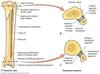
The lower leg is made up of 2 bones: tibia and fibula
- The tibia (medial) is weight bearing
- The fibula (lateral) is purely for muscle attachements and bares no force; it stabilises the ankle
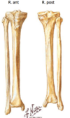
The tibia and the fibula are attached to each other in 2 positions (proximally and distally)
What is a consequence to this?
Because they are two bnes held together by ligaments, a fracture of one is most commonly associated with fractures of the other.
What part of the tibia is most susceptible to fracture?
The junction between the medial and inferior thirds of the tibia (ie. 2/3rds of the way down the bone)
This is because it has the narrowest and more cylindrical shape and has the poorest blood supply (vessels from above and vessels from below contribute but not a lot)
Describe the superior tibiofibular joint
- Is a plane synovial joint that is intracapsular.
- It has no movement.
- Allows some gliding movement but very limited
- it is reinforced by ligaments in the front and back.
Describe the inferior tibiofibular joint
- It is a fibrous joint (syndesmosis) held together by fibrous tissue
- It is important in preventing the tibia and fibula from separating during weight bearing times - also important in shock absorption
- It has an anterior, posterior and inetrosseus ligament attachment
- Separation is called diastasis

How common is disolcation or subluxation in the tibiofibular joints?
Very rare. They are very stable joints
What is the interosseus membrane in the lower limb? What are some of its functions?
It is also called the middle tibiofibular ligament. It extends between the interosseous crests of the tibia and fibula, helps stabilize their relationship
- It also separates the muscles on the front from those on the back of the leg (compartments)
- Is a site of attachment for muscles

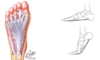
What are the bones of the foot?
- Tarsal bones (5 types)
- Metatarsals
- Phalanges

What is important about the first two (distal most) tarsal bones?
They are vertically orientated (while the remaining are horizontal)
Describe the first tarsal bone
Talus
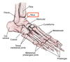
This bone articulates with with tibula and fibular above forming the ankle.
It also articulates with the calcaneus below and the other tarsal bones (navicular) in front
Describe the Calcaneus bone
It is the important weight bearer on the ground of the leg.
It articulates with the talus bone on top

What are the other tarsal bones that make up the foot?
- Navicular (medially, attached to the talus)
- Cuboid (laterally, attached to the calcaneus)
- Medial, intermediate and lateral cuneiforms

How many metatarsal bones are there? What is special about the 2nd?
There are 5: one each coming off the cuneiforms, and two coming off the cuboid
The 2nd metatarsal is slender in contrast to the others and hence is susceptible to fractures “root marching fracture”
Describe the phalanges (14 of them)
There are 2 phalanges in the hallux (the big toe) and then there are three in each of the other digits..
They are the same as in the hand.
Describe the trabeculae of the foot
They are arrange in weight bearing forces
There are numerous accessory and sesamoid bones in the foot (with tendons)
What are their importance?
There are 2 under the first metatarsal that create a tunnel
Common cause of avulsion fractures: particularly the Os trigonum

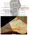
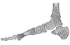
The talus is the key bone of the foot
What are the [3] main joints in relation to the talus?
- The ankle joint above it (with the tibia and fibula)
- The subtalar joint below it (with the calcaneus)
- The midtarsal joint in front of it (talocalcaneonavicular joint and the calcaneocuboid joint)
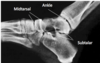
What type of joint is the ankle joint?
What are the movements?
A hinge joint
Thus there is flexion and extension (plnatr and dorsiflexion)
There is very little lateral movement
The body of the talus bone fits into a “mortice” between the malleloli of the bones of the leg.
Describe what is meant by this
The tibia forms the medial malleolus that extends down
The fibula forms the lateral malleolus extending downwards.
The tarlis bone fits in between these two extensions of bone (this space is referred to as the mortice). This gap is deepened posteriorly by the inferior transverse ligament

Describe the joint axis of the ankle in relation to the movements of the ankle
The lateral mallelous extends further down distally than does the medial. This means that the axis of movement of the ankle is not horizontal (it is at an oblique angle)
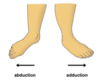
- Plantar flexion causes the sole of the foot to move medially
- Dorsiflexion causes the sole of the foot to sway out laterally

Describe the capsule of the ankle joint
It extends anteriorly into the neck of the talus
What are the supports of the ankle joint?
Being a Hinge joint, it has collateral ligaments passing from the malleoli to the nearest bones of the foot
There is also the ligaments form the distal tibiofibular syndesmosis (omplex fibrous joint composed of multiple ligaments and a broad fibrous membrane that spans between the tibia and fibula throughout the length of both bones)
Describe the medial (deltoid) collateral ligament of the ankle
They are not very strong ligaments that is rarely injured

Describe the lateral collateral ligament of the ankle
It is not as strong as the medial collateral
It is made up of 3 distinct bands:
- Anterior talofibular ligament: fibula to tarlis
- Posterior talofibular ligament: fibula to tarlis
- Calcaneofibular ligament: fibula to calcaneus