Brain & Meninges (Part 1) Flashcards
(61 cards)
cranial meninges
The outermost covering of the brain Consist of three connective tissue membranes that envelop the brain and spinal cord Membranes lie between the nervous tissue and bone
cranial meninges: primary functions
Primary function is to protect the CNS Form a supporting framework for arteries, veins, and venous sinuses Enclose the subarachnoid space, which is vital for the normal function of the brain
cranial meninges: 3 connective tissue layers
- Dura mater: tough thick external fibrous layer 2. Arachnoid mater: thin intermediate layer 3.Pia mater: delicate internal vascularized layer
meninges: dura mater
Tough outer covering of the brain Consists of 2 layers Innervated mostly by CN 5 Very well vascularized
meninges: dura mater (2 layers)
- Periosteal: pressed closely against cranial bones 2. Meningeal
Periosteal layer
Firmly attached to skull Continuous with skull periosteum at foramen magnum
Meningeal layer
In close contact with arachnoid mater Continuous with spinal cord meningeal dural covering
Two layers of dura are closely associated in all areas except where?
venous sinuses and dural infoldings
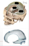
dural infoldings (4)
- Falx cerebri 2. Tentorium cerebelli 3. Falx cerebelli 4. Diaphragma sellae
dural infoldings: falx cerebri
Largest dural infolding Lies in the longitudinal cerebral fissure Separates right and left cerebral hemispheres
dural infoldings: falx cerebri (attachment site and end site )
Attached to crista galli anteriorly, and internal occipital protuberance posteriorly Ends by becoming continuous with tentorium cerebelli
dural infoldings: tentorium cerebelli
Second largest dural infolding Separates occipital lobes from the cerebellum
dural infoldings: tentorium cerebelli (attachments)
Anterior clinoid process Petrous part of temporal bone Internal surface of occipital bone
dural infoldings: falx cerebelli
Vertical dural infolding Inferior to tentorium cerebelli Separates cerebellar hemispheres
dural infoldings: Diaphragma sellae
Smallest dural infolding Suspended between anterior and posterior clinoid processes Forms roof over hypophysial fossa
meninges: arachnoid mater
Thin, avascular membrane Filled with a “web” of collagen – shock absorber Only enters the longitudinal fissure, otherwise not present in any other sulci or fissures
meninges: arachnoid mater (contains)
Contains cerebrospinal fluid in the subarachnoid space
meninges: pia mater
Thin, delicate membrane Completely lines all fissures and sulci of the brain Highly vascularized Tightly adherent to brain surface and cranial nerve roots
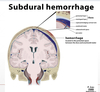
meninges: pia mater (tear/rupturing)
Location of all intracerebral hemorrhages leads to intracranial hemorrhage (aka cerebrovascular accident)
clinical correlation: meningitis
Inflammation of the meninges
clinical correlation: meningitis (symptoms)
Severe headache Fever Photophobia Stiff neck
clinical correlation: meningitis (cause)
Can be caused by bacteria or viruses Most cases are viral and relatively benign Bacterial meningitis is extremely serious and fatal if not treated promptly
dura mater: vascular supply( 3 aa.)
Middle meningeal artery: largest supplier of blood Anterior meningeal arteries: branches from ethmoidal arteries Accessory meningeal artery: branch of maxillary artery
leptomeninges
pis & arachnoid mater