Breast Disease Flashcards
(37 cards)
What are the 4 different methods used to perform a biopsy?
U/S-guided, stereotactic (take mammogram at 2 slightly different angles, see shadow, use software program to figure out where it is) or MRI-guided FNAC
FNAB
VAC
FNAC
Fine needle aspiration cytology
Limitation of FNAC
Cannot differentiate between invasive cancer and DCIS
Limitation of FNAC
Where is it used?
Cannot differentiate between invasive cancer and DCIS
Used in setting of known cancer of the breast to determine if lymph nodes affected
VAC
Bigger core biopsy; uses vacuum to suck tissue into larger needle
Can remove 3-4mm lesions completely
What causes peau d’orange?
Obstructed lymphatics
Triple test
Examination
Mammogram + U/S
Pathology
23 year old woman who commenced the OCP 6/12 ago presents with 3/12 Hx of L breast lump which is slightly tender; it does not vary with menstrual cycle
What does this Hx suggest?
No fluctuation with menstrual cycle so not likely to be hormonal change
Fibroadenoma is most common in young women
How does a fibroadenoma present clinically?
Mobile (“breast mouse”)
Smooth and well defined
Firm but not rock hard
May be slightly lobulated
Can be very large and can grow rapidly
Describe the natural Hx of a fibroadenoma
1/3 get smaller, most stay the same (they do all their growing in the patient’s 20s), small percentage get bigger
What is this U/S appearance consistent with?

Fibroadenoma
Benita, 38 year old woman, presents with a 3/12 Hx of breast tenderness with presence of a mass
Further r/v suggests that the pain fluctuates and is worst immediately prior to her period when the lump she describes is quite obvious; with onset of menses the pain and nodularity settles
Dx?
Probably fibrocystic disease or natural variation in breast tissue density with hormonal changes
Due to effect of oestrogen and progesterone on breast glandular tissue and lining of intramammary ducts (more common in women in 30s and 40s)
Benita, 38 year old woman, presents with a 3/12 Hx of breast tenderness with presence of a mass
Further r/v suggests that the pain fluctuates and is worst immediately prior to her period when the lump she describes is quite obvious; with onset of menses the pain and nodularity settles
Ix?
Benita, 38 year old woman, presents with a 3/12 Hx of breast tenderness with presence of a mass
Further r/v suggests that the pain fluctuates and is worst immediately prior to her period when the lump she describes is quite obvious; with onset of menses the pain and nodularity settles
Mx?
OCP may help
Evening primerose oil is effective in 60-70% (believed to change fluid retention; also prescribed for PMS and pelvic pain)
Consider denisol (but beware AEs e.g. hirsutism, which may not disappear on ceasing Rx)
FU in 3-4 months then discharge
NB Sometimes resolves spontaneously following child-birth
Connie, 43, presents with a very short Hx of sudden onset of a breast lump in the R breast; she examines her breasts regularly and was devastated to find this new lump developed over a matter of days
Had noticed some mild tenderness in this area prior to the development of the lump
DDx?
Benign breast disease: hormonal mastopathy, cystic disease of the breast (fibrocystic disease is the histological descriptor), mammary dysplasia, aberrations of normal development and involution
Cyst may be complicated (e.g. bleeding); this would account for the sudden onset
Need to rule out underlying DCIS
Connie, 43, presents with a very short Hx of sudden onset of a breast lump in the R breast; she examines her breasts regularly and was devastated to find this new lump developed over a matter of days
Had noticed some mild tenderness in this area prior to the development of the lump
U/S performed; interpret the findings
When would this require referral to breast clinic?
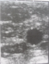
Simple cyst: black in the middle, well-defined, hyper-reflective posteriorly, circular
Would only require referral to breast clinic if palpable lump, complex or complicated cyst
Deanne, 49, presents with 2/12 Hx of nipple discharge from the L breast only
What features of the presentation are important in evaluating the cause of the nipple discharge?
Bilateral or unilateral: if related to hormonal change (e.g. lactation), would expect from both breasts
Does it come from multiple openings in the nipple (there are 15-20) or just from one spot: if it comes from many, this suggests the discharge is physiological
Deanne, 49, presents with 2/12 Hx of nipple discharge from the L breast only
What are some common causes of nipple discharge?
Hormonal (e.g. pituitary tumour)
Cancer (relatively uncommon)
Infection (mastitis; not common, usually comes from around the areola)
Papillary lesions, e.g. warty growth (usually within large duct systems, often singular but may be multiple, with multiple having a greater association with cancer risk), papilloma (most common cause of bloody discharge)
Cyst draining into duct system (usually clear not bloody discharge)
Deanne, 49, presents with 2/12 Hx of nipple discharge from the L breast only
Ix?
Mammogram
Focussed U/S of nipple
Deanne, 49, presents with 2/12 Hx of nipple discharge from the L breast only
Mx?
Biopsy (usually excisional)
What is the main cause of a serous nipple discharge? What are some less common causes?
Hyperplastic lesions (papillary lesions including hyperplasia, papilloma, carcinoma-in-situ and invasive ductal carcinoma)
Less common: duct ectasis
What is the main cause of a bloody nipple discharge? What are some less common causes?
Hyperplastic lesions (papillary lesions including hyperplasia, papilloma, carcinoma-in-situ, invasive ductal carcinoma)
Less common: duct ectasia, pregnancy
What is the main cause of a watery nipple discharge? What are some less common causes?
Hyperplastic lesions (papillary lesions including hyperplasia, papilloma, carcinoma-in-situ and invasive ductal carcinoma)
Less common: duct ectasia

