cardiovascular Flashcards
(189 cards)
define arterial ulcer
- localised area of damage and breakdown of skin
- due to inadequate arterial blood supply
- typically feet of patients with sever atheromatous narrowing of arteries supplying leg
aetiology of arterial ulcers
- caused by lack of blood flow to capillary beds of lower extremities
- prevalence increases with age and obesity
risk factors:
- coronary heart disease
- Hx of stroke/TIA
- DM
- peripheral arterial disease
- immobility
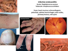
symptoms + signs of of arterial ulcers
- punched out appearance with clearly defined edges
- eliptical shape
- mainly on foot dorsum/toes
- grey granulomatous tissue
- ischaemic signs: hairlessness, pale skin, absent pulses, nail dystrophy, calf muscle wasting
- night pain
- pain is worse in supine because arterial blood flow is further reduced

investigations for arterial ulcers
1. doppler US of lower limbs
- assess latency of arteries
- assess potential for revascularisation/bypass surgery
- ABPI - <0.9= PAD, <0.5- critical limb ischaemia
- percutaneous angiography
- ECG
- fasting serum lipids
- fasting blood glucose + HbA1c
- FBC (anaemia can worsen ischaemia)
management of arterial ulcer
Immediate:
- pain relief
surgery
- angioplasty (balloon => widen arteries in atherosclerosis)
- stenting
- bypass grafts
- amputate
define cardiac arrest
acute cessation of cardiac function
aetiology and risk factors of cardiac arrest
reversible:
- hypothermia
- hypoxia
- hypovolaemia
- hypo/hyperkalaemia
- toxins
- thromboembolic
- tamponade
- tension PTX
presenting symptoms of cardiac arrest
sudden; management precede/concurrent to Hx
preceding symptoms:
- fatigue
- fainting
- blackouts
- dizziness
physical examination findings of cardiac arrest
unconscious
absent breathing
absent carotid pulses
investigations for cardiac arrest case
cardiac monitor
- classification of rhythm
bloods:
- FBC
- ABG
- U&E
- cross match
- clotting screen
- toxicology screen
- blood glucose
management of cardiac arrest
approach arrest scene with caution
* cause of arrest may pose threat
BLS
- if arrest is witnessed, consider precordial thump
- clear and maintain airway
- assess breathing, if absent, 2 rescue breaths
- assess carotid pulse for 10 seconds, if absent, 30 chest compressions
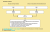
advanced life support
advanced life support management of cardiac arrest with shockable rhythm
cardiac monitor + defibrillator
assess rhythm
shockable rhythms: pulseless VT/VF
- defibrillates once (150-360J biphasic, 360J monophasic)
- resume CPR for 2 mins
- reassess and shock again if no change
- 1mg IV adrenaline after 2nd defibrillation
- 1mg IV adrenaline every 3-5 mins
*persistant shockable rhythm after 3rd shock
- 300mg IV bolus amiodarone
advanced life support management of cardiac arrest with asystole/PEA
cardiac monitor + defibrillator
assess rhythm
pulseless electrical activity (PEA)/asystole:
- CPR for 2 mins
- reassess
- 1mg IV adrenaline every 3-5 mins
*asystole or PEA + <60bpm, 3mg IV atropine once only
during CPR for cardiac arrest
check electrodes, paddle positions, and contacts
secure airway
consider magnesium, bicarbonate, and external pacing
stop CPR and check pulse ONLY IF change in rhythm or signs of life
treatment of reversible causes of cardiac arrest
hypothermia
- warm slowly
hypovolaemia
- IV colloids
- IV crystalloids
- blood products
hypo/hyperkalaemia
- give insulin (+dextrose) increase K+uptake
toxins
- toxin antidote
thromboembolic
- treat as PE/MI
tamponade
- pericardiocentesis
tension PTX
- aspiration/chest drain
complications of cardiac arrest
irreversible hypoxic brain damage
death
prognosis of cardiac arrest
resus less successful if cardiac arrest occurs outside hospital
increased duration of inadequate effective CO = poor prognosis
define DVT
thrombus formation within deep veins of usually calf or thigh
deep veins in leg more prone due to blood stasis (Virchow’s triad)
DVT risk factors
- polycythaemia
- thrombophilia
- OCP
- post surgery
- prolonged immobility/ long flights
- obesity
- pregnancy
- dehydration
- smoking
- malignancy
presenting symptoms of DVT
- asymmetrical swollen leg
- may be painless
examination findings of DVT
- local erythema, warmth, and swelling
- varicosities (dilated superficial veins)
- skin colour changes
- +/- unilateral leg pain
- Homan’s sign
what is Homan’s sign
seen in patients with DVT
forced passive dorsiflexion of ankle causes deep calf pain
how to stratify risk of PE in case of DVT
stratified using Well’s PE criteria
2 or more = high risk
- history: breathlessness, cough, haemoptysis
- check RR, pulse oximetry, and pulse rate
investigations for DVT
Use Wells score for DVT (<2 = low risk)
doppler US - gold standard
bloods:
- *- D dimer** (if low = unlikely to be DVT)
- thrombophilia screen if indicated
impedance plethysmography
- changes in blood volume causes changes in electrical resistance
if suspected PE:
- ECG
- CXR
- ABG