Cardiovascular pathology Flashcards
(101 cards)
What kind of tissue is this and what are some key characteristics of this tissue type?

This is the normal appearance of myocardial fibers in longitudinal section. Note the central nuclei and the syncytial arrangement of the fibers, some of which have pale pink intercalated disks.
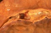
What is this a cross-section of?

This is a normal coronary artery. The lumen is large, without any narrowing by atheromatous plaque. The muscular arterial wall is of normal proportion.
Which valve is this?

This is the tricuspid valve. The leaflets and thin and delicate. Just like the mitral valve, the leaflets have thin chordae tendineae that attach the leaflet margins to the papillary muscles of the ventricular wall below.
What valve is this?

The aortic valve shows three thin and delicate cusps. The coronary artery orifices can be seen just above.The endocardium is smooth, beneath which can be seen a red-brown myocardium. The aorta above the valve displays a smooth intima with no atherosclerosis.
What is in this image?

This is the external appearance of a normal heart.The epicardial surface is smooth and glistening.The amount of epicardial fat is usual.The left anterior descending coronary artery extends down from the aortic root to the apex.
What is shown here?

The coronary artery shown here has narrowing of the lumen due to build up of atherosclerotic plaque.
What is occurring in this coronary artery?

This section of coronary artery demonstrates remote thrombosis with recanalization to leave only two small, narrow channels.
What is occurring in this coronary artery?

There is a severe degree of narrowing in this coronary artery. It is “complex” in that there is a large area of calcification on the lower right, which appears bluish on this H&E stain.
What is shown here?

This distal portion of coronary artery shows significant narrowing. Such distal involvement is typical of severe coronary atherosclerosis, such as can appear with diabetes mellitus or familial hypercholesterolemia. This would make a coronary bypass operation difficult.
What is the arrow pointing at?

There is a pink to red recent thrombosis in this narrowed coronary artery. The open, needle-like spaces in the atheromatous plaque are cholesterol clefts.
This is a magnified atheroma. What are some histological characteristics seen here?

This high magnification of the atheroma shows numerous foam cells and an occasional cholesterol cleft. A few dark blue inflammatory cells (lymphocytes) are scattered within the atheroma.
What is the white arrow pointing at?

A fatty streak in the aorta.
What disease is seen in these aortas? Rank them from least to most severe.

These three aortas demonstrate mild, moderate, and severe atherosclerosis from bottom to top. At the bottom, the mild atherosclerosis shows only scattered lipid plaques. The aorta in the middle shows many more larger plaques. The severe atherosclerosis in the aorta at the top shows extensive ulceration in the plaques.
What is seen here?

This high magnification microscopic view of an aortic atheroma shows prominent foam cells as well as cholesterol clefts.
This cross section shows…

…a large overlying atheroma on the left. Cholesterol clefts are numerous in this atheroma. The surface on the far left shows ulceration and hemorrhage. Despite this ulceration, atheromatous emboli are rare (or at least, complications of them are rare).
What pathological process led to this gross appearance?

This is severe atherosclerosis of the aorta in which the atheromatous plaques have undergone ulceration along with formation of overlying mural thrombus.
What is seen in the vessel on the far right?

This is the left coronary artery from the aortic root on the left. Extending across the middle of the picture to the right is the anterior descending branch. This coronary shows severe atherosclerosis with extensive calcification. At the far right, there is an area of significantnarrowing.

At high magnification, the dark red thrombus is apparent in the lumen of the coronary. The yellow tan plaques of atheroma narrow this coronary significantly, and the thrombus occludes it completely.

A thrombosis of a coronary artery is shown here in cross section. This acute thrombosis diminishes blood flow and leads to ischemia and/or infarction with damage to the myocardial fibers. This can be evidenced clinically by the onset of chest pain–angina.
Coronary atherosclerosis is shown here complicated by hemorrhage into the atheromatous plaque. Such hemorrhage acutely may narrow the arterial lumen.

Cross sections of this anterior descending coronary artery demonstrate marked atherosclerosis with narrowing. This is most pronounced at the left in the more proximal portion of this artery. In general, the worst atherosclerosis is proximal, where arterial blood flow is more turbulent.

Within the lumen of the LAD can be seen a dark red recent coronary thrombosis. The dull red color to the myocardium as seen below the glistening epicardium to the lower right of the thrombus is consistent with underlying myocardial infarction.
This is the left ventricular wall which has been sectioned lengthwise to reveal…….?

This is a large recent myocardial infarction. The center of the infarct contains necrotic muscle that appears yellow-tan. Surrounding this is a zone of red hyperemia. Remaining viable myocardium is reddish- brown.
Location? How old?

Anteroseptal, ~1 week. The center is tan with surrounding hyperemia.


