Case Studies - Exam I Flashcards
(22 cards)
Horse:
Is this a post-mortem change or an actual lesion?
What is the MDx and Etiology (Etx)?
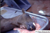
MDx: Post-mortem Artifactual Nasal Froth
Etx: Common artifact of dying
Lesion or Disease name: Nasal Froth
*Side note: this occurs when there is pulmonary edema, which then leaks out after death
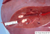
Cat:
Is this a post-mortem change or an actual lesion?
What is the MDx?

Post-mortem change
MDx: Pleural cavity, euthanasia solution artifact
*side note: dark brown surfaces with odor of alcohol*
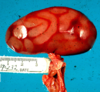
Cow:
Is this a post-mortem change or an actual lesion?
What is the MDx and EDx?
Post-mortem change
MDx: Liver, artifact reverse flow of brain
EDx: Increased air pressure result during slaughter
Description: Pale white soft tissue in the hepatic vein
Caribou calf:
Is this a post-mortem change or an actual lesion?
What is the MDx?

Post-mortem change
MDx: Carcass, post-mortem caused carcass damage
*Remarks: failure to see blood around the carcass damage suggests that this finding is only an artifact (animal predation) caused after death*
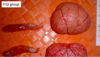
Pig:
Is the black, outlines a post-mortem change or an actual lesion?
What is the name of the black outlines and what causes it?
What is the *MDx?

Pseudomelanosis (black outlines) is a Postmortem change
MDx: Liver, bile ducts subacute purulent (suppurative) choangitis
Pseudomelanosis (black outlines): is considered a postmortem change due to the bacterial action on the blood producing disulfides
*at this point we are only supposed to know pseudomelanosis as we have not been taught about choangitis
What is pseudomelanosis?
Pseudomelanosis (black outlines): is considered a postmortem change due to the bacterial action on the blood producing disulfides
Horse:
Is the grey appearing mucosa a post-mortem change or an actual lesion?
What is the MDx?

Grey mucosa: a post-mortem artifactual change
MDx: Colon, acute necrotizing colitis
*the grey appearing mucosa surrounding bowel is primarily autolytic change and artifactual*
Normal mucosa should be pink or pale pink
Dog:
Is this a post-mortem artifact or an actual lesion?
What is the MDx?

Post-mortem artifact (Livor mortis / hypostatic congestion)
MDx: Brain, meninges, unilateral darkened (Hypostatic congestion)
*the R. side of the brain is darker red than the L. side; animal was lying on it’s right side at the time of death resulting in Hypostatic Congestion*
Pig:
Is this a post-mortem artifact or an actual lesion?
What is the MDx?
Post-mortem artifact / change
MDx: Kidney, terminal congestion with intestinal loop pressure artifacts of no blood.
*terminal congestion = hypostatic congestion/livor mortis*
Cow:
Are the brown/red dots a post-mortem artifact or an actual lesion?
What is the MDx?

Post-mortem artifact
MDx: Liver, normal (hemorrhage) artifact during slaughtering process
Dog:
Are the large black areas post-mortem artifact or an actual lesion?
What is the MDx?

Post-mortem artifact
MDx: Spleen, multifocal unequal explusion of blood
Result of some scattered areas of smooth muscle contraction preventing blood escape at death or soon after.
Calf:
Is the eye discoloration a post-mortem artifact or an actual lesion?
What is the MDx?

Post-mortem artifact
MDx: Eye, corneal clouding (opacity)
Cow:
Is this a post-mortem artifact or an actual lesion?
What is the MDx?
What is the etiology (Etx)?

An actual lesion (see EDx)
MDx: Larynx, trachea: severe, diffuse, fibrinonecrotic, laryngitis and tracheitis.
Etx: Bovine Herpesvirus-1
*on Destany’s exam
Pig:
What is the MDx?
What are 3 possible causes (Etx)?

MDx: Abdominal serosal surfaces: severe, acute, diffuse, fibrinous peritonitis.
Haemophilus parasuis:
Etx1: Streptococcus suis
Etx2: Mycoplasma hyorhinis
Etx3: Escherichia coli
Feline Tongue:
What is the pathogenesis of these lesions?

Pathogenesis:
Renal insufficiency -> elevated levels of circulating BUN -> breakdown by oral bacteria produces ammonia -> caustic burns -> ulcerative glossitis
*side note: Glossitis = inflammation of the tongue*
*on two of Destany’s exams
Feline:
What is the MDx?
What is the Etiology (Etx)?

MDx: Kidney: multifocal to coalescing, pyogranulamatous nephritis
Etx: Feline coronavirus (mutated)
*side note: the difference between this and lymphoma is that this travels along the blood vessels; look at the R. kidney to see this*
*on Destany’s exams twice
Feline:
What is the pathogenesis of enlarged parathyroid glands?
What are 2 possible sequelae (a condition that is the consequence of a previous dz or injury)?
Pathogenesis of renal secondary hyperparathyroidism:
Chronic renal disease -> ↓ GFR -> inadequate phosphorus secretion-> hyperphosphatemia -> excess phosphorus binds with calcium in serum -> ↓ ionized calcium -> ↑ parathyroid hormone (PTH) secretion -> ↑ bone resorption
Results in Parathyroid gland hyperplasia
2 Possible Sequelae:
1. Fibrous Osteodystrophy
2. Soft tissue mineralization
Dog:
What is the MDx?
What is the Etiology (Etx)?

MDx: Kidney, severe multifocal to coalescing, petechial cortical hemorrhages
Etx: Canine Herpes Virus
*on Destany’s exam*
Sheep:
What is the MDx?
What is the Etiology (Etx)?
What is the Disease name?
What is the name of the Special stain?

MDx: Intestine, diffuse granulomatous enteritis
Etx: Mycobacerium avium, subspecies paratuberculosis
Disease name: Ovine Johne’s Disease
Special Stain: Ziehl-Neelsen Acid Fast
*classical lesion; on Destany’s exam
Horse, aorta:
What is the EDx?
What is the Cause (Etx)?

EDx: Verminous endarteritis (with aneurysm and thrombosis)
Cause (Etx): Strongylus vulgaris 4th stage larvae
Feline:
MDx?
Associated lesion?

MDx: aorta, internal iliac arteries: occlusive fibrinous thromboembolus (saddle thrombus)
Associated lesions:
1. Hypertrophic cardiomyopathy
2. Renal/ other tissue infarcts
Feline:
MDx?
Etiology (Etx)?

MDx: Cerebellum: diffuse congenital hypoplasia
Etx: in-utero Feline Panleukopenis virus infection (feline parvovirus)
