Cell and Electrical Signalling, including immune system and muscles (4) + (5) Flashcards

1
Q

what is the autonomic nervous system

A

the part of the peripheral nervous system associated with involuntary responses and involuntary muscle movement

2
Q

give two examples of auto-immune disease

A

DIABETES TYPE 1

MULTIPLE SCLEROSIS

3
Q

give a description of the structure: motor neurone

A

a nerve cell that carries impulses away from the CNS to an effector eg. muscle or gland

4
Q

give a description of the structure: Schwann cell

A

glial cells in the peripheral nervous system that produce the myelin sheath around neuronal axons

5
Q

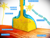
what is a synapse (1 mark)

A

a junction between two nerve cells, consisting of a minute gap across which impulses pass by diffusion of a neurotransmitter

6
Q

what is the nervous system

A

a system for coordination and control in animals, along with the endocrine system

7
Q

which specialised cells make up the nervous system + how does this process occur (2 marks)

A

stimuli are received from the internal/external environment by modified nerve cells called receptors in the sense organs or internal receptors

receptors convert the energy of the stimuli into electrical energy, a process called transduction

the electrical energy is transmitted as electrical impulses along a sequence of neurones/nerve cells- SENSORY NEURONE → INTER/RELAY NEURONE (CNS) → MOTOR NEURONE

motor neurones connect to effectors that carry out a response

8
Q

outline the NS pathway

A

STIMULUSRECEPTORSENSORY NEURONE- dendrites, dendron, soma, axon, axon terminals ⇒ INTERNEURONE (CNS) using relay neuroneMOTOR NEURONE- dendrites*, axon, axon terminalsEFFECTOR (MUSCLE OR GLAND) → RESPONSE

*containing soma; motor neurone have soma in CNS, sensory neurones have it near CNS

9
Q

what does the functioning of the NS require

A

an electrical potential difference across the plasma membrane; a membrane potential

10
Q

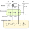
how do the characteristics of a SENSORY neurone, INTERNEURONE and MOTOR neurone differ

A

Sensory neurone: from the effector a dendrite leads to the cell body and away via an axon

Interneurone: very short dendrites lead to the cell body and away via a very short axon, non-myelinated

Motor neurone: cell body with little dendrites branching off, a myelinated axon takes the electrical impulse to the effector

11
Q

how is a membrane potential set up (4 marks)

A
  1. SODIUM/POTASSIUM PUMP: a transmembrane protein pump requiring ATP. It pumps 3 Na+ out of the cell for every 2 K+ pumped into the cell. Therefore there is more Na+ outside and K+ inside, polarising the cell to a membrane potential of -70mV.
  2. SODIUM CHANNELS ARE CLOSED (2nd pump)
  3. POTASSIUM CHANNELS ARE LEAKING (3rd pump) K+ does not all diffuse out of the cell because it is attracted to the negative region inside the cell.
12
Q

what is a dendrite (2 marks)

A

(same as dendron) a short branched extension of a nerve cell, along which impulses received from other nerve cells at synapses are transmitted to the cell body

13
Q

what are glial cells

A

non-neuronal cells that maintain homeostasis, form myelin and provide support and protection for neurones in the CNS and PNS eg. SCHWANN CELLS

14
Q

what do reflex actions involve (1 mark)

+ what is their purpose (3 marks)

A

They are responses that do not involve conscious thought; involve involuntary movement and a reflex arc.

  • response to danger
  • to avoid damage to the body
  • to maintain balance
15
Q

what is the pathway for a reflex arc

A

RECEPTOR ⇒ SENSORY NEURONE ⇒ RELAY NEURONE in spinal cord or unconscious part of brain ⇒ MOTOR NEURONE ⇒ EFFECTOR

in a monosynaptic reflex, the sensory neurone connects directly to a motor neurone eg. the knee jerk reflex

16
Q

give an example of a reflex arc and its purpose (3 marks)

A

KNEE JERK REFLEX

used by body to maintain balance and posture, allowing the person to stand with little effort or conscious thought

used by doctors to detect nervous problems eg. cerebellar disease

17
Q

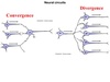
INTERNEURONES: compare structure and position in NS

A
  • same structural components as motor and sensory neurones- dendrites, soma, axon, synaptic endings NO MYELIN SHEATH- no need for accelerated speed of conduction along axon/dendron bc they do not extend across long distances*
  • found entirely in CNS and tf do not need extended axons or dendrons*
18
Q

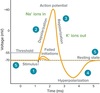
what is an action potential

A

a momentary change in the membrane potential caused by a transient change in the membrane’s permeability to sodium and potassium ions

can be observed with an oscilloscope that monitors the electrical potential or voltage, recorded by a microelectrode

19
Q

what is a membrane potential (3 marks)

A

In the neurone it is a resting potential, which is an electrical potential difference across the plasma membrane, typically between -60mV and -80mV. The inside is negative relative to the outside, the membrane is polarised

20
Q

what causes an action potential (3 marks)

A
  1. It is initiated when a neurone or muscle cell (an excitable cell) is depolarised to threshold value (approx. -50mV)
  2. Initiated by a change in permeability of the membrane to Na+ and K+ ions; Na+ ions flow into the cell.
21
Q

what happens after depolarisation (4 marks)

A
  1. During the AP, the inside of the cell becomes positively charged relative to the outside.
  2. Repolarisation/downstroke is caused by a change in the membrane permeability to Na+ and K+ ions; potassium ions flow out of the cell.
  3. The cell becomes negatively charged inside relative to the outside again.
22
Q

what occurs at the end of an action potential (2 marks)

A

The membrane becomes more negative on the inside than at a resting potential; HYPERPOLARISATION. It is a necessary refractory period.

23
Q

what is a voltage-gated ion channel?

A

Nerve axons contain two types of channels: Na+ specific and K+ specific, that are opened and closed by voltage dependent gates.

These enable neurones to produce action potentials because these are contained in their membranes.

24
Q

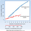
what is a Node of Ranvier (1 mark)

A

a gap in the myelin sheath of a nerve, between adjacent Schwann cells

25
Q

what is an axon (1 mark)

A

a projection of the neurone that conducts electrical impulses away from the neurone’s cell body/soma

26
Q

what is a dendron (1 mark)

A

a projection of the neurone which conducts nerve impulses from a synapse to the cell body (synonymous with dendrites)

27
Q

what is a synaptic knob (1 mark)

A

a swelling at the very end of the axon which communicates with a dendrite or cell body of another neurone across a synapse

28
Q

what is a sensory neurone (1 mark)

A

a nerve cell that transmits impulses from receptors to the CNS

29
Q

what are the structural differences between SENSORY and motor neurones (6 marks)

A
  1. CELL BODY IN PNS cell body in CNS
  2. CELL BODY IN THE MIDDLE OF THE NEURONE cell body at the start of the neurone
  3. SHORTER AXON longer axon
  4. DENDRON PRESENT dendron absent
  5. STARTS AT/ CONNECTS TO SENSORY NEURONE ends at neuromuscular junction/ motor-end plate/ effector
  6. DENDRITES AT THE END OF DENDRON (DO NOT CONNECT DIRECTLY TO CELL BODY) dendrites connect directly to cell body
30
Q

give one main difference between sensory and motor neurones

A

Sensory neurones transmit impulses received from a receptor and transmit them to the CNS.

Motor neurones: from CNS to effector.

31
Q

how is an electrical impulse created in a sensory receptor (4 marks)

A

DEPOLARISATION:

Stimuli received by sensory receptors eg. Pacinian corpuscle- pressure receptors, reverse the polarisation in the membrane of the receptor cells. This causes depolarisation in the sensory neurone.

ENERGY TRANSDUCTION:

Stimulus energy is converted to electrical energy in the receptor cells of sense organs, triggering an electrical impulse/action potential in the sensory neurone.

32
Q

what is the difference between a resting potential and action potential

A

Resting potential: when the membrane is polarised

Action potential: when the membrane is depolarised

33
Q

name a type of sensory receptor and where it is found, the nature of its stimulus and the type of stimulus energy that is transduced (x2)

A

EYE- rods and cones of the retina- photoreceptor- LIGHT- electromagnetic spectrum/light

SKIN- Pacinian corpuscle/mechanoreceptor- PRESSURE- mechanical

34
Q

how do stimuli trigger an action potential in sensory receptors (5 marks)

A

Stimulus energy is transduced/converted to electrical energy in the receptors, triggering an electrical impulse/action potential in the sensory neurone.

  1. Stimulus energy causes sodium channels to open in the membrane of the receptor cell. (closed at resting potential)
  2. Concentration of sodium is higher outside the cell relative to inside. Sodium diffuses in.
  3. Membrane is depolarised. This creates a GENERATOR POTENTIAL.
35
Q

how does the size/strength of stimulus affect generator potential and action potential

A

The larger the stimulus strength, the greater the diffusion of Na+ into the cell

the greater the depolarisation of the membrane

the larger the generator potential

WHEN THE GENERATOR POTENTIAL IN THE RECEPTOR REACHES A THRESHOLD VALUE approx. -50mV, IT TRIGGERS AN ACTION POTENTIAL IN THE SENSORY NEURONE

36
Q

what is myelin sheath (2 marks)

A

a mixture of proteins and phospholipids forming a whitish insulating sheath around many nerve fibres, made by Schwann cells, which increases the speed at which impulses are conducted. It is impermeable to Na+ and K+ ions

37
Q

what is the cell body/soma (1 mark)

A

the portion of a neurone that contains the nucleus but does not incorporate the dendrites or axon

38
Q

what is a neuro-muscular junction (1 mark)

A

the synapse formed by the contact between a motor neurone and a muscle fibre

39
Q

why is an action potential described as an all or nothing response

A
  • an action potential is not produced unless a stimulus depolarises the cell membrane to threshold value: -60mV to -50mV usually.
  • all action potentials along the neurone have the same amplitude and duration
40
Q

Stage: RESTING

describe:

  1. INSIDE THE MEMBRANE
  2. OUTSIDE THE MEMBRANE
  3. POTENTIAL
  4. NA/K PUMP WORKING
  5. V-G SODIUM CHANNELS
  6. V-G POTASSIUM CHANNELS
A
  1. HIGH K+ LOW Na+
  2. LOW K+ HIGH Na+
  3. -70mV
  4. yes
  5. closed
  6. closed but leaking
41
Q

Stage: DEPOLARISATION

describe:

  1. INSIDE THE MEMBRANE
  2. OUTSIDE THE MEMBRANE
  3. POTENTIAL
  4. NA/K PUMP WORKING
  5. V-G SODIUM CHANNELS
  6. V-G POTASSIUM CHANNELS
A
  1. HIGH K+ HIGH Na+
  2. LOW K+ Decreasing Na+
  3. Rises from -70mV to +35mV
  4. yes
  5. open
  6. closed but leaking
42
Q

Stage: REPOLARISATION

describe:

  1. INSIDE THE MEMBRANE
  2. OUTSIDE THE MEMBRANE
  3. POTENTIAL
  4. NA/K PUMP WORKING
  5. V-G SODIUM CHANNELS
  6. V-G POTASSIUM CHANNELS
A
  1. Decreasing K+ HIGH Na+
  2. Increasing K+ relatively low Na+
  3. decreases from +35mV to -70mV
  4. yes
  5. closing or closed
  6. open
43
Q

Stage: HYPERPOLARISATION

describe:

  1. INSIDE THE MEMBRANE
  2. OUTSIDE THE MEMBRANE
  3. POTENTIAL
  4. NA/K PUMP WORKING
  5. V-G SODIUM CHANNELS
  6. V-G POTASSIUM CHANNELS
A
  1. Decreasing K+ Na+ starting to decrease
  2. LOW K+ Na+ increasing
  3. -70mV to 80mV
  4. yes
  5. closed
  6. closing
44
Q

what is the difference between passive and active immunity, giving examples of each

A

active immunity = we have produced antibodies by our own B cells eg. due to a vaccination, chicken pox

passive immunity = we are given antibodies but do not produce them ourselves eg. from a mother’s breastmilk

45
Q

which factors affect the speed of conduction of an action potential

A
  1. DIAMETER OF AXON
  2. MYELINATION- SALTATORY CONDUCTION
46
Q

how does DIAMETER affect speed of conduction of an action potential (2 marks)

A

the wider the diameter of the axon, the faster the speed of conduction.

The resistance to flow of an electrical current is inversely proportional to the cross-sectional area of conductor eg. a wire or axon

47
Q

how does myelination affect speed of conduction of an action potential (5 marks)

A

Myelination is lipid-based myelin sheath produced by schwann cells that prevents electrical conduction i.e. of electrical impulses/action potentials. Gaps where the axon membrane is exposed are nodes of Ranvier.

ACTION POTENTIALS are only generated at the NODES OF RANVIER.

The current of Na+ ions during depolarisation of the action potential at one node of Ranvier diffuses along the interior of the axon to the next node.

Sodium channels will only open at nodes. Therefore conduction is faster because fewer channels are opened and each region does not need to be depolarised, repolarised and hyperpolarised. This is saltatory conduction.

48
Q

what is myelination (4 marks)

A

myelinated neurones contain myelin sheaths along their axons formed by schwann cells - myelin is closer to the axon, nucleus+cytoplasm on outside. It is mostly made of lipid, a poor conductor of electrical currents, providing electrical insulation of the axon.

49
Q

why is electrical conduction slower in non-myelinated axons

A

Action potentials occur on each continuous patch of membrane- it takes time for each part to depolarise, repolarise, hyperpolarise and channels to open/close.

50
Q

what is multiple sclerosis (3 marks)

A
  • a progressive auto-immune disease
  • the myelin sheath gradually breaks down
  • the speed of nervous transmission along the axon is reduced
  • impulses may be stopped completely if V-G channels only occur at nodes
51
Q

how are electrical impulses conducted between neurones (5 marks)

A

SYNAPTIC TRANSMISSION

  • Junctions between neurones or neurone/muscle cell are chemical synapses.
  • Presynaptic neurones synthesise and package the NT into synaptic vesicles, stored in the synaptic terminals (knobs).
  • release of neurotransmitters by the presynaptic neurone,
  • An action potential reaches the synaptic knob, releasing neurotransmitters into the synaptic cleft. NT bind to receptor sites on the post-synaptic neurone.
  • If enough NT are released, an action potential is initiated in the postsynaptic neurone.
52
Q

what is the mechanism for synaptic transmission (6 marks)

A
  1. An action potential depolarises the pre-synaptic membrane of the synaptic terminal, opening V-G Ca2+ channels in the pre-synaptic neurone.
  2. Ca2+ ions diffuse into the synaptic cleft. The high concentration of Ca2+ causes synaptic vesicles to fuse with the pre-synaptic membrane, releasing the neurotransmitters by exocytosis into the cleft.
  3. Neurotransmitter diffuses across the synaptic cleft and bind to the receptor on ligand-gated Na+ channels in the post-synaptic membrane, opening channels through which sodium diffuse into the post-synaptic neurone across the membrane.
  4. A post-synaptic generator potential is generated. If enough NT is released to bind and open sodium channels, enough generator potentials combine and create an action potential in the post-synaptic neurone.
  5. The neurotransmitter is released from the receptors and synaptic transmission ends.
53
Q

what is a cholinergic synapse and where do they occur

A

Synapses using the neurotransmitter acetylcholine (ACh)

When ACh binds to the receptors on the ligand-gated Na+ channels on the post-synaptic membrane, it opens and Na+ diffuses in, initiating a post-synaptic action potential

They occur in the central NS, autonomic NS and at neuromuscular junctions (between a motor neurone and skeletal muscle cell)

54
Q

what happens to acetlycholine after synaptic transmission

A

ACh is hydrolysed to acetic/ethanoic acid and choline in the synaptic cleft by enzyme ACETYL CHOLINESTERASE.

These molecules are reabsorbed into the pre-synaptic neurone and recycled to ACh by a condensation reaction, and packaged into vesicles in the synaptic terminals.

55
Q

what are the two branches of the nervous system

A

CENTRAL nervous system and PERIPHERAL nervous system

56
Q

what are the two branches of the CENTRAL nervous system and what are their FUNCTIONS

A

BRAIN ——- SPINAL CHORD

receives and processes sensory information, initiates responses, memories, thoughts, emotions

conducts signals to and from the brain, controls reflex activities

57
Q

what are the two branches of the PERIPHERAL nervous system and what are their FUNCTIONS

A

MOTOR neurones & SENSORY neurones

CNS to muscles and glands & sensory organs to CNS

58
Q

what are the two branches of the motor neurone branch of the PNS and what are their FUNCTIONS

A

SOMATIC nervous system vs. AUTONOMIC nervous system

somatic controls voluntary/conscious movements and autonomic involuntary/unconscious responses

59
Q

what are the two branches of the AUTONOMIC (involuntary responses) branch of the motor neurones/PNS and what are their FUNCTIONS

A

SYMPATHETIC division: fight or flight

PARASYMPATHETIC division: rest or digest

60
Q

what is the location of SKELETAL muscle in the body and what is its role/what is it controlled by

A

-attached to bones/skeleton

part of the somatic nervous system, voluntary movement of body and limbs, controlled by the cerebrum + cerebellum

61
Q

what is the location of CARDIAC muscle in the body and what is its role/what is it controlled by

A

-only found in the heart. -MYOGENIC

involuntary continuous rythmic contractions from formation of heart to end of life, controlled by medulla oblongata

62
Q

what is the location of SMOOTH muscle in the body and what is its role/what is it controlled by

A

-in arteries and arterioles, bronchi and bronchioles, alimentary canal, visceral organs

part of the autonomic nervous system, involuntary contractions

63
Q

describe the contractions of SKELETAL muscle

and how their cells are arranged

A

RAPID & SHORT

multinucleate cells lie end to end in tubular parallel fibres

64
Q

describe the contractions of CARDIAC muscle

and how their cells are arranged

A

INTERMEDIATE CONTRACTION SPEED & INTERMEDIATE PERIOD OF CONTRACTION

uninucleate cells, high quantity of mitochondria, respiration remains aerobic, no fatigue

cells branch and interconnect

65
Q

describe the contractions of SMOOTH muscle

and how their cells are arranged

A

SLOW & can REMAIN contracted for PROLONGED period of time

uninucleate cells that can contract in different directions

66
Q

what is the appearance and structure of the muscle fibres of skeletal muscle

A

shows CROSS STRIATIONS/stripes

cells are PARALLEL to each other and contract in ONE DIRECTION

67
Q

what is the appearance and structure of the muscle fibres of cardiac muscle

A

FAINT STRIATIONS

INTER-CALATED DISCS

BRANCHED fibres resulting in SIMULTANEOUS contractions (squeezing action)

68
Q

what is the appearance and structure of the muscle fibres of smooth muscle

A

NON-STRIATED

SPINDLE-SHAPED

IRREGULARLY arranged, contraction NOT in one direction

69
Q

what is the structure of voluntary muscle (3 marks)

A

Voluntary muscle consists of bundles of striated multinucleaste muscle fibres (muscle cell) surrounded by connective tissue

the bundles contain myofibrils

70
Q

how do motor neurones (somatic and autonomic NSs) connect to muscle cells

A

fibres are innervated by a motor neurone nerve ending; a motor end plate

71
Q

what is sarcolemma (1 mark)

A

muscle cell membrane; has many folds at the neuromuscular junction (transverse tubules = deep invaginations)

72
Q

what is a myofibril (1 mark)

A

a contractile organelle made up of units of sarcomeres

(contains repeating dark and light bands, I and A, which are responsible for striated appearance)

73
Q

what is a motor-end plate (1 mark)

A

divisions of the axon of the motor neurone forming fine branches ending in synaptic knobs

74
Q

what is a neuro-muscular junction (1 mark)

A

the motor-end plate + synaptic gap

where a motor neurone connects to a muscle fibre by a chemical synapse, with the diffusion of acetylcholine across the synaptic cleft

75
Q

what is a sarcomere (1 mark)

A

contractile unit of a myofibril, made of actin and myosin filaments

76
Q

what is sarcoplasmic reticulum (1 mark)

A

specialised membrane system- store of calcium ions

surrounded by sarcoplasm

77
Q

what are the events that occur at the motor end plate in the excitation of muscle fibres

A
  • action potential reaches synaptic knob of the motor-end plate, depolarising the pre-synaptic membrane and opening voltage-gated Ca2+ channels
  • Ca2+ diffuses in and vesicles containing ACh bind to the pre-synaptic membrane, where ACh is released into the synaptic cleft by exocytosis
  • ACh diffuses across the cleft and binds to complementary receptors on the sarcolemma, opening Na+ ion channels in the post-synaptic membrane/sarcolemma
  • the membrane is depolarised and it it reaches threshold value an action potential passes along the sarcolemma and into the muscle fibre through the transverse tubules
  • T-tubules connect to the sarcoplasmic reticulum surrounding the myofibrils
  • the wave of depolarisation triggers the release of Ca2+ ions from the sarcoplasmic reticulum. The ions initiate the sliding of filaments by allowing myosin to bind to actin in the myofibrils
  • ACh in the synaptic cleft is hydrolysed by acetyl cholinesterase, re-enters the pre-synaptic motor neurone to prevent overstimulation of the muscle fibre, and is recycled
78
Q

where does the trigger for muscle fibre contraction come from

A

an action potential (wave of depolarisation) is distributed to the sarcoplasmic reticulum by the T-tubule system

Ca2+ ions released from the sarcoplasmic reticulum trigger muscle fibre contraction by allowing myosin to bind to actin in the myofibrils

79
Q

name the 2 bands, 2 lines, 2 filaments and 1 zone and what they correspond to

A

I band = z line + region of actin only, where myosin does not overlap light band

A band = M line, H zone and region of myosin (with actin) dark band

M line = very centre of myosin, keeps myosin tethered

Z line = centre of actin filaments a sarcomere is from Z line to Z line

actin = thin filament

myosin = thick filament

H zone = myosin only

80
Q

what happens to the sarcomere when muscle fibres contract

A

Actin + myosin do not change length.

Actin moves towards the centre of the sarcomere (M line) during muscle contraction. The filaments slide over one another/overlap

The widths of the I band and H zone are reduced

A band is unchanged

Sarcomere shortens

After a full contraction, they disappear. Ends of thick filaments touch Z lines. Sarcomere shortens further.

81
Q

what are the sources of ATP for muscle contraction

A

quantity of ATP stored in muscle is low

ATP can be supplied by:

aerobic respiration by many mitochondria in muscle cell. Through the respiratory pigment MYOGLOBIN, which stores O2 in the sarcoplasm

phosphocreatine/ creatine phosphate

a store of energy, it can phosphorylate ADP

creatineP + ADP ⇒ creatine + ATP

anaerobic respiration

when muscle activity is high, produces 2 ATP and lactate, leading to oxygen debt (in order to convert lactate to pyruvate, NAD is required, supplied by etc/oxphos only when O2 present)

82
Q

what is the sliding filament model for muscle contraction

A

In order for the thick myosin filament to ‘slide over’ the thin actin, ATP binds to the myosin head and is hydrolysed to ADP and inorganic phosphate.

The myosin head straightens and forms a cross-bridge by binding to actin.

The head bends towards the centre of the sarcomere by releasing ADP and P: this is the power stroke.

Actin moves towards the centre of the sarcomere and ATP binds to the head to release it.

83
Q

what is the role of calcium in muscle contraction

A
  1. CALCIUM IONS BIND TO TROPONIN
  2. troponin and tropomyosin move away from myosin-binding sites on actin, allowing the myosin head to bind

when excitation finishes, calcium ions are pumped back into the sarcoplasmic reticulum by active transport ; muscle relaxes

84
Q

what happens to the heart rate in times of danger or exercise (nervous control of heart rate) 4 marks

A

The heart rate increases (from an average 70bpm approx.) in order to provide extra oxygen required for increased aerobic respiration.

The change is controlled by the cardiovascular centre in the medulla oblongata (base of the brain). It uses the autonomic NS to innervate the SA node, therefore influencing the frequency of the waves of excitation/heart rate.

85
Q

what are the two regions of the CARDIOVASCULAR CENTRE

A

EXCITATORY CENTRE: increases heart rate by sending impulses to the SA node via the accelerator nerve of the sympathetic branch of the autonomic NS.

INHIBITORY CENTRE: decreases heart rate by sending impulses via the vagus nerve of the parasympathetic branch of the autonomic NS.

86
Q

what are the two types of receptor in the cardiovascular system for the regulation of heart rate + where are they located

A

baroreceptors & chemoreceptors

in the aorta, carotid arteries and vena cava (blood vessels)

87
Q

what is the function of chemoreceptors (how do they affect heart rate)

A

-detect levels of CO2 and pH in the blood an dsend impulses to the cardiovascular centre

If CO2 is high (tf pH is low), the accelerator nerve of the sympathetic branch of the autonomic NS in excitatory increases heart rate

88
Q

what is the function of baroreceptors (how do they affect heart rate)

A

-they detect changes in blood pressure and send impulses to the cardiovascular centre

If blood pressure is high, the vagus nerve of the parasympathetic branch of the NS decreases heart rate

If blood pressure is low, the accelerator nerve of the sympathetic branch of the autonomic NS increases heart rate

89
Q

what is the difference between natural and artificial immunity, giving examples of each

A

natural = we naturally develop immunity, by being given antibodies or being infected by pathogens and developing an immune response eg. passive- from a mother’s breastmilk, active- chicken pox

artificial = injected with …. eg. passive- injected with monoclonal antibodies, active- vaccination

90
Q

how is the movement of limbs detected

A

by stretch receptors in the skeletal muscles of limbs, which send impulses to the CV centre to increase heart rate

91
Q

what is the function of the hypothalamus in the regulation of heart rate

A

under ‘fight or flight’ conditions or stress, the hypothalamus sends signals to the CV centre to increase heart rate. The CV sends impulses via the sympathetic branch of the autonomic NS to the adrenal glands to secrete the hormone adrenaline.

92
Q

what is the function of the cerebral cortex in the regulation of heart rate

A

impulses may be sent from the cerebral cortex to the CV centre to affect heart rate eg. meditation, sexual arousal, cognitive factors

93
Q

what is the overall structure of the autonomic NS

A
  • consists only of motor neurones
  • innervates cardiac and smooth muscle and glands (exocrine, endocrine)
  • does not involve voluntary/conscious control
  • controlled by the medulla oblongata and the hypothalamus in the brain
  • there are two divisions: sympathetic and parasympathetic; each division has two neurones from the CNS to the effectors: PRE-GANGLIONIC + POST-GANGLIONIC
94
Q

what is a ganglion and what is the pre-ganglionic neurone and post-ganglionic neurone

A

GANGLION- a group of nerve cell bodies outside the CNS (ganglia)

PRE: neurone which leaves the CNS in the ventral root and has its synaptic terminals in a ganglion

POST: neurone which has its cell body in the ganglion and goes to the effector

95
Q

what is a Schwann cell (1 mark)

A

A glial (non-neuronal) cell in the PSN that produces the myelin sheath around neuronal axons

96
Q

what is a leucocyte

A

WHITE BLOOD CELLS, including lymphocytes, macrophages and other phagocytes.

97
Q

name two antibiotic resistant bacteria

A

MRSA

causes serious wound infections, resistant to several antibiotics, including meticillin

Clostridium difficile

infects the digestive system

98
Q

suggest three ways a doctor can reduce the likelihood of antibiotic resistance developing

A
  • not prescribing antibiotics for minor infections

    • not prescribing to prevent infections, except patients who are elderly/have HIV*
    • advise patients to take all of the antibiotics to make sure the infection is fully killed*
99
Q

what happens in a fight or flight response

A
  • SYMPATHETIC NS
  • quickly run out of the way = electrical response from receptors in eyes, brain, muscles
  • Hypothalamus
  • Neurosecretory cells- adrenal medulla- adrenaline = non-steroid hormone i.e. binds to CSM
  • Vasodilation of muscles, inc. breathing rate in lungs, inc. heart rate in heart, decrease rate of digestion by vasoconstriction of intestines, glycogenolysis in liver releases glucose into bloodstream, dilation of pupils
100
Q

name the channels on the post-synaptic neurone

A

Ligand-gated ion channels

101
Q

how is the electrical impulse inhibited at a synapse after it has ‘diffused across’

A

Acetylcholinesterase enzyme in pre-synaptic cleft hydrolyses acetylcholine to acetate + choline.

  • prevents transmission continuing forever
  • ensures AP is only transmitted in one direction
102
Q

what is convergence & divergence in neurones

A

CONVERGENCE- many neurones releasing neurotransmitters for one post-synaptic neurone

DIVERGENCE- one pre-synaptic neurone releases neurotransmitters to many post-synaptic transmitters

103
Q

what is spacial summation

A

summation = combination of APs

where multiple small action potentials add up to one large action potential that depolarises to threshold in the post-synaptic neurone

104
Q

what is temporal summation

A

summation = combination of APs

multiple action potentials firing simultaneously (each one becoming larger) depolarise the post-synaptic membrane

105
Q

How does the medulla oblongata of the brain control HR?

Which neurotransmitter do they release?

A

Accelerator nerve increases HR by releasing noradrenaline at the SAN. eg. during exercise

Vagus nerve decreases HR by releasing acetylcholine at the SAN. - increases delay at the AVN. eg. after exercise

These are regulated by the cardioacceleratory + cardioinhibitory centres in the brain.