Ch 7 - Skeletal System Flashcards

(95 cards)

1
Q

Skeletal System functions (5)

A
  1. support
  2. protection
  3. movement
  4. storage(calcium+phos,fat,min) 5. blood cell production(marrow-rise to RBC) - hematopoiesis
2
Q

Bone, cartilage, tendons ligaments are what type of tissue

A

CT

3
Q

extracellular matrix contains:

A
  1. collagen (glue producing tough ropelike protein) 2. proteoglycans (protein+polysaccharides) 3. water 4. minerals
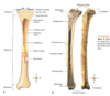
4
Q

4 types of bone

A
  1. long bone (longer than are wide, levers for muscles; femur, humerus, ulna/radius)
  2. short bone (approx. as broad as they are long, glide; wrist, ankle, carpal bone, talus)
  3. flat bone (thin, flat, proect organs; skull, ribs, scapula, sternum, internal and extrernal table(compact) with dipole (calncellous bone)
  4. irregular bone (vertebra, facial, sphenoid)
5
Q

5 parts of large bone - with description and label diagram

A
  1. diaphysis
  2. epiphyses
  3. articular cartilage
  4. periosteum
  5. medullary cavity
  6. endosteum
6
Q

diaphysis

A

long central shaft, provides support, hollow so not as heavy

7
Q

epiphysis & process

A

ends of long bone, spongy/cancellous filled with red marrow, upon development, seperated from diaphysis by epiphyseal line

  1. epiphyses are seperated from diaphysis by epiphyseal plate (growth plate) which is made of cartilage (until it fills in) and allows for bone growth-in kids,teens
  2. cartilage later is replaced by bone (ossification) and forms epiphyseal line-adults
  3. region is now called metaphysis
8
Q

articular cartilage

A

covers the ends pf epiphysis, where bone connects to bone (joint)

9
Q

periosteum

A

outer surface of bone (except for joing surfaces)

dense CT

contains blood vessels and nerves – PAIN ex shin splints

contains enthesis - where tendons imbed themselves into the periosteum of bone, connected by Sharpy’s fibers (stitch tendons into the p of bone)

contains osteoblasts (repair/remodeling of formation of bone = dynamic)

10
Q

medullary cavity

A

tubelike, hollow space in diaphysis

“marrow cavity” - adults bone, filled with CT rich in fat,WBC = yellow marrow

in youth, starts as mainly red marrow, blood cells

in adult, red marrow confined to proximal ends of long bone
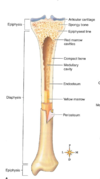
11
Q

endosteum

A

lines medullary cavity and spongy bone

is a thinner CT membrane

contains osteoblasts (repair/remodeling of formation of bone = dynamic)

12
Q

Compact bone

A

mostly solid matrix

blood vessels enter and exit bone

waste and nutrients in/out through the haversian canal system

13
Q

cancellous bone

A

aka spongy bone, trabeculae beams filled with marrow

lacy network of bone, small marrow-filled spaces

Location: mainly epiphysis of long bone

Forms: interior/intter part of bone

Gives stregnth without added withed

14
Q

osteoblasts

A

small “bone-forming” cells

cells that secrete osteoid (matrix)

osteogeneic stems cells in endosteum undergo cell division – form osteoblasts

ossification - formation of bone by osteoblasts

remove calcium from blood and gives to forming bone

15
Q

osteoclasts

A

large “bone-reabsorbing” cells

responsible for active erosion of bone minerals

as minerals and calcium are dissolved during bone erosion, they are reabsorbed back into blood (original source) - osteoclasts returns to blood

16
Q

cartilage

A

CT (hyaline - rings of ribs, lungs, bronchi; elastic - ear,opening respitory tract; fibro - dense CT)

sustains great weight when covering articulating bone surgaces

shock absorbant

NO canal system of blood vessels penetrate

17
Q

chondrocytes

A

scatter mutrients and O2 to cartilage via diffusion through the perichondrium

only healthy cells in cartilage matrix that makes collegan and proteglycans

18
Q

appositional vs interstital(longitudal) growth in cartilage

A

longitudal/intersistial, “enodenous growth” - occurs WITHIN cartilage tissue, childhood/adolescence, chondrocytes divide and secrete additional matrix, which can happen b/c soft nature of cartilage tissue.

appositional growth, “exogenous growth” - occurs OUTER surfaces of cartilage tissue. beyond adolescence and throughout adult life. chondrocytes in deep layer of perichondrium divide and secrete additional matrix, which is placed on surface of cartilage, wihch causes it to increase in size. Bone grows in DIAMETER, where scoliosis is imporant to manage

19
Q

bones develop baby 2 processes

A

intermembranous and endochondral ossification - result in compact and cancellous bone

20
Q

intramembranous ossification

A

within CT membrane

where flat bones and formed within fibrous membrane

21
Q

endochondral ossification

A

“bone formation in cartilage”

22
Q

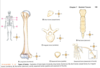
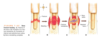
4 phases of bone healing

A
  1. blood vessels ruptured, swelling
  2. bleeding
  3. fracture hematomas, fibrocartilage splints bone
  4. develops into granulation tissue containing inflammatory cells, fibroblasts, bone/cartilage forming cells, capillaries
  5. Formation of bony callus tissue
  6. Replaced with normal bone
23
Q

PTH vs calcitonin

A

hormones secreted by PTG and thyroid gland

primary homestatic mechanisms for blood regulation fo Ca+

High blood Ca+ level(hypercalcemia) - calcitonin released, breakdown bone matrix decreases, Ca+ in blood decreases

Low blood Ca+ level(hypo) - PTH released, breakdown increases, Ca+ in blood rises

24
Q

avascular necrosis

A

death of bone tissue due to lack of blood flow

  • can lead to breaks in bones and sometimes collapse

“snow cap sign”, “bite sign”, sclerosis

can happen when there is fracure or dislocation

predisposing factors: cortisone shots

25
legg-calve-perthes disease
epiphyseal necrosis (avascular necrosis) in kids, occurs before closure of growth plate creates abnormal femoral angle
26
Paget's disease
27
soft drinks..
can lead to inhibition of ossification of bone, cause deminerilization of bone
28
Rickets
in children where bones fail to calcify, bowing deformity in weight bearing bones Lack of Ca and/or Vit D
29
open fracture/compound fracture
- Occurs when there is small cut in skin or severe soft tissue injuries that threaten survival of limb
30
Gustilo-Anderson classification system
Grade 1-3, increases with number
31
Comminuted fracture
bone breaks into many fragments
32
compression fx
bone is crushed, common osteoporotic bones
33
depressed fx
broken bone portion pressed inward ex: skull
34
impacted fx
broken bone ends are forced into each other ex: trying to break a fall
35
spiral fx
ragged break occurs when excessive twising forces -common in sports
36
greenstick fx
bones break incompletely, much in the way a green twig breaks - common children
37
pathological fracture
break over a tumor site
38
simple fracture
clean break
39
oblique fracture
oblique direction
40
Colles fracture
break the distal radius and ulna wrist happens when we fall wrists are hit on inside
41
smith's fracture
opposite of Colles, when you fall and wrist is hit on outside (posterior or extensor aspect)
42
inversion fracture
ankle inverts and rolls outwards
43
ACL avulsion fracture
avulsion - bone was pulled off of itself when muscle is contracted to hard, it tears and pulls a chunk of bone off
44
stress fracture
45
repeated overpronation, plantar flexion, exceissive weight shin splints
46
shoulder dislocation vs subluxation
**dislocation**: ball and socket of joint have fully seperated (humerus fully detached from glenoid fossa). Happens when fall and try to stop fall. **subluxation**: type of dislocation, but ball and socket NOT fully seperated. Can occur by damage to muscles/tendons.
47
Bones in child vs adult spine
33 vertabrae at birth, eventually they fuse to sacrum and coccyx and 24 remain
48
How find heart using bone anatomy
right below jugular notch(T3), behind sternal angle, ends at xiphisternal joint(T9)
49
invertebral disc
- fibrocartilage - allow movement of spine - fluid filled (h20), is lost with subluxation patterns and age - if slipped can cause: herniation, LBP, stiffness, deg.
50
anulus fibrosus
- most common tissue of origin for back pain is the outer layer annulus fibrosus - tough circular exterior of disc that surrounds soft inner for (nucleus) - collagen fibers, highly ordered in 10-20 sheets called circumferential **lamallae** **-** these are arranged in concentric rings that surround the nucleus pulposus - flexion: is distracted, can resist force, but less able to resist axial torsion
51
nucleus pulposus
posterior central axis, soft part of disc, gelly, mainly h20 - Fluids are passed in/out of annulus and NP by diffusion - nutrients derived to tissues and cells through diffusion -
52
sinuvertabral nerves
meningeal branches of spinal nerves that branch form segmental spinal nerve
53
nociception
sensory nervous system's response to certain harmful stimuli - small afferents - pain depends on how active mechanoreceptors are (large afferents) and nociceptors - stimulation travels along nerves via spinal cord to brain, then results in pain sensation - acute musculoskeletal injury affects the activity of the cutaneous and vasomoter symp neurons. So there is a relation btw acute ms tissue injury and autonomic NS
54
mechanoreceptor
sensory receptor that responds to mechanical pressure or distortion
55
sympathetics
sympathetic action modulates 1. skeletal muscle fibers 2. metabolism 3. ionic transport accross membrane 4. contractility
56
through ear lobe, shoulder joint, center of hip joint, midline of knee, center of ankle joint
57
tuberosity
large, rounded projection (no tibia)
58
crest
narrow ridge of bone
59
trochanter
very large, blunt, irregular process (ONLY on femur, 2 of them)
60
tubercle
small, rounded projection or process (abducter tubercle)
61
process
any bony prominence
62
facet
smooth, nearly flat articular surface
63
meatus
canal-like passageway
64
fossa
depression in a bone
65
foramen
round or oval opening through a bone
66
sinus
cavity within a bone, filled with air and lined with mucous membrane
67
lordosis vs kyphosis vs vs scoliosis vs 2ndry curvatures
L - "sway back", the spine of person curves significantly inward at lower back K - abnormally rounded upper back more than 50 degrees S- sideways curve, S or C shaped 2 - develops during head raising, crawling, walking
68
structural vs functional classification
S - joints named according to type of CT that joins bones together( fibrous vs cartilaginous) or by presence of fluid filled capsules(synvoinal joints) F - joints diveided into 3 classes according to degree of movement: **synarthroses, amphiarthroses, diathroses**
69
synarthroses
fibrous, immovable, sutures of skull
70
amphiathroses
cartilaginous, slightly movable, pubic of symphysis
71
diathroses
synovial, freely movable, shoulder joint
72
bursae
in synvoinal joint - pillow-like structure which consists of a synvonial membrane filled with synovial fluid. Tend to be associated with bony prominences (ex knee or elbow), where they function to cushion the joint and facilalte movement of tendons - reduces friction wear and tear ex: shoulder or knee
73
fibrous joints (synarthoses)
immovable three types: syndesmoses, sutures gomphoses
74
syndesmoses
joints in which fibrous bands (ligs) connects two bones. ex: btw radius and ulna (interosseous ligament)
75
sutures
found only in skull teethlike, fibrous cartilage joints
76
gomphoses
unique joints that occur btw the root fo a tooth and the alveolar process of the mandible or maxilla
77
cartilaginous joints
amphiarthroses joined by hyaline cartilage/fibrocartilage
78
synovial joints
freely movable, diathroses, ex: shoulder or hip joint
79
tendon sheath
wraps completely around tendon - aids in a reduction of friction - like a bun around a hotdog
80
saddle joint
- biaxial synovial joint - ex: thumb joint btw 1st mc and c bone - saddle shaped fits into socket - flexion/extension in one plane, abd/add in other
81
condyloid (ellipsodial) joint
- biaxial synovial joint - ex: joint btw radius and carpal bones - oval condyle fits into socket - flex/ext one plane, add/abd in other
82
plane joint
"gliding" joint - multiaxial synovial joint - ex: joints btw articular facets of vertabraes, joints btw carpal and tasal bones - flat articulating surfaces - gliding movements w/out angular or circular movements
83
OA
Osteoarthritis wear and tear of cartilage, mechanical collagen matrix unorganized \> less proteoglycans \> breakdown colleges fibers \> decrease osmotic pull \> increase water content
84
RA
Autoimmune Noniceptive inflammation of synovial membrane
85
DJD
Mechanical wear and tear
86
Degenerative disc disease
I wear and fair of normal and minor injuries and IVD start to lose water as the annulus fibrouses weakens
87
Herniation
Tear in outer fibrous ring of disk allows soft central part of,disc to extend past damaged rings cause pain when compressed on nerves
88
Spondylosis
Cause of normal bad posture, subluxation of joints, sports degeneration of spinal column with abnormal pressure, creates abnormal bones to compensate, weight from abnormal boon causes condition
89
Osgood schlatters disease
Aphphytis of tibial tubercle -inflammation of patellar ligament and tuberle repeated tension growth plate, running jumping Painful lump below knee cap- children teens young A
90
Hill-Sachs lesion
cortical depression in the posterolateral head of the humerus. It results from forceful impaction of the humeral head against the anteroinferior glenoid rim when the shoulder is dislocated anteriorly.
91
Bunion
swollen bursal sac or an osseous (bony) anomaly on the metatarsophalangeal joint. The larger part of the bump is a normal part of the head of the first metatarsal bone that has tilted sideways to stick out
92
March fracture
Fracture of distal metatarsal due to recurrent stress common soldiers
93
Corns
Abnormal anatomy of foot that causes pressure at points and then thickening of skin
94
Morton’s neuroma
benign neuroma of an intermetatarsal plantar nerve, most commonly of the second and third intermetatarsal spaces (between 2nd−3rd and 3rd−4th metatarsal heads), which results in the entrapment of the affected nerve.
95
Plantar fascitis
Policeman’s heal most common causes of heel pain. It involves inflammation of a thick band of tissue that runs across the bottom of your foot and connects your heel bone to your toes