Chapter 20: The Kidney - Congenital, Cystic Dz, Urinary Obstr. and Neoplasms Flashcards

1
Q

What occurs to the solitary kidney as a result of unilateral agenesis?

Some pts eventually develop what later in life?

A
  • Solitary kidney enlarges = compensatory hypertrophy, therefore increased risk for HTN
  • Some pts eventually develop progressive glomerular sclerosis —> CKD
2
Q

True renal hypoplasia is most often observed in?

Bilateral or unilateral?

May contribute to an increased lifetime risk for?

A
  • Low birth weight infants
  • May be bilateral but is more often unilateral
  • Increased risk for CKD
3
Q

Where is the most common location of Ectopic Kidneys?

May lead to what issues?

A
  • Just above the pelvic brim or within the pelvis
  • Ureteral abnormalities may cause obstruction/infection
4
Q

What are the 3 major pathologic features of ADPKD?

Unilateral or bilateral?

A
  • Large, mutlicystic kidneys, bilaterally
  • Liver cysts
  • Berry aneurysms
5
Q

Mutation in which gene and on which chromosome account for 85% of ADPKD cases?

A

PKD1 on chromosome 16

6
Q

Mutation in which gene and on which chromosome account for 15% of ADPKD cases?

A

PKD2 on chromosome 4

7
Q

How does the severity of disease and progression to complications differ between ADPKD pts with a PKD1 vs. PKD2 mutation?

A
  • PKD1 = more severe, increased risk of earlier onset renal failure (95% by 70 yo)
  • PKD2 = somewhat better prognosis; less risk of renal failure, at least earlier on.
8
Q

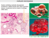
What are the cysts seen in ADPKD filled with, describe its morphology

A

Clear serous fluid or turbid red-brown, hemorrhagic fluid

9
Q

What is the typical clinical course of ADPKD and how does it typically present?

A
  • Generally asymptomatic w/ insidious onset in 4th-6th decade w/ renal insufficiency (HTN, azotemia)
  • Some exhibit abd. pain due to cyst enlargement and hemorrhage, thus hematuria may be manifestation
10
Q

There is a more aggressive (earlier onset, more severe) clinical course in which patients w/ ADPKD?

A
  • Blacks (particularly those w/ sickle cell trait)
  • M>F
  • Those w/ concomitant HTN
11
Q

What are the clinically significant extra-renal manifestations of ADPKD?

A
  • Hepatic cysts; less common in spleen, pancreas, and lung
  • Berry aneurysms –> subarachnoid hemorrhage
  • Mitral valve prolapse
  • Many have diverticular dz of colon
12
Q

How is diagnosis of ADPKD made?

A

Radiologic imaging

13
Q

How do the majority of pts w/ ADPKD ultimately end up dying?

A

Coronary or hypertensive heart disease

14
Q

Most cases of ARPKD are due to mutation in which gene and on what chromosome?

A
  • PKHD1 on chromosome 6
  • Encodes fibrocystin
15
Q

What are the 2 most common clinical subtypes of ARPKD?

Type of hepatic fibrosis in each?

A
  1. Perinatal (most common) –> minimal hepatic fibrosis
  2. Neonatal –> mild hepatic fibrosis
16
Q

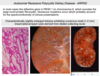
What is the gross morphology of the surface of the kidneys in ARPKD?

What does a cut section of the kidney show?

A
  • Enlarged w/ SMOOTH surface (contast to cystic surface of ADPKD)
  • Cut section shows numerous small cysts linearly arrayed in cortex ans medulla w/ spongelike appearance
17
Q

Where are the cysts seen in ARPKD derived from?

A

Dilated collecting ducts

18
Q

What is the typical clinical course of ARPKD?

A
  • Perinatal form = most common; survival only a few hours; death due to pulmonary hypoplasia
  • If survive infancy develop congenital hepatic fibrosis —> portal HTN and splenomegaly
19
Q

In almost all cases of ARPKD the liver contains what?

A

Cysts associated w/ portal fibrosis and proliferation of bile ducts

20
Q

What are the 3 major subtypes of medullary cystic disease?

A
  1. Medullary sponge kidney
  2. Nephronophthisis
  3. Adult-onset medullary cystic disease
21
Q

What is seen in Medullary Sponge Kidney?

Affected population?

Renal function?

A
  • Mutliple cystic dilations of the collecting ducts and medulla
  • Occurs in adults and is usally discovered radiographically
  • Renal function is usually normal
22
Q

What are the disorders characterized as ciliopathies or abnormalities of the cilium-centrosome complex?

A
  • Polycystic kidney diseases (both AD and AR)
  • Medullary Cystic Kidney
  • Nephronophtisis
23
Q

In Nephronophthisis-Medullary Cystic Disease where are the cysts localized?

Gross morphology of the kidneys and on microscopy of the cortex?

A
  • Localized to corticomedullary junction and medulla
  • Kidneys are small w/ contracted granular surfaces
  • Cortex has diffuse atrophy and thickening of tubular BM w/ interstitial fibrosis
24
Q

What are the 3 clinically recognized variants of the Nephronopthisis-Disease complex?

Which is most common?

A
  1. Sporadic, non-familial
  2. Familial juvenile nephronophthisis (most common)
  3. Renal-retinal dysplasia
25
Q

Although the medullary cysts in Nephronophthisis-Medullary Cystic Disease Complex are important, what is the cause of the eventual renal insufficiency?

A

Cortical tubulointerstitial damage

26
Q

Inheritance pattern of familial juvenile nephronophthisis?

A

Autosomal Recessive

27
Q

How do children w/ familial juvenile nephronophthisis typically present?

A
  • First w/ polyuria and polydipsia
  • Sodium wasting and tubular acidosis are also prominent
  • Expected course is progresion to ESRD in 5-10 years
28
Q

What is the most common genetic cause of ESRD in children and young adults?

A

Nephronophthisis complex

29
Q

What is the inheritance pattern of Adult-onset Medullary Cystic Disease?

Which genes mutated?

A
  • Autosomal Dominant
  • MCKD1** and **MCKD2
30
Q

How does Familial Juvenile Nephronophthisis differ from Adult-onset Nephronophthisis (medullary cystic disease)?

A
  • Juvenile is marked by progressive renal failure beginning in childhood
  • Adult-onset is marked by chronic renal failure beginning in adulthood

*Both present w/ corticomedullary cysts, shrunken kindeys, salt wasting and polyuria

31
Q

Since the medullary cysts associated w/ nephronophthisis are usually to small to visualize, this disease should be considered in children presenting how?

A
  • Unexplained chronic renal failure
  • A positive family hx
  • Chronic tubulointerstitial nephritis on biopsy
32
Q

How is Multicystic Renal Dysplasia acquired and what is it characterized by?

A
  • Sporadic disorder; common mass in perinatal period
  • Unilateral or b/l kidney(s) usually enlarged, extremely irregular, and multicystic
33
Q

What is the characteristic histological finding of Multicystic Renal Dysplasia?

A

ISLANDS of undifferentiated mesenchyme, often with CARTILAGE and immature collecting ducts

34
Q

Most cases of Multicystic Renal Dysplasia are associated what other renal/GU anomalies?

A
  • Ureteropelvic obstruction
  • Ureteral agensis or atresia
  • Other lower GU anomalies
35
Q

What is the significance of unilateral Multicystic Renal Dysplasia, especially in a child and its prognosis?

Bilateral?

A
  • Unilateral = abd. mass in child may mimic a neoplasm. Surgically removed via nephrectomy = excellent prognosis
  • Bilateral will eventually progress to renal failure
36
Q

What type of cysts are associated with pts w/ ESRD who have undergone prolonged dialysis and sx’s?

What is the significance of these cysts long-term?

A
  • Numerous cortical and medullary cysts w/ calcium oxalate crystals
  • Often asymptomatic, but may bleed = hematuria
  • ↑↑↑ risk of renal cell carcinoma
37
Q

What are Simple Cysts of the kidney?

Significance?

A
  • Single or multiple cysts on renal surface (cortex)
  • Generally filled w/ clear fluid
  • Common postmortem finding
  • May be a bizarre radiograph finding requiring differentiation by observer from a tumor
38
Q

Dilation of the renal pelvis and calyces associated with progressive atrophy of the kidney due to obstruction to the outflow of urine is known as?

A

Hydronephrosis

39
Q

The initial functional alterations caused by obstruction are primarily manifested as what?

Eventually progresses to?

A
  • 1st sign = impaired concentrating ability
  • Later on the GFR decreases
40
Q

What are the early vs. late mophological features of Hydronephrosis?

A
  • Early = simply dilation of the pelvis and calyces, may be significant interstitial inflammation
  • Later = cortical tubular atrophy + marked diffuse interstitial fibrosis
41
Q

In advanced cases of Hydronephrosis what morphological changes are seen in the kidney?

A
  • Kidney = thin-walled cystic structure
  • Striking parenchymal atrophy
  • Total obliteration of pyramids + thinning of cortex
42
Q

Which imaging technique is useful for diagnosis of obstructive uropathy?

A

Ultrasound

43
Q

In hydronephrosis due to bilateral partial obstruction, what are the common signs/symptoms?

A
  • Earliest = inability to concentrate, polyuria and nocturia
  • Some pts develop distal tubule acidosis, salt wasting, 2’ renal calculi and chronic tubulointerstital nephritis
  • HTN is common
44
Q

In hydronephrosis due to complete bilateral obstruction, what are the common signs/symptoms?

How must this be dealt with and what is commonly seen after treatment?

A
  • Rapid onset of oliguria or anuria = INCOMPATIBLE w/ survival unless obstruction is relieved
  • After removal there is postobstructive diuresis = massive amts of urine that is rich in NaCl
45
Q

Is Nephrolithiasis (aka kidney stones) typicall bi- or unilateral?

Sex most often affected and at what age?

A
  • 80% are unilateral
  • M>F
  • Peak age at onset = 20-30 yo
46
Q

Is there a familial or hereditary predisposition to developing nephrolithiasis?

A

Yes

47
Q

What are the 4 main types of renal calculi?

A
  1. Calcium stones (70%)
  2. Struvite stone/Triple stone (magnesium-ammonia-phosphate)
  3. Uric acid stones
  4. Cystine
48
Q

Magnesium ammonium phosphate stones are formed largely due to?

Size of stone and form what?

A
  • Following infection by urea-splitting bacteria (i.e., Proteus and some staphylococci)
  • Some of the largest stones form staghorn calculi
49
Q

What is important to take into consideration about the development of Uric Acid stones?

A
  • Common in ppl w/ gout, and diseases such as leukemia
  • BUT, more than 1/2 of pts have neither hyperuricemia nor increased excretion of uric acid
50
Q

What are 4 factors that influence the development of calculi (stones) in the urinary tract?

A
  1. Increased concentration of stone constituents
  2. Changes in urinary pH
  3. Decreased urine volume
  4. Presence of bacteria
51
Q

What is the favored sites within the urinary tract for the formation of stones?

A
  • Renal calyces and pelves
  • Bladder
52
Q

What is the most common malignant tumor of the kidney in both adults and in children?

A
  • Adults = Renal Cell Carcinoma (1st) and Urothelial Carcinomas of the Calyces and Pelvis (2nd)
  • Children = Wilms Tumor
53
Q

What is the most common benign neoplasm of the kidney?

Arise from what?

A
  • Renal papillary adenomas
  • Arise from renal tubular epithelium
54
Q

Describe the morphology (i.e., size, shape, location, and color) of Renal Papillary Adenomas?

A
  • Small, usually <0.5 cm within cortex
  • Discrete, cortical, pale yellow-gray, well-circumscribed nodules
  • Branching, papillomatous structures w/ cuboidal to polygonal cells
55
Q

Renal papillary adenomas share what similar cytogenetic features that are also present in low-grade RCC?

A

Trisomies 7 and 17

56
Q

Which feature of Renal Papillary Adenomas are related to its prognosis?

A
  • Size
  • Benign = <1.0 cm or less in diameter
  • Neoplasma >1.0 cm w/ adenoma histomorph. = low grade RCC
57
Q

Which benign neoplasm of the kidney is highly associated with Tuberous Sclerosis?

A

Angiomyolipomas

58
Q

Angiomyolipomas usually present in which sex and when?

Associated with what genes?

A
  • Adults, middle-aged, F>M
  • Loss of TSC1 or TSC2 tumor suppressor genes
59
Q

What is the clinical significance of Angiomyolipomas as far as significant complications which may arise?

A

May spontaneously rupture w/ massive hemorrhage, initialy presentation may be shock secondary to massive retroperitoneal and/or intra-abdominal hemorrhage

60
Q

Tuberous Sclerosis has what type of inheritance?

Manifests as a complex of what type of disorders?

A
  • Autosomal Dominant
  • Tumors or lesions of: brain, skin, kidney, lungs, and eyes
61
Q

Renal Oncocytomas arise from where and what are their characterisitcs?

A
  • Benign epithelial neoplasm w/ large eosinophilic cells that have small, round, benign-appearing nuclei w/ large nucleoli
  • Arise from intercalated cells of collecting ducts
  • Grossly, appear tan or mahogany brown, usually well encapsulated w/ a central scar
62
Q

On EM, Renal Oncocytomas have eosinophilic cells packed with what?

A

Mitochondria

63
Q

Whos is most often affected by RCC and when does it arise?

A
  • M > F
  • Arise in older adults; 6th-8th decade
64
Q

What is the major risk factor and other risk factors for RCC?

A
  • Major = smoking
  • Obesity (particularly woman)
  • HTN
  • Estrogen therapy
  • Asbestos, petroleum prods., and heavy metals
  • Tuberous Sclerosis
65
Q

Most RCC arises how?

A

Sporadic

66
Q

The VHL gene is implicated in what types of RCC?

A

Both familial and sporadic clear cell carcinomas

67
Q

Pts w/ Von Hippel-Lindau syndrome, most often develop what type of renal malignancies?

A
  • Hereditary clear cell carcinoma
  • Hereditary papillary carcinoma

*Often bilateral and multiple

68
Q

Hereditary leiomyomatosis and renal cells cancer syndrome is inherited how and due to what gene?

This disease is characterized by what?

A
  • Autosomal dominant due to mutations in FH gene
  • Characterized by: cutaneous and uterine leiomyomata and an aggressive type of papillary carcinoma w/ ↑ propensity for metastasis
69
Q

Hereditary papillary carcinoma has what type of inheritence and due to what gene?

Characterized by?

A
  • Autosomal dominant w/ mutations in MET proto-oncogene
  • Multiple bilateral tumors w/ papillary histology
70
Q

What is the inheritance of Birt-Hogg-Dube syndrome?

Manifested by what?

A
  • Autosomal dominant mutation BHD gene
  • Skin (fibrofolliculomas, trichodiscomas, and acrochordons), pulmonary (cysts or blebs), and renal tumors
71
Q

What is the most common type of Renal Cell Carcinoma?

Describe its histological characteristics.

A
  • Clear cell carcinoma
  • Non-papillary cells w/ clear cytoplasm (some granular)
72
Q

How do the majority of the clear cell type of renal cell carcinoma arise?

Majority contain which mutation and on what chromosome?

A
  • Sporadic (95%), but can be familial
  • Short arm deletions/translocations of chromosome 3(harbors theVHL tumor suppressor gene)
73
Q

The loss of VHL in RCC causes the inappropriate expression of what genes?

A
  • HIF-1 induced genes
  • VEGF
  • IGF-1 (stimulates growth)
  • PDGF
74
Q

Papillary carcinomas (10-15% of renal cancers) are associated w/ what cytogenetic abnormalities in both the sporadic and familial form?

A
  • Trisomies 7 and 17 and loss of Y in males = Sporadic
  • Trisomy 7 = Familial
75
Q

Which gene located on chromosome 7 is associated with Renal Papillary Carcinoma?

Encodes what?

A
  • MET a proto-oncogene
  • Encodes RTK for HGF = Scatter Factor
76
Q

Renal Chromophobe Carcinoma (5% of renal cancers) arise from where and are composed of what kind of cells?

i.e., describe their distinct morphology

A
  • Arise from intercalated cells (difficult to distinguish from oncocytoma)
  • Pale eosinophilic cells, with a perinuclear halo, arranged in solid sheets w/ large concentration around blood vessels
77
Q

What is the prognosis of renal chromophobe carcinoma?

A

Excellent

78
Q

Cytogenetic examination of which renal carcinoma shows mutliple chromosome losses and extreme hypodiploidy?

A

Chromophobe Carcinoma

79
Q

Xp11 translocation carcinoma is a distinct subtype of RCC, why?

Defined by translocation of what gene?

A
  • Often occurs in young patients
  • Defined by translocations of the TFE3 gene w/ a number of partner genes
80
Q

Collecting duct (Bellini duct) carcinoma is a rare and is defined by what histological characteristics?

A
  • Malignant cells forming glands enmeshed within prominent fibrotic stroma
  • Typically in medullary location
81
Q

Medullary carcinoma is a rare tumor most often seen in whom?

A

Patients w/ sickle-cell trait

82
Q

Renal clear cell carcinomas usually occurs as what type of lesion?

Often occuring where in kidney?

A
  • Most commonly upper pole
  • Occur as solitary UNILATERAL lesions that are bright yellow-gray-white spherical masses
83
Q

Which renal tumor is this and how can you tell?

A
  • RCC –> Clear cell type
  • Rounded or polygonal shaped cells w/ adundant clear or granular cytoplasm, containing glycogen and lipids
84
Q

What are some of the most striking characterisitcs as far as growth and invasion go for RCC?

A
  • Tendency to reach large size and have widespread metastases before local signs/sx’s
  • Tendency to invade the renal vein and can grow upwards to IVC, sometimes all the way to right side of heart
85
Q

Papillary type of RCC arise from where in the kidney and commonly grows how, unilateral or bilateral?

What are the major morphological characteristics and major cell types found?

A
  • Arise from DCT and can be multifocal and BILATERAL
  • Typically hemorrhagic and cystic
  • Contain interstitial FOAM cells and may also have psammoma bodies
86
Q

What morphological change arises infrequently in all types of RCC and is an ominous feature?

A

Sarcomatoid changes –> spindle cells simulating mesenchymal neoplasm

87
Q

What is the classic triad of sx’s for Renal Cell Carcinoma?

Which is the most reliable clue?

A
  1. Costovertebral pain
  2. Palpable mass
  3. Hematuria = most reliable clue
88
Q

Why is RCC considered one of the great mimics in medicine?

A
  • Tends to produce diversity of systemic sx’s not related to kidney
  • Abnormal hormone prod, may cause: polycythemia, hypercalcemia, HTN, hepatic dysf., feminization or masculinization, Cushing’s, eosinophilia, leukemoid rxns, and amyloidosis
89
Q

What is the most common mode of spread from RCC and where are the most common sites?

A
  • Hematogenous
  • Lungs and bones –> regional LN’s, liver, adrenal, and brain
90
Q

Tx options of choice for RCC?

A
  • Radical nephrectomy
  • VEGF inhibitors and various tyrosine kinases
91
Q

Urothelial Carcinoma of the Renal Pelvis usually becomes clinically apparent when?

Produce what signs/sx’s?

A
  • Clinically apparent in relatively short time
  • Lie within pelvis and by fragmentation, produce noticeable hematuria
  • May block urinary outflow and causes palpable hydronephrosis and flank pain
92
Q

In 50% of urothelial carcinomas of the renal pelvis there exists a concomitant tumor where?

A

Bladder urothelial tumor

93
Q

In which 2 disorders is there a higher incidence of Urothelial Carcinoma of the Renal Pelvis?

A
  • Lynch syndrome (HNPCC)
  • Analgesic nephropathy
94
Q

What is the prognosis of Urothelial Carcinoma of the Renal Pelvis?

Are they infiltrative?

A
  • Commonly infiltrate wall of renal pelvis and calyces
  • Poor prognosis