Chapter 8 Flashcards
(234 cards)
sensory receptors
- connect to the cortex through a sequence of intervening relaying neurons that allow each sensory system to mediate different responses and to interact with other sensory systems
- transduce or convert energy to neural activity, light, photons

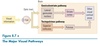
vision sensory receptors
light energy is converted to chemical energy into further receptors of the retina, which actually is part of the brain, and this chemical energy is in turn converted to action potentials
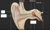
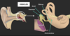
auditory system conversion
air pressure waves are converted first to mechanical energy, which eventually activates the auditory receptors that produce action potentials
somatosensory sensory system
mechanical energy activates receptor cells that are sensitive to touch or pressure → receptors generate action potentials
pain
tissue damage releases chemicals that act like neurotransmitter to activate pain fibers and produce action potentials
taste and olfaction
chemical molecules carried on the air or contained in food, fit themselves into receptors of various shapes to activate action potentials
*
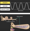
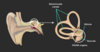
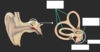
auditory receptors
- respond to sound wave frequencies between 20 and 20,000 Hertz
- elephants can actually hear and produce sounds below 20 Hertz
- bats can hear and produce sounds as high as 120,000 Hertz
color deficient
lack one or more types of photoreceptors for color vision, the red, blue, and green cones
can see many colors, but not the same colors as people with all three cones can
action potentials
- dendrite of a somatosensory neuron is wrapped around the base of the hair
- when the hair is displaced in a certain direction, the dendrite is stretched by the displacement
- sodium channels in the dendrites’ membrane are stretch sensitive, so they open in response to scratching
- if the influx of sodium ions in the stretch-sensitive channels is sufficient, the resulting voltage change will depolarize the dendrite to its threshold, or an action potential, and the voltage-gated, sensitive, K plus, and sodium channels, will open, resulting in a nerve impulse heading to the brain

receptive fields
and ex
- area from which a stimulus can activate a sensory receptor
- not only sample sensory information, but also help locate sensory events in space and facilitate different actions in space
- localize sensations
- ex: Our lower visual receptive field facilitates the use of our hands in making skilled actions. Whereas, our upper visual field facilitates our movements through our more distant surroundings.
rapidly adapting receptors
+ ex x2
- receptor that responds at the onset of stimulus on the body
- easy to activate, but stop responding after very short time
- ex: if you touch your arm very lightly, you will immediately detect a touch, but if you keep your finger still, the sensation will fade as receptors adapt → detect the movement of objects
- ex: rods - respond to visible light of any wavelength and have lower response threshold than do the slowly adapting cone shaped receptors which are instead sensitive to color and position
slowly adapting receptors
ex
- receptor that responds for the duration of a stimulus on the body
- react to stimulation slowly
- ex: if you push a little harder when you first touch your arm, you will feel the touch longer
exteroceptive receptors
ex
receptor that responds to external stimuli
ex: optic and auditory flow -> useful in telling us how fast we are going, whether we are going in a straight line, or up or down, and whether we are moving, or an object in the world is moving
optic flow
(exteroceptive receptors)
stimulus configuration - when you run, visual stimuli appear to stream past
auditory flow
(exteroceptive receptors)
when you move past the sound source, you hear changes in sound intensity that take place because of your changing location
interoceptive receptor
ex
- receptor that responds to internal stimuli
- position and movement of our bodies
- interpret meaning from external stimuli
- ex: learn from interoceptive receptors in our muscles and joints, and in the vestibular organs of the inner ear
receptor density
- receptor density determines a sensory system’s sensitivity
- ex: tactile receptors on the fingers are numerous compared with those on the arm
two point sensitivity
You can prove this by moving the tips of two pencils apart to different degrees, as you touch different parts of your body. The ability to recognize the presence of two pencil points close together, is highest on the parts of the body having the most touch receptors
interneurons
(sensory system)
- all receptors connect to cortex through sequence of 3 or 4 interneurons
first relay for pain receptors
- first relay for pain receptors in the spinal cord is related to reflexes that produce withdrawal from a painful stimulus
- Even after damage to the spinal cord that cuts it off from the brain, a limb will still withdraw from a painful stimulus, why? Because rapidly drawing your fingers from a hot stove is a reflex produced at the spinal level.
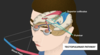
pain pathway
- spinal cord - reflex
- relays in the brainstem, esp in midbrain PAG
- neocortex
- ex: pain you feel
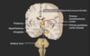
periaqueductal gray matter
- surround cerebral aqueduct
- prompt many complex responses to pain stimuli
- behavioral activation and emotional responses
- enduring pain that you feel long after touching a hot stove may be related to neural activity in the periaqueductal gray matter nuclei
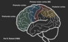
neocortex
- not only localize pain in a body part, but also identify the felt pain, its external cause, and possible remedies
- cortex can also adapt to our experience with hot stoves so that we know in advance not to touch one
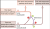
gating
with ex
- inhibition of sensory information produced by descending signals from the cortex
- the messages sensory systems carry can be modified at neural relays
- descending impulses from the cortex can block or amplify pain signals at the level of the brainstem and at the level of the spinal cord
- ex: when excited or playing a sport
- can also amplify sensory signal
- ex: when we think about the injury, it might feel much more painful because a descending signal from the brain now amplifies the pain signal from the spinal cord
- ex: attention: form of gating that takes place in the cortex, one that allows us to move efficiently from one action to another
- hierarchical code sent from sensory receptors, through neural relays, is interpreted in the brain, especially in the neocortex, and eventually translated into perception, memory, and action