chest ct Flashcards
(41 cards)
1
Q
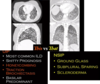
uip distribution
A
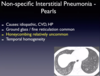
worse in lower lobes
2
Q

A
3
Q
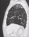
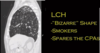
lung disorder with sparing of the costophrenic angles
A
PLCH
4
Q

A

5
Q
A
6
Q

A
7
Q

A

8
Q

A

9
Q
A
10
Q
smoker with cysts and nodules
A
pulmonary LCH
11
Q

A
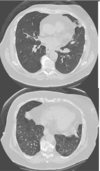
Pulmonary alveolar proteinosis
ground glass plus crazy paving
treatment = bronchoalveolar lavage
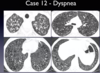
12
Q
middle aged woman with chronic mild SOB. No smoking history
expiratory HRCT images
A
Diffuse idiopathic neuroendocrine cell hyperplasia (DIPNECH)
multiple small pulmonary nodules and mosaic attenuating from air trapping due to constrictive bronchiolitis
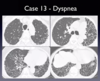
13
Q

A

14
Q

A
bronchiolar inflammation

15
Q
A
16
Q

A

17
Q

A

18
Q

A

19
Q

A
Ritalin lung can look like this too
20
Q
A

21
Q
A
22
Q

A
23
Q

A
24
Q

A

25

26

CF
27

lymphangitis carcinomatosis
stage IV cancer
28

acute HSP
29

pulmonary edema
big heart + ggos + pleural effusions + smooth septal thickening
30

bronchial atresia
"don't touch lesion"
31
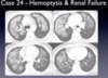
pulmonary hemorrhage 2/2 goodpasture's syndrome
32

septic emboli
peripheral nodules in different stages of cavitation
typically gram positive
33

achalasia
this degree of patulousness won't be seen in a pt with esophageal cancer
34

sarcoidosis
35

silicosis - progressive massive fibrosis
not eligible for lung transplantation
36

ABPA
37
18yo annorexic chick. Next step?

no further workup.
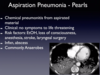
LLL has gross fat and represents lipoid pneumonia from chronic aspiration
38
56 yo persistent cough

mucinous adenocarcioma
39
82 yoM with cardiac history

amiodarone
hyperdense peripheral opacities
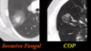
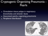
40

COP
41
76yF with cough and failed to impoved on two different ABX for PNA diagnosed on CXR.

Eosinophilic pneunomia
upper lobe, peripheral
