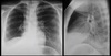
Chest pathology Flashcards
alveolar, interstitial and pneumonia (64 cards)
Define alveolar disease 2
filling alveolar spaces with abonormal material; blood , pus, water, protein, cell debris or combination.
Define interstitial disease 2m
effects supporting tissue of lung parenchyma, interstitium. . including the alveolar walls
State 5 visible features of alveolar disease 5m
- fluffly/blobby
- ill defined margins
- Coalescing/ merging
- segmental/ lobar
- sometimes - air bronchogram
state 6 visible appearences of interstitial disease 6
- Small Nodules
- linear/reticular
- linear/reticular with septal lines
- reticular nodular
- sometimes - reduced lung volume (extensive disease)
- Honeycomb pattern (End stage disease)
State 10 differential diagnosis for alveolar airspace patterns 7
- Pulmonary oedema - cardiac or non cardiac
- Lobar pneumonia
- Haemorrhage
- lymphoma
- Bronchoalveolar cell carcinoma
- Adult respiratory distress syndrome (early)
- Aspiration pneumonia
State 9 differental diagnosis for interstitial patterns
- Pulmonary oedema
- pneumonia - viral or pneumocytis carinii
- TB
- Sarcoid
- idiopathic pulmonary fibrosis
- Rheumatoid lung
- sclerodema
- lymphangitis carcinomatosa
- crack smoking
Sate how lobar pneumonia would illuatrate as chest shadows and the organism 6
- homogeneous throughout/ most of the lobe and may show air bronchogram or pleural effusion- streptococcus pneumoniae (pneumococcus)
- non segmental patchy but confined to one lobe- may swell or expand the effected lobe/ may cavitate - klebsiella pneumoniae
State the chest shadowing difference with bronchopneumonia
mild peribronchial thickening - ill-defined nodules but generall scattered and diffused.
how many segments of the rt and left lung? 1
rt 10
lt 9
State the complications associated with CVC misplacement?
State the age range responsible for 80% of sarcoidosis diagnosis? 1m
20-50
What radiographic appearences are there associated to coccidioidomycosis? + clinical symtoms 5m
inc time to develope
- Acute mild symtoms 1 -4 weeks after exposure
- Radiographically similar to Histoplasmosis
- single or multiple foci nodules
- or consolidation
- Hilar/ lymphone node involvement
- Pleural eff 20% of the time
- In chronic
- 5% cavitation
State acute clinical and radiographic appearences linked with acute histoplasmosis? 4m
- Asymtomatic or flu like symtoms
- Solitary/ multiple nodules + lymphadenopathjy in symtomatic
- Bilateral consolidation in symtomatic
State chronic clinical symtoms and radiographic appearences associated to histoplasmosis 3m (rare)
- Usually in emphysema patients
- Bilateral opper lobe opacities that extend into the plaura
- cavitation can develope
What is the differential diasnosis to histoplasmosis fungal infection? 1m
Coccidioidomyosis
Likely timeframe from exposure to symtoms for coccidioidomyosis? 1
1-4 weeks
Radiographic appearences for coccidiodomyosis? 4
- Single or multiple foci nodules
- consolidation
- Hilar / lymphadenopathy
- 20% pleural eff
- In chronic possible cavitation
List two of the five organisms that can cause aspergilliosis in humans? 2 m
- Saprophysic aspergilliosis
- Allergic bronchopulmonary aspergilliosis
Exposure to what things can cause aspergilliosis - not the organisms! 2m
Sand or decaying matter
List the constituents of fungus ball? 5m
fungal cells
fibrin
tissue debris
inflamatory cells
mucus
What is also known as a fungus ball? 1
Saprophytic aspergilliosis
What radiographic sign best hallmarks a saprophytic fungall ball? 1m
Air cresent sign around the fungal oval mass
Where does a fungal ball usually develope? 1m
Inside another cavity
Name the likely reaction to fungal antigen in patients with asthma or cystic fibrosis? 1m
Allergic bronchipulmonary aspergilliosis