Child with Breathing Difficulties Flashcards

1
Q

Definition of pneumonia?

A
  • Generally referring to community acquired pneumonia (CAP) as distinct from Hospital / Nosocomial acquired infection.
  • Pneumonia: a lung infection of the pulmonary parenchyma that affects the air sacs (alveoli) at the end of the airways.
  • The infection interferes with the delivery of oxygen from the air sacs into the blood and the removal of carbon dioxide from the blood.
2
Q

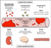
Pathophysiology of pneumococcal pneumonia?

A
  • Pneumococci enter the airway, the capsule protects the bacteria against phagocytosis by alveolar macrophages and multiple rapidly in the alveolar spaces and produce extensive oedema.
  • ‘RED HEPATISATION’:
    • Marked capillary congestion and outpouring of polymorphonuclear leukocytes with intra-alveolar haemorrhage.
  • ‘GREY HEPATISATION’
    • As the inflammatory process progresses, macrophages replace the PMN and ingest debris. The process usually resolves but complications may ensue.
    • The alveolar exudate is then removed and the lung gradually returns to normal. (Usually … but complications may ensue).
3
Q

Risk factors for pneumonia?

A
  • AGE; highest risk <5y also severe disease more likely in children <5y (OR 1.5, 95% CI 1.07 to 2.11)
  • GENDER; males had higher incidence rates at all ages.
  • INDIGENOUS BACKGROUND; 10-20 fold higher risk of hospitalisation compared to non-Indigenous children
  • Predisposing Condition:
    • MALNUTRITION
    • PREMATURITY; children born at 24-28 weeks more likely severe disease (OR 4.02 95%CI 1.2 to 13.9)2.
    • CHRONIC LUNG DISEASE; bronchopulmonary dysplasia | cystic fibrosis | bronchiectasis | primary ciliary dyskinesia
    • IMMUNODEFICIENCY
    • NEURODISABILITY
    • COMORBID INFECTION; measles | varicella | diarrhoeal illness
4
Q

Describe the microbiology of pneumonia.

A
  • Age is the most important factor to determine aetiology (<5yo viral is more common)
  • Viral pathogens more common in children than bacterial.
    • Predominant virus Respiratory Syncitial Virus (RSV).
    • Others include Parainfluenza, Human meta-pneumovirus, Influenza, Adenovirus, Coronavirus, Measles, Varicella.
  • Presence of a viral pathogen does not preclude a bacteria.
    • Coinfection common.
  • Bacteria: Streptococcus pneumoniae is the most common.
    • Disease varies by serotype; the serotypes most commonly associated with invasive pneumococcal disease (IPD) are; 1, 14, 6B, 19F and 23F.
  • Less frequent but important bacteria;
    • Haemophilus influenzae type b (HiB); uncommon in high income countries following vaccination. Vaccine studies from Bangladesh, Chile and the Gambia suggest causes 20% of severe pneumonia cases.
    • Streptococcus pyogenes, associated with rapid onset illness and more severe disease requiring ICU or empyema intervention. May be associated with toxic shock syndrome.
    • Staphylococcus aureus, more common in Indigenous communities, more common in infants, associated post viral infections particularly following influenza and varicella, higher rates of necrotising pneumoniae and bronchopleural fistulae. Panton Valentine Leukocidin (endotoxin made by some staph which causes holes in cells)
  • Atypical bacteria: named so around radiological appearance. These include Chlamydia pneumoniae and more commonly Mycoplasma pneumoniae.
    • These are more common in school age but not unusual in <5y.
    • Consider Bordetella pertussis as important pathogen in infants/preschool age.
5
Q

Describe the differences in causative agents between children <5yo and >5yo

A
6
Q

What primary prevention/intervention is available for pneumonia?

A
  • Adequate Nutrition: Undernourished children are at substantially higher risk of severe disease. Zinc . Low protein state impairs immune system | Weakened respiratory muscles impair secretion clearance.
  • Breastfeeding: exclusive breastfeeding associated with fewer and less severe infections. Infants <6 months have x5 increased risk of pneumonia death c/w breastfed infants.
  • Immunisation : direct and indirect benefits (Pneumococcus | HiB | Measles | Pertussis | Varicella)
7
Q

Clinical features for pneumonia?

A
  • Fever: abrupt onset ± rigors
  • Cough: may have rusty sputum
  • Chest Pain
  • Abdominal Pain
  • Tachypnoea
  • Hypoxia
  • Headache
  • Arthralgia
8
Q

Examination findings for pneumonia?

A
  • Inspection: Cyanosis, Signs of increased work of breathing
  • Asymmetric chest expansion occurs in large pleural effusions.
  • Tactile fremitus: palpable vibrations transmitted through the lungs to the chest wall. In health these are symmetric. With pleural effusion or pneumothorax this is decreased. With consolisation this is increased.
  • Percussion: helps examiner establish whether the underlying tissues are air-filled, fluid filled or consolidated.
  • Auscultation: symmetry and quality of air entry, Bronchial breath sounds: louder | harsher | higher pitch. Inspiratory crackles, Pleural rub.
9
Q

What were the strongest indicators of pneumonia on clinical assessment?

A
  • No single symptom was strongly associated with pneumonia.
  • The strongest associations between clinical features and radiographic pneumonia;
    • Chest Pain
    • Fever (temp > 37.5°C)
    • Tachypnoea
    • Hypoxaemia (SaO2 ≤96%)
    • Increased work of breathing
  • The presence of wheeze and / or normal oxygen saturation (SaO2 > 96%) decreased the likelihood of radiographic pneumonia.
10
Q

Should CXR be considered routine when assessing for pneumonia?

A
  • No. Only order when it will change management
11
Q

What are the indications for a CXR?

A
  • Patients with suspected complicated pneumonia SHOULD have CXR
  • Recommend PA and Lateral CXR for:
    • Hypoxaemic patients
    • Patients whom have failed oral Abx
  • Follow up if recurrent lobar involvement | complicated pneumonia.
12
Q

Why does pneumonia produce CXR changes?

A
  • Most pneumonias produce airspace disease, either lobar or segmental.
    • The alveoli fill with fluid / exudate and appear denser (whiter) than the surrounding normally aerated lung.
    • May contain ‘air-bronchograms’ if the bronchi themselves are not filled with fluid.
    • Except for the presence of air-bronchograms, airspace pneumonia is usually homogenous in density,
    • Where pneumonia abuts a pleural surface (fissure or chest wall) it will be sharply marginated.
  • Describe:
    • Size | Site
    • Character of parenchymal infiltrate
    • Presence of effusion
    • Presence of air/fluid level
13
Q

What is round pneumonia?

A
  • More common in children <8y (but 15% occur between 8- 12).
  • Poorly formed pores of Kohn which allow collateral ventilation between alveolar units.
    • Infection is localised to developed connection channels
  • This leads to an advancing front of inflammatory change sharply demarcated against unaffected lung parenchyma causing a focal round mass.
  • Most resolve without progression to lobar pneumonia.
  • Most are posteriorly located in lower lobes.
  • May be confused as posterior mediastinal mass, this can be differentiated using US.
14
Q

Describe interstitial (atypical) pneumonia

A
  • Most commonly associated with viral pneumonia and Mycoplasma pneumoniae as well as Pneumocystis pneumonia in patients with AIDS.
  • Early changes: tends to involve airway walls and alveolar septa giving fine reticular pattern in the lungs. (may mimic appearance of pulmonary oedema).
  • Progression to adjacent alveoli may produce patchy or confluent airspace disease.
  • Typically bilateral
15
Q

Match patterns of radiological disease with most likely causative organism

A
16
Q

Which pathogen will most likely cause upper lobe cavitary pneumonia with spread to the opposite lower lobe?

A

Mycobacterium tuberculosis

17
Q

Which pathogen will most likely cause upper lobe lobar pneumonia with bulging interlobar fissure?

A

Klebsiella pneumoniae

18
Q

Which pathogen will most likely cause lower lobe cavitary pneumonia?

A

Pseudomonas aeruginosa or anaerobic organisms (Bacteroides)

19
Q

Which pathogen will most likely cause perihilar interstitial disease or perihilar airspace disease?

A

Pneumocystis carinii (jiroveci)

20
Q

Which pathogen will most likely cause thin-walled upper lobe cavity?

A

Coccidioides (Coccidiomycosis) or TB

21
Q

Which pathogen will most likely cause airspace disease with effusion?

A

Streptococci, staphylococci, TB

22
Q

Which pathogen will most likely cause diffuse nodules?

A

Histoplasma, Coccidioides, Mycobacterium tuberculosis (histoplasmosis, coccidiomycosis, TB)

23
Q

Which pathogen will most likely cause soft tissue, finger-like shadows in upper lobes?

A

Aspergillus (Allergic bronchopulmonary aspergillosis)

24
Q

Which pathogen will most likely cause solitary pulmonary nodule?

A

Cryptococcus (Cryptococcosis)

25
Q

Which pathogen will most likely cause spherical soft-tissue mass in a thin-walled upper lobe cavity?

A

Aspergillus (Aspergilloma)

26
Q

What is the silhouette sign?

A
  • The Silhouette sign: if two objects of the same radiographic density touch each other then the edge between them disappears.
  • Can be valuable in localizing disease.
  • Structure that is no longer visible and disease location
    • Ascending aorta: right upper lobe
    • Right heart border: right middle lobe
    • Right hemidiaphragm: right lower lobe
    • Descending aorta: Left upper or lower lobe
    • Left heart border: Lingula of left upper lobe
    • Left hemidiaphragm: left lower lobe
27
Q

What are the key aspects of pneumonia management?

A
  • Determine Severity
  • Antibiotic Treatment
  • Supportive care
  • Assessment / Monitoring of complications
  • Follow up
28
Q

What are factors indicating severe pneumonia?

A
  • Temp > 38.5C
  • Tachypnoea
    • >70 in infants
    • >50 in older children
  • Severe breathing difficulty
  • Not feeding in infants
  • Nasal flaring
  • Cyanosis
  • Apnoea
  • Grunting
  • Tachycardia
  • Signs of dehydration
  • Cap refill >2s
29
Q

What is the antibiotic therapy for pneumonia?

A
  • Amoxicillin recommended as 1st line, by BTS and IDSA.
  • Addition of a macrolide at 48h if there is no improvement. Macrolide is 1st line in a child with penicillin allergy.
  • Penicillin resistance to S.pneumoniae uncommon in UK and AUS. Higher rates in mainland Europe, South Africa.
  • Treatment duration generally for 7-10 days.
  • Australian eTG suggests; if good clinical improvement by day 3 treat for 5 days, if slower response then to continue for 7 days. Except for Azithromycin where treatment is 5 days.
30
Q

How do you manage severe pneumonia?

A
  • Management priorities:
    • A| B | C | D
    • Consider differential diagnoses
    • Appropriate Investigation; CXR | Blood Culture | EUC | VBG
  • Pneumonia:
    • Supportive treatments
    • Parenteral Antibiotics
    • Third generation cephalosporin. (If penicillin allergy ciprofloxacin or moxifloxacin)
    • Antistaphylococcal : clindamycin | lincomycin | vancomycin.
    • Consider Oseltamivir (‘Tamiflu’)
31
Q

Describe the antibiotic choice algorithm for pneumonia.

A
32
Q

What are suggested drug treatments for severe community acquired pneumonia in developed countries?

A
33
Q

What are some complications of pneumonia?

A
  • Pulmonary
    • Pleural effusion
    • Empyema
    • Necrotising pneumonia
    • Bronchopleural fistulae
    • Lung abscess
    • Bronchiectasis
    • Respiratory Failure
  • Extrapulmonary
    • Sepsis
    • SIADH; Syndrome of Inappropriate Anti-Diuretic Hormone
    • HUS; Haemolytic Uraemic Syndrome
34
Q

Describe empyema (definition, most commonly found in which group, most common pathogens)

A
  • Definition: presence of purulent fluid in the pleural space.
  • The most common complication of paediatric CAP; estimated prevalence 2-7% of hospitalised patients.
  • Most common in preschool age children; peak incidence in <4yr age group
  • Most common pathogens;
    • S.pneumoniae: the most common,
    • S.pyogenes: patients more often present with severe disease, high rates of ICU admission, consider associated toxic shock syndrome.
    • S.aureus: more common in infants, indigenous patients, associated with necrotising pneumonia and bronchopleural fistulae.
35
Q

When to suspect empyema?

A
  • Persisting clinical symptoms despite appropriate antibiotic treatment
  • Pleuritic chest or abdominal pain | Prolonged disease course | Clinical examination suggestive of effusion
  • CXR | Chest US
36
Q

How do you manage an empyema?

A
  • Treatment aims;
    • i). remove infected fluid (may also identify pathogen) and sterilisation of the pleural cavity.
    • ii). to prevent organising collection impeding lung expansion ‘Trapped Lung’
  • Treatment;
    • Prolonged antibiotic treatment
    • Either;
      • Pleural drain with fibrinolytics
      • Video assisted thoracoscopic surgery (VATS)
37
Q

Describe necrotising pneumonia (definition, most common pathogens, potential complications)

A
  • Pathological term describing the formation of abscess and cavitation within the lung parenchyma
    • Liquefactive necrosis of lung parenchyma with destruction of the normal lung architecture. Liquifaction transitions to cavitation.
  • Most cases are restricted to a single lobe.
    • Lower lobar involvement is more common.
  • As for empyema, rates of necrotising pneumonia have been increasing during the last 20 years.
  • May be complicated by intrapulmonary abscesses or bronchopleural fistulae (especially when lobar segment is peripheral).
  • Most common pathogens associated are S.pneumoniae, S.aureus (PVL) or less commonly with Pseudomonas aeruginosa.
  • Despite causing severe lung damage, necrotising pneumonia generally fully resolves with excellent long term outcomes
38
Q

When to suspect necrotising pneumonia?

A
  • Persisting clinical symptoms despite appropriate antibiotic treatment or presentation with severe pneumonia.
  • Respiratory distress | haemoptysis | sepsis. Frequently coexist with empyema.
  • Suspected by CXR demonstrating liquid or gas filled caviatations.
  • CT imaging is the preferred modality;
39
Q

Management of necrotising pneumonia?

A
  • HRCT (with contrast) imaging: loss of normal lung architecture | decreased areas of parenchymal enhancement | multiple small air or fluid filled cavities.
  • Prolonged antibiotics
  • Consider image guided drainage
40
Q

Describe haemolytic uraemic syndrome (pathophys)

A
  • Haemolytic Uraemic Syndrome; is the most common cause of acute kidney injury in children
  • Destruction of erythrocytes as a result of 2 mechanisms;
    • 1) mechanical haemolysis as a result of narrowed microvasculature caused by microthrombi.
    • 2) complement mediated opsonisation. With pneumococcus there is a removal of sialic acid from vascular endothelial cell surfaces with a procoagulant effect.
  • S.pneumonia is a leading cause thought to represent 5%-15% of all cases.
41
Q

What are the lab and clinical findings for haemolytic uraemic syndrome?

A
42
Q

When to suspect haemolytic uraemic syndrome and how to treat?

A
  • Pallor, hypertension and reduced urine output
  • Diagnostic criteria;
    • Microangiopathic haemolytic anaemia (Hb< 100g/L) with fragmented erythrocytes,
    • Thrombocytopenia (PLT< 130x109/L)
    • Acute renal impairment; (elevated creatinine and oliguria) -Proven or suspected pneumococcal infection.
  • Rx: Renal replacement | Antihypertensives
43
Q

Should clindamycin be added for severe pneumonia?

A

Yes. Clindamycin has anti-toxin properties and should be added in any septic child with pneumonia or severe pneumonia not responding to first line treatments

44
Q

How do you initially assess asthma exacerbation severity?

A
45
Q

What is the initial management of asthma exacerbation?

A
46
Q

Aside from salbutamol, what other medications can be used in the initial management of asthma exacerbation?

A
47
Q

What investigations and observations can be considered for asthma exacerbations?

A
48
Q

What medications and doses are used in acute asthma?

A
  • Salbutamol inhaled
    • < 6yrs 6 puffs/2.5mg neb.; > 6yrs12 puffs/5mg neb
  • Salbutamol neb continuous
    • < 6yrs 2x2.5mg neb; > 6yrs12 2x5mg neb
  • Salbutamol intravenous
    • commence at 5mcg/kg/min (max. 50kg) then titrate1-5mcg/kg/min
  • Steroids
    • 1-2mg/kg initial dose (max50mg), 1mg/kg/day oral prednisolone daily for 3 -5 days
    • 1mg/kg IV methylprednislone (or 4mg/kg hydrocort.) q4-6h
  • I.V Magnesium sulphate
    • Bolus: 5–54 mg/kg over 20–30 min
  • Aminophylline (IV)
    • 5mg/kg over 30 mins; 1.0mg/kg/hr
49
Q

In acute asthma, is there a difference between spacer (holding chamber) and nebulised salutamol?

A

No difference in outcomes except in severe cases

50
Q

What is the evidence for oral steroids in acute asthma?

A
  • OCS effective
    • Children hospitalised for asthma
    • ED treatment of acute asthma
    • 3 days = 5 days for ED presentations going home
  • OCS not effective
    • ED treatment of pre-schoolers with viral induced wheezing
  • PIOCS effective for 6-14 year olds with acute asthma
  • PIOCS not effective for childhood intermittent wheezing
51
Q

What is the evidence for Intermittent Inhaled Corticosteroids (ICS) vs LTRA for Acute Asthma?

A
  • High Dose ICS
    • marginal benefit
  • Intermittent LTRA
    • reduced ED visits & HC utilisation and improved QOL
  • 5 days LTRA vs prednisolone in 2-17 year old children with mild-moderate acute asthma
    • treatment failure pred 7.9% vs mont 22.4%
    • oral pred has better effect than montelukast
52
Q

What is the discharge criteria for acute asthma?

A
  • Children with acute asthma be considered ready for discharge when clinically stable on 3rd hourly bronchodilator.
  • Oximetry should not be used as the primary criteria for discharge as it remains unclear what is a “satisfactory” level.
  • Involvement of both nursing and medical staff in management decisions is likely to speed up the discharge process
  • At time of discharge all patients/parents should receive:
    • (a) Discharge Summary
    • (b) Asthma Management Plan (with post discharge plan)
    • (c) Discharge Medications
    • (d) Follow up Arrangements
  • During admission or consultation:
    • Check patient’s/parents’ asthma knowledge and inhaler technique
    • Provide additional education as required
    • Reassess asthma control and prescribe or adjust preventer if required
53
Q

Which asthma devices are appropriate for different ages?

A
  • Pressurised Metered Dose Inhaler (pMDI/ puffer) with spacer
    • Small volume spacer ( with face mask) < 4 years / school age
    • Small & or Large volume spacer (without face mask) > 4 years/school age
    • Some school age children who may require a face mask eg: if struggling with maintain the seal around the mouthpiece due to illness or developing technquie
  • Puffer: > 8 years Cipla Puffer: > 12 years and or Autohaler: > 7 years
  • Dry Powder Inhalers(DPI):
    • Accuhaler > 8 years
    • Turbuhaler > 12 years
    • Ellipta > 12 years
  • Nebuliser: Can be used across all age ranges
54
Q

What are some common errors with puffers and spacers?

A
  • Puffers: lack of hand-breath coordination by not exhaling before placing device in the mouth, inhaling too fast, not taking a deep enough breath
  • Spacers: loading more than 1 puff medication at time, not taking enough breaths per medication puff, breathing to quickly in/out spacer, blocked spacer valve/ spacer thickly coated with medication
55
Q

What are some barriers to good technique with asthma medications?

A
  • Nil & or minimal explanation/education
  • Cost and access
  • Lack of understanding/confidence, NESB and or literacy difficulties
  • Firm beliefs in what device work best for their child or have differing opinions from clinicians eg: spacers vs. nebulisers
  • Unable to engage families &/or child in an education intervention
56
Q

Do you have an approach to check asthma device technique?

A
  • Tailor: Individualise the device
  • Educate: Educate and demonstrate correct technquie
  • Assess: Ability for using the device
  • Correct: Any issues with technique
  • Have another go: After correction to reassess
  • Evaluate: Is this the correct device
  • Reduce: Minimize the number of devices/medications prescribed
  • Age and Adherence
  • Body image
  • Cost and Current trends
  • Device aids eg: Puffer Haleraid or Turbuhaler Aid
57
Q

Advantages of a puffer?

A
  • Placebo trainer
  • Size/portable
  • Available reliever & preventer
  • Attach haleraid for both standard reliever & preventer
  • Wash in soapy water/air dry as needed
58
Q

Disadvantages of a puffer?

A
  • Poor coordination
  • Post inhalation cough
  • Dose counters on Seretide puffer , Flixotide & Seretide Cipla puffers
  • Intal Forte casing washed daily to avoid clogging
59
Q

Advantages of spacers?

A
  • Placebo trainer
  • As effective as nebuliser
  • Reduce potential side effects
  • Reduced co-ordination
  • Portable/ Independence
  • Inexpensive (as compared to nebuliser)
  • Used with reliever & preventer puffers
  • Used at childcare, school, sporting settings
60
Q

How do you clean and prime a spacer?

A
  • Cleaning
    • At home washed in warm soapy water/ air dry
    • Wash regularly ( app. 2 - 4 weeks) this depends on use
    • Note: Intal Forte clogs up the spacer, should be washed once per week
  • Priming
    • If not washed before first use: prime spacer with salbutamol
    • Pre-primed spacer eg: e- chamber range, Lite Aire, Breath eazy, Able spacer 2
61
Q

Disadvantages of spacers?

A
  • Size large volume spacer
  • Static charge/ not cleaning correctly/missing or damaged valve
  • Face mask can absorb about 20% of the medication so it important:
    • Mask gives a good seal, cover from top of nose to chin
    • Different styles and sizes available so ensure that the mask fits the spacer and child
62
Q

Advantages of an autohaler?

A
  • Breath activated, less coordination than a puffer
  • Alternative to using puffer alone
  • Available as preventer(consider about technquie & potential oral side effects )
63
Q

Disadvantages of an autohaler?

A
  • Lever can be difficult to pull into position for younger children
  • Children stop inhaling after “puff” sound OR may not be able to inhale enough to trigger the medication release esp. during an asthma flare-up
  • No dose counter
  • More expensive than standard salbutamol puffer
64
Q

Advantages of a turbuhaler?

A
  • Size/portable
  • Dose counter on Symbicort
  • Dose indication window on Bricanyl & Pulimcort
  • Turbuhaler aid & placebo trainer
  • Cleaning: wipe mouthpiece as needed
65
Q

Disadvantages of turbuhaler?

A
  • Device must be upright for loading
  • May be difficult to use in asthma flare-up
  • Post inhalation cough
  • Mistaking the sound drying agent as active medication
66
Q

Advantages of the accuhaler?

A
  • Placebo trainer
  • Size
  • Dose counter
  • Available as preventer & symptom controller( in paediatrics not recommended to use symptom controller as a stand alone medication)
  • Cleaning: wipe mouthpiece as needed
67
Q

Disadvantages of accuhaler?

A
  • Post inhalation cough
  • Medication back of mouth and throat
68
Q

Advantages of nebuliser?

A
  • Minimal coordination
  • Multiple medication delivery at once( pre measured vials)
  • Face mask & or mouthpiece
  • Cleaning: Wash face mask/mouthpiece as needed
69
Q

Disadvantages of nebuliser?

A
  • Time consuming, expensive, still nebulisers need electrical access, modern neb that chargeable via USB ports
  • Air driven device and easily mistaken to deliver oxygen
  • Service/ filter change 6 to 12 months( depending on use/age of device)
  • Dose delivered depends on flow rate & nebuliser efficiency
70
Q

What is the definition of asthma?

A
  • Asthma is a heterogeneous disease, usually characterised by chronic airway inflammation. It is defined by the history of respiratory symptoms such as wheeze, shortness of breath, chest tightness and cough that vary over time and in intensity, together with variable airflow limitation
  • Wheeze, shortness of breath, chest tightness and/or cough:
    • More than 1 symptom
    • That vary over time (worse at night)
    • That vary in intensity
    • Symptoms often worse at night or on waking
    • Symptoms often triggered by exercise, laughter, allergens, cold air
    • Symptoms often appear or worsen with viral infections
  • Variable expiratory airflow limitation
  • Aspecific and allergenic triggers
  • Associated with AHR and chronic airway inflammation
71
Q

What are the 3 patterns of asthma?

A
  • Infrequent intermittent
  • Frequent intermittent
  • Persistent
    • Mild
    • Moderate
    • Severe
72
Q

What increases the probabiltiy of asthma over viral induced wheeze?

A
  • Age over 5yo
  • Symptoms lasting for longer than during viral induced wheeze
  • Triggers becoming more non-viral
73
Q

How do you assess asthma control?

A
  • In the past 4 weeks, has the patient had
    • Daytime asthma symptoms more than twice a week
    • Any night waking due to asthma
    • Reliever needed for symptoms more than twice a week
    • Any activity limitation due to asthma?
  • Level of asthma symptom control
    • Well controlled: 0
    • Partly controlled: 1-2
    • Uncontrolled: 3-4
74
Q

In children under 5yo, what are the risk factors for asthma exacerbation within the next few months?

A
  • Uncontrolled asthma symptoms
  • One or more severe asthma exacerbations in the previous year
  • Start of the child’s usual flare-up season (esp if Autumn)
  • Exposures: tobacco smoke indoor or outdoor, air pollution, indoor allergens (e.g. house dust mite, cockroach, pets, mold), esp in combination with viral infection
  • Major psychological or socioeconomic problems for child or family
  • Poor adherence with controller medication, or incorrect inhaler technique
75
Q

In children under 5yo, what are the risk factors for fixed airflow limitation?

A
  • Severe asthma with several hospitalisations
  • History of bronchiolitis
76
Q

In children under 5yo, what are the risk factors for medication side effects?

A
  • Systemic:
    • frequent courses of oral corticosteroids;
    • high dose and/or potent inhaled corticosteroids
  • Local:
    • moderate/high dose or potent inhaled corticosteroids;
    • incorrect inhaler technique;
    • failure to protect skin or eyes when using inhaled corticosteroids by nebuliser or spacer with face mask
77
Q

What is the stepwise approach to management for asthma control?

A
  • Preferred controller choice
    • Step 1: Nil (reliever PRN)
    • Step 2: Low dose ICS
    • Step 3: Low dose ICS/LABA (for 6-11yo, prefer medium dose ICS)
    • Step 4: Med/high dose ICS/LABA
    • Step 5: Specialist referral
  • Other controller options
    • Step 1: Consider low dose ICS
    • Step 2: Leukotriene receptor antagonist, low dose theophylline
    • Step 3: Med/high dose ICS; Low dose ICS + LRTA (or + theophylline)
    • Step 4: Add tiotropium; Med/high dose ICS + LTRA (or + theoph*)
    • Step 5: Add lose dose OCS
  • Reliever
    • PRN saba - Step 1 and 2
    • PRN saba or low dose ICS/formoterol - Step 3-5
78
Q

What is the stepwise management approach to asthma control in children <5yo?

A
79
Q

How would you distinguish between frequent intermittent and infrequent intermittent?

A
  • Frequent - episodes within every 6 weeks
  • Infrequent - episodes more than 6 weeks apart
80
Q

Are leukotriene receptor antagonists better than ICS for exercise induced asthma?

A

Yes

81
Q

Differentials for breathing difficulties or other than asthma?

A
  • Conditions characterised by cough
    • Pertussis (whooping cough)
    • Cystic fibrosis
    • Airway abnormalities (e.g. tracheomalacia, bronchomalacia)
    • Protracted bacterial bronchitis in young children
    • Habit-cough syndrome
  • Conditions characterised by wheezing
    • Upper airway dysfunction
    • Inhaled foreign body causing partial airway obstruction
    • Tracheomalacia
  • Conditions characterised by difficulty breathing
    • Hyperventilation
    • Anxiety
    • Breathlessness on exertion due to poor cardiopulmonary fitness
82
Q
A