Chronic inflammation Flashcards

1
Q

chronic inflammatory conditions are

A

incredibly common - not usually life threatening - debilitating (IBD and RA)

2
Q

definition

A

prolonged inflammation with associated repair

3
Q

characterised by

A
  • Delayed onset - Variable duration (days years) - Variable appearances • No 5 cardinal features - Limits damage, initiates repair - Can cause debilitating symptoms
4
Q

how does chronic inflammation arise

A
  1. Takes over from acute inflammation- if resolution not possible with acute inflammation
  2. Develops alongside acute inflammation- severe/persistent irritation
  3. Arises “de novo”- without preceding acute inflammation e.g. autoimmune conditions - Rheumatoid arthritis/ IBD/ diabetes
5
Q

sometimes the proportion of cell types can

A

indicate diagnosis

6
Q

which cells are indicative of rheumatoid arthritis

A

mainly plasma cells

7
Q

which cells are indicative of chronic gastritis

A

mainly lymphocytes

8
Q

which cells are indicative of of leishmaniasis (protozoal infections)

A

mainly macrophages

9
Q

which cells are involved in chronic inflammation

A
  1. Macrophages 2. Lymphocytes- T/B cells 3. Mast cells 4. Eosinophils 5. Fibroblasts 6. Giant cells
10
Q

macrophages in circulation

A

monocytes

11
Q

macrophages once entering tissue space

A

macrophage (histiocyte)

12
Q

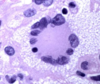
features of macrophages

A
  • big cells with a large cytoplasm - abundant foamy cytoplasm - slipper shaped nucleus - can look like a cancer cell
13
Q

primary role of maxcrophage

A

phagocytosis - removal of pathogens/ necrosis/ debris - antigen presentation to immune system

14
Q

secondary role of macrophages

A
  • Also produce inflammatory mediators- controls and regulates inflammatory response
15
Q

macrophages can look different depending on

A

what stimulus they are eating
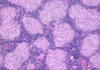
16
Q

lymphocyte features

A
  • large, spherical stained nucleus
  • thin rim of cytoplasm
  • small

subdivided into T and B lymphocytes (cant distinguish appearance- have to use immunohistochemistry)

17
Q

T lymphocytes

A

CD8+ (protein on surface)- Cytotoxic (MHC I)

CD4+- helper T cells (MHC II)

18
Q

B lymphocyte

A

Mature into plasma cells

Produce antibodies (immunoglobulins)

Neutralises pathogens

19
Q

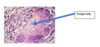
plasma cell features

A
  • Nucleus pushed off to one side (eccentric)
  • Chromatin in the nucleus clumps into spheres (clock-face)
  • Next to the nucleus there is slightly paler staining- peri- nuclear cytoplasmic clearing –> Golgi due to antibody synthesis
20
Q

function of plasma cells

A

fully differentiated B lymphocytes

Produce antibodies

21
Q

features of eosinophils

A
  • 2 lobes (bilobed nucleus)
  • cytolasm stains bright red and granular
22
Q

why is the cytoplasm of an eosinophil bright red and granuklar

A
  • Full of chemical mediators- Histamine, heparin and prostaglandins
  • E.g. Release during hypersensitivity reactions and parasitic infections
23
Q

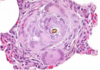
features of fibroblasts/myofibroblasts

A
  • webbed cytoplasm
  • stretched nucleus
24
Q

role of fibroblast/myofibroblast

A
  • ‘Prolonged inflammation with associated repair’
  • Role in generation and repair
  • Produce and secrete and lay down collagen- helping to reconstruct tissue
25
Q

giant cells features

A
  • Multinucleate
  • HUGE
  • Fusion of multiple macrophages
26
Q

role of giant cells

A
  • “Frustrated phagocytosis”
  • Increasing effectiveness of phagocytosis
  • E.g. if there are any resistant foreign body e.g. suture, resistant parasite of bacteria
27
Q

how many types of giant cell

A

3

28
Q

name the 3 giant cells

A

1) Foreign body giant cells
2) Langhans giant cells
3) Touton giant cells

29
Q

1) Foreign body giant cells

A

To destroy foreign bodies

One big cytoplasm

Nuclei randomly assorted

30
Q

2) Langhans giant cells

A
  • Nuclei line up around the periphery- horseshoe
  • Important in TB
31
Q

3) Touton giant cell

A
  • Nuclei line up in a concentric circle in the middle of the cell
  • Can be seen in fat necrosis
32
Q
A
33
Q

which giant cell associated with TB

A

langhans giant cell

34
Q

which giant cell associated with fat necrosis

A

touton

35
Q

which type of giant cell

A
36
Q

which type of giant cell

A

Langhans

37
Q

which type of giant cell

A

Touton

38
Q

Effect of chronic inflammation

A
  1. fibrosis
  2. impaired function
  3. atrophy
  4. stimulation of immune response
39
Q
  1. fibrosis
A
  • a.Deposition of collagen
  • b.E.g. chronic cholecystitis, liver cirrhosis
40
Q

impaired function

A

a. E,g, IBD
b. Rarely increased function (e.g. thyrotoxicosis in Graves disease- increase production of T3/T4)

41
Q

atrophy

A

atrophic gastritis

42
Q

stimulation of immune repsosne

A

Antigen presentation- chronic –> brought about by macrophages

43
Q

fibrosis of the gall bladder

A

thickened and pale- collagen

44
Q

impaired functione xample

A

idiopathic IBD

  • Crohns disease
  • UC
45
Q

crohns disease

A
  • can affect all of the GI tract (mouth to anus)
  • discontinous patches of inflammation (skip lesions)
  • inflammation affects full thickness of the bowel (transmural)
  • sometimes find granulomata
  • less likely to have rectal bleeding
46
Q

ulcerative colitis

A
  • affects large boewl only
  • continous inflammation
  • inflammation affects superficial bowel wall only (mucosa and submucosa only)
  • no granulomata
  • more likely to have rectal bleeding
47
Q

cirrhosis

A

causes impaired function- too much fibrosis and attempted regeneration

e. g. Cirrhoic liver
- alcohol
- hepatitis
- drugs and toxins
- fatty liver disease

48
Q

granulomatous inflammation found in

A

chronic inflammation

49
Q

granuloma form

A

A collection of epithelioid histiocytes (macrophages) with surrounding lymphocytes

May also see giant cells within granuloma

50
Q

causes of granulomatous inflammation

A
  1. forgin body reaction
  2. infection
  3. idiopathic
51
Q

foreign body granulomatous inflammation

A
52
Q

granuloma caused by infection

A

e. g. tuberculosis
e. g. leprae
- dififcult to destory
- thick cell walls
- mycolic acids

53
Q

idiopathic granuloma

A

e. g. crohns disease
e. g. sacroidosis (mulitple well formed granulomas form on lymph nodes, lungs and skin - shortness of breath, lumps in lymph and skin lumps)