circulatory disorders part 1 Flashcards
hydrostatic pressure
pressure driving blood into capillaries (blood pressure)
oncotic pressure
protein pressure, moves fluid into vessels
inflammatory edema
increased vascular permeability
exudate
non inflammatory edema
transudate
ie liver failure, CHF

edema
wet, gelatinous/heavy, swollen organs,fluid weeps from cut surfaces, may be yellow

histological appearance of edema
clear or pale eosinophilic staining depending on whether it is non inflammatory or inflammatory edeam.
spaces are distended
blood vessels may be filled with red blood cells
lymphatics are dilated
collagen bundles are separted.
only look at image of gelding

pitting edema
pressure is applied to an area of edema a depression or dent results as result of excessive intersitial fluid that is forced into adjacent areas

hydrothorax
fluid in thoracic cavity

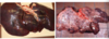
pericardia effusion
mulberry heart disease (inflammatory edema).
note fibrin strands and cloudy appearance of the pericardial fluid

ascites or hydroperitoneum
fluid (transudate) within peritoneal cavity
dog with CHF

ascites
horse with CHF

anasarca
generalized edema with profuse. accumulation of fluid within subcutaneous tissue

submandibular edema (bottle jaw)
commonly associated with severe GI parasitism and hypoproteinemia in sheep

protein losing enteropathy
horse forelimb, this animal has generalized edema
clinical significance of edema
dependent upon: extent location and duration
tissue may become firm and distorted due to an increase in fibrous connective tissue after prolonged edema
pulmonary edema
inflamm and non inflammatory
non inflammatory edema: associated to left-sided congestion heart failure
inflammatory edema: damage to pulmonary capillary endothelium (pneumonia or acute respiratory distress syndrome)

pulmonary edema, pig

pulmonary edema
chronic pulmonary edema
most commonly associated with cardiac failure
alveolar walls become thickened–> may lead to fibrosis
congestion, micro-hemorrhages –> accumulation of heart failure cells
hyperemia and congestion
bother terms indicate local increase in blood volume and flow within vascular bed
hyperemia: increase of arteriole mediated engorgment of vascular bed. blood is oxygenated (red)
congestion indicates passive, venous engorgment. blood is not oxygenated (blue)
types of hyperemia
physiological hyperemia: digestion, exercise, dissipate heat, neurovascular (red cheeks)
pathological hyperemia: usually caused by inflammation

gastric volvulus in dog

intestinal volvulus in horse

pulmonary congestion
usually result of heart failure and associated with edema