CN IV Flashcards
(28 cards)
describe the functional components of CN VII
- SVE = branchiomotor to muscles of facial expression, etc
- SVA = TASTE from anterior 2/3 of tongue
- GVE = parasympathetic to glands
- GSA = general sensation from skin in posterior ear, pinna, external auditory meatus
- GVA = visceral sensation from nasal cavity,s oft palate, adjacent phayngeal wall
- Carries its own MOTOR, PARASYMPATHETIC and SENSORY FIBERS
What does the facial nerve proper contains
- Consists of axons of nerve cells whose cell bodies are located in facial (motor) nucleus
- Motor root, SVE
what are the contents of nervus intermedius root
- central proceses of SVA, GSA and GVA neurons whose cell bodies reside in the geniculate ganglion (general and visceral sensation and Taste)
- GVE fibers arising from superior salivatory nucleus (parasymapthetic)
descrive geniculate ganglion
- Sensory ganglion
- contains cell bodies of PSEUDOUNIPOLAR NEURONS (GVA, SVA, GSA)
- NO synpases
describe pterygopalatine ganglion
- contains cell bodies of POSTGANGLIONIC Parasympathetics (GVE) neurons whose axons terminate in the:
–> lacrimal gland = tear formation - provides moisture and cleans eye
–> nasal mucous membrane = nasal secretions - moistrue to nasal mucosa
–> Minor salivary glands in palate = salivary secretion
**Alligator tears - phenomenon during eating
describe submandibular ganglion
- contains cell bodies of postganglionic parasympathetics (GVE) neurons whose axons termiante in the:
–> submandibular gland (salivation)
–> sublingual gland (salivation)
define facial nucleus
- Motor nuclei
- Contains cell bodies of motor neurons (LMN’s)
Define superior salivatory nucleus
- Parasymapthetic nuclei
- contains the cell bodies of PREGANGLIONIC parasympathetic neurons whose axons synapse in pterygopalatine or submandibular ganglia
define solitary nucleus
- sensory nuclei
- facial nerve shares this nucleus with glossopharyngeal and vagus nerves
- receives the central processes of SVA (Taste) and GVA neurons (visceral sensation)
describe the solitary tract
- carries central processes of SVA and GVA neurons to solitary nucleus
- solitary nucleus proceses taste sensation and projects to the hypothalamus which mediates the visceral respones to unpleasant sensation (vomiting)
Desribe spinal tract of the trigeminal nerve
- carries central proceses of GSA neurons to spinal nucleus of trigeminal nerve
- facial nerve DOESN’T have a pain nucleus so it sends it nociceptive fibers to SPINAL TRACT OF V that terminates in the pain nucleus of V
What type of fibers does the upper half of facial motor nucleus receive
- Receives BILATERAL CORTICONUCLEAR projectons (UMN projections)

What type of fibers does the lower half of facial motor nucleus receive
- receives ONLY CONTRALATERAL CORTICONUCLEAR projections (UMN projections
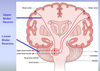
describe the pattern of innervation of the muslces of facial expression by LMN of facial motor nucleus
- LMN whose cell bodies reside in upper half of facial motor nucleus, innervate muscles of upper half of face
- LMN whose cell bodies reside in LOWER HALF of facial motor nucleus, INNERVATE muscles of LOWER HALF of face
describe the corneal blinke reflex
1) Afferent (sensory) limb of reflex arc = opthalmic division of V
2) Efferent (motor) limb of reflex arc = facial nerve to orbicularis oculi
- Direct corenal reflex = response on side stimualted
- Consensual corneal reflex = response on the other side

Describe the Pyramid system
- CONSCIOUS, voluntary control of movement
- Influences movement via the UMN of corticonuclear tract
- UMN of corticonuclear tract –> facial nucleus –> LMN of facial nerve –> muscles of facial expression
What is the result of an UMN (corticonuclear fibers) to pyramid system
- Result is an Asymmetrical smile

describe an alternate pathway to facial nucleus
1) accessory motor areas (frontal lobe) and basal ganglia
2) reticular formation (bilateral projections)
3) facial nucleus
4) LMN carry fibers to muscles of facial expression (smiling: duchenne smile)
–> results in a symmetrical smile despite an UMN lesion
What will result in a lesion of first order neuron (trigeminal afferent) of the corneal blink reflex
- If you stimulate cornea of eye 1 (eye with lesion), neither eye will blink
- If you stimulate cornea of eye 2 (normal), BOTH eyes will blink

What is the result of a lesion in the Facial nerve (efferent limb of reflex arc) of corneal blink reflex? (eye 1 has lesion)
- If you stimulate cornea of eye 1, ONLY eye 2 will blink
- If you stimulate cornea of eye 2, ONLY EYE 2 WILL BLINK

Describe the cause of Bell’s Palsy
- Due to LMN lesion
–> the ENTIRE HALF of the face is usually paralyzed, IPSILATERAL TO LESION
If a patient is experiencing: Dry eye, hyperacusis, diminished taste sensation, weakness/paralysis in one side of face. Where is the lesion?
- Lesion is in the FACIAL CANAL
- Greater petrosal nerve (GVE) is affected causing dry eye
- Nerve to stapedius (SVE) is affected causing Hyperacusis (acute sense of hearing)
- Geniculate ganglion was affected cuasing pain behind the ear (GSA)
- Chorda tympai nerve is affected (loss of taste (SVA) in anterior 2/3 of tongue)
What do you expect the patient to experience if lesion is located distal to facial canal (after it gives off branches)
- Patient will ONLY Have paralysis in the IPSILATERAL HALF OF FACE
- facial palsy is chronic, slow and progressive suggesting a slow-gorwing tumor affecting facial nerce branches
What are the causes of Bell’s palsy
- viral infection of facial nerve’s connective tissue causes Inflamamtion, edema and consequent swelling of nerve leading to compressing the enclosed nerve fibers
–> resulst in ischemia and compromised conduction of facial nerve fibers
- Bell’s palsy can have a degree of facial nerve injury from mild to severe (80% make a full recovery)